Back to Journals » International Journal of Nanomedicine » Volume 20
Bond-Centric Modifications of Hyaluronic Acid: Synthesis, Processing, and Biomedical Applications
Authors Yang A, Yang P, Shen N, Wu R, Liu X, Ju Y, Lei L ![]() , Fang B
, Fang B
Received 15 August 2025
Accepted for publication 29 November 2025
Published 15 December 2025 Volume 2025:20 Pages 15063—15108
DOI https://doi.org/10.2147/IJN.S560798
Checked for plagiarism Yes
Review by Single anonymous peer review
Peer reviewer comments 2
Editor who approved publication: Professor Lijie Grace Zhang
Anqi Yang,1 Pu Yang,1 Naisi Shen,1 Rui Wu,1 Xiangjun Liu,1 Yikun Ju,1 Lanjie Lei,2 Bairong Fang1
1Department of Plastic and Aesthetic (Burn) Surgery, The Second Xiangya Hospital, Central South University, Changsha, Hunan, 410011, People’s Republic of China; 2Key Laboratory of Artificial Organs and Computational Medicine of Zhejiang Province, Institute of Translational Medicine, Zhejiang Shuren University, Hangzhou, 310015, People’s Republic of China
Correspondence: Lanjie Lei, Key Laboratory of Artificial Organs and Computational Medicine of Zhejiang Province, Institute of Translational Medicine, Zhejiang Shuren University, Hangzhou, 310015, People’s Republic of China, Email [email protected] Bairong Fang, Department of Plastic and Aesthetic (Burn) Surgery, The Second Xiangya Hospital, Central South University, Changsha, Hunan, 410011, People’s Republic of China, Email [email protected]
Abstract: Hyaluronic acid (HA), a natural polysaccharide present in human connective tissues, is widely used in biomedicine because of its excellent biocompatibility and biodegradability. However, products based on natural HA have several drawbacks, leading to widespread studies on the modification and processing of HA to improve its clinical use. This review discusses common methods of modifying HA, including physical and chemical modification as well as crosslinking. It focuses in detail on various chemical modification strategies from the perspective of the resultant chemical bonds, systematically organizes HA chemistry according to bond types, and refines the design rules for linking chemistry in relation to degradability, mechanical properties, responsiveness, and safety. It then summarizes the latest applications of HA-based products in the fields of ophthalmology, bone and joint treatment, aesthetic medicine, wound healing, and drug delivery. Finally, it explores challenges for the clinical application of HA and provides an outlook on future research directions. By summarizing the applications of HA across distinct biomedical domains, we hope to provide new ideas and directions for its further development and use.
Keywords: hyaluronic acid, modification, clinical application, biocompatibility, drug delivery
Introduction
Hyaluronic acid (HA) is a linear polysaccharide composed of
As a natural polymeric material, HA has excellent water retention, low immunogenicity, and good biocompatibility. Thus, it has drawn considerable interest in the biomedical field. However, products based on natural HA have several drawbacks, such as rapid physiological degradation, inadequate mechanical characteristics, which limit their use in clinical application.5,9 Consequently, a variety of HA modification methods have emerged, including physical and chemical modification. Physical modification aims to alter the properties of HA through processing methods such as heat treatment, ultrasonic treatment, electrospinning, and physical crosslinking. Chemical modification mainly involves grafting or chemical crosslinking by targeting the carboxyl, hydroxyl, or N-acetyl groups of HA via amidation, esterification, etherification, and free radical polymerization.
Previous reviews have mainly focused on the chemically reactive sites of HA and the various reactions that take place. However, few have provided a general framework based on the chemical bonds formed during HA modification. Therefore, this review aims to outline particular strategies for the chemical modification of HA-based materials from the perspective of the resulting chemical bonds, summarizing their properties and corresponding clinical applications. For example, the borate ester bonds formed between HA and boronic acid derivatives have excellent dynamic reversibility and are highly sensitive to environmental changes (eg, pH, sugar molecules, and peroxides), which render borate ester-modified HA highly applicable in various biomedical applications, including biosensing platforms, controlled drug release formulations, and stimuli-responsive hydrogel matrices.9 Furthermore, the disulfide bonds formed through the crosslinking of thiolated HA are dynamic, degradable, and redox-responsive, which is highly beneficial for uses in drug delivery systems, tissue engineering, and related fields.10–12
HA and its derivatives exhibit a diverse array of clinical applications. In ophthalmology, in addition to acting as a lubricant to alleviate dry eye symptoms, HA and its derivatives are used to protect the corneal endothelium, improve surgical safety, and minimize inflammation in cataract surgery. In addition, they can be used as a vitreous substitute in vitrectomy and have a wide range of prospects for use as contact lens materials to improve the wearing experience. Clinical studies have demonstrated that HA exhibits significant therapeutic potential for managing osteoarthritis and various articular disorders, as its viscous properties contribute to joint lubrication and cartilage protection. Injecting HA can improve joint lubrication, reduce pain, and promote cartilage repair and bone healing.13,14 In aesthetic medicine, HA is widely used as a dermal filler to enhance the aesthetic qualities of facial wrinkles and skin indentations. HA fillers are effective in boosting the moisture content and elasticity of the skin, achieving cosmetic results.15,16 Finally, HA serves as an exemplary vehicle for drug delivery systems, as it can enhance drug targeting, increase bioavailability, and improve drug stability. Therefore, it is increasingly studied for the targeted delivery of antitumor drugs, anti-inflammatory drugs, and vaccines. However, its clinical translation requires addressing the potential toxicity of residual cross-linking agents (eg, butane-1,4-diol diglycidyl ether [BDDE]) and ensuring compliance with ISO 10993–5/-10 standards for cytotoxicity, skin sensitization, and implantation testing before progressing to the next stage of registration and clinical application.
In this review, we delve into the modification and processing methods of HA and the current status of its clinical application, aiming to provide references and insights for future research. By summarizing the applications of HA in different biomedical fields, we hope to provide new ideas and directions for its further development and use (Scheme 1)
 |
Scheme 1 Modification, processing, and biomedical applications of hyaluronic acid. |
In order to enhance the scientific validity and credibility of the research, this review covered relevant studies since January 1, 2005 in PubMed, Web of Science, Scopus and other databases. Using “hyaluronic acid” as the core term, Boolean combinations were constructed with keywords such as “crosslink,” “ester,” “borate,” “amide,” “ether,” “Schiff base,” “disulfide bond,” “microneedle,” and “drug delivery.” The inclusion criteria comprised original experimental, preclinical, and clinical studies containing details of chemical covalent modification or physical processing, while conference abstracts and studies with missing data were excluded. Evidence was prioritized from randomized controlled trials, in vivo animal studies, cell-material composites, and pure in vitro physicochemical studies, with the aim of providing high-quality, reproducible, and comprehensive evidence for HA-based research and translation over a 20-year period.
Overview of HA
Molecular Structure and Function
HA is an important biopolymer belonging to the glycosaminoglycan family. It comprises alternating disaccharide subunits, specifically N-acetylgalactosamine and
 |
Figure 1 Structure of the disaccharide repeat unit of hyaluronic acid. Adapted reprinted with permission from Ref.6 based on CC BY License, Copyright © 2018 by the authors. Licensee MDPI, Basel, Switzerland. |
The structural characteristics of HA correlate directly with its functional versatility, with different molecular weight ranges providing distinct biological activities.7 HA is highly hydrophilic and biocompatible and can form stable colloids with water molecules, which gives it good lubricity and viscoelasticity. Consequently, it plays important roles in biological systems such as synovial fluid, skin tissue, and ocular environments.20 Furthermore, HA is essential for cellular communication and modulates various physiological activities including cell growth, motility, and specialization through its interaction with specific membrane receptors.21 Currently, at least six distinct types of HA receptors have been recognized in the human organism, each exhibiting unique tissue distributions and functionalities. CD44 is recognized as the receptor with the highest level of expression across various tissues; it mediates HA endocytosis, signaling, migration, and proliferation on the epithelial surface, immune, and tumor cells, and its clustering can be induced by high molecular weight HA.22 Receptor for hyaluronic acid-mediated motility (RHAMM/CD168) is predominantly found in macrophages, fibroblasts, and tumor cells, where it contributes to cell proliferation, migration, invasion, and drug resistance across various tumors, as well as to spindle checkpoint regulation; its intracellular variant can also bind HA.23 Lymphatic vessel endothelial hyaluronan receptor 1 (LYVE-1) is localized to the endothelium of lymphatic vessels and hepatic sinusoids, where it facilitates HA clearance from lymphatic fluid and has been associated with tumor lymphatic metastasis. HA receptor for endocytosis [hyaluronan and receptor for endocytosis (HARE)], also present in hepatic and lymph node medullary sinusoids, efficiently removes HA fragments from the blood through lattice protein-mediated endocytosis.24 Layilin is found to be expressed across various types of epithelial and mesenchymal cells, mediating HA-induced adhesion and morphological changes, although it remains less extensively studied.25 Toll-like receptor (TLR)-4, predominantly found in immune cells, recognizes low-molecular-weight HA fragments and triggers the NF-κB pathway, promoting inflammatory reactions. Moreover, molecules such as TLR-2, sialic acid-binding immunoglobulin-like lectin (Siglec)-9, cell migration-inducing hyaluronidase 1 (CEMIP), and transmembrane protein (TMEM2) can act as co-receptors for HA or its fragments, participating in immunomodulation and tissue repair.26 Overall, CD44 and RHAMM have the broadest functional roles; LYVE-1 and HARE are key to humoral clearance; and TLR-4 and related receptors mediate damage-associated signaling. Different receptors exhibit selectivity for HA molecular weight, tissue microenvironment, and pathological state, providing potential targets for drug delivery, tumor diagnosis and therapy, and inflammation regulation. In contemporary biomedical research, HA has been recognized as a potential agent for the regeneration of tissues. Its exceptional biophysical characteristics render it particularly suitable as a biological scaffold material, leading to growing interest in its utilization for soft tissue reconstruction and cutaneous wound management.27
Biosynthesis and Metabolism
The biosynthesis and degradation of HA are dependent on two key enzymes, hyaluronan synthase and hyaluronidase.4–6 Hyaluronan synthase is located in cell membranes and is responsible for synthesizing HA and releasing it into the extracellular environment, whereas hyaluronidase plays a crucial role in the degradation of HA to produce low-molecular-weight fragments. The synthesis and degradation of HA are regulated by factors including cell type, microenvironment, and physiological or pathological state. For example, HA synthesis typically increases during inflammatory responses, promoting healing and repair.27 In addition, recent research has indicated that HA metabolism is strongly associated with the development of pathological conditions such as arthritis, diabetes, and tumor development. Therefore, acquiring a comprehensive insight into the mechanisms that govern the synthesis and metabolism of HA is important for developing new therapeutic strategies.
Tissue Distribution and Physiological Role
HA is present in all human anatomical systems, with particularly high concentrations observed in cutaneous tissues, articular structures, ocular components, and diverse connective tissues. It serves essential functions in tissue hydration and structural integrity. It is mainly produced by synoviocytes, fibroblasts, and chondrocyte.28 In the skin, HA is concentrated in the dermis and holds significant importance in retaining moisture, promoting cell migration, and regulating inflammatory responses.29 It is also a principal constituent of synovial fluid, where it provides lubrication and cushioning, reduces friction, and protects the articular cartilage. HA is closely related to joint health, with low levels of HA linked to the onset of conditions like arthritis.30 In addition, HA plays an important physiological function in the eye and is involved in maintaining the stability and clarity of intraocular fluid.31 In conclusion, HA is not only crucial in physiological processes but also exhibits various regulatory functions in a range of pathological conditions, making it an important target for regenerative medicine and biomaterials research.
Extraction and Preparation
The synthesis of HA has long been of interest to both research and clinical medicine. Since Meyer and Palmer made the initial observation of HA within the vitreous humor of bovine eyes in 1934,32 it has been identified and isolated from numerous animal tissues, including rooster combs, human umbilical cords, and animal eyeballs. The complete extraction process involves pretreatment, extraction, separation and purification, and drying. Currently, commonly used extraction methods include salt extraction and enzymatic extraction. The addition of inorganic salts and enzymes helps dissociate HA–protein complexes in animal tissues, while enzymes also hydrolyze proteins, nucleic acids, and other impurities, facilitating HA extraction.33 Kang et al34 successfully extracted HA from chicken combs by degreasing tissue homogenates with acetone and performing multiple extractions with sodium acetate solution, yielding approximately 500 mg of dry HA from 500 g of frozen rooster combs. Tissue extraction is complex and has low efficiency; enzymatic extraction has become a major research focus because of its high efficiency. Commonly used enzymes include neutral protease, pepsin, trypsin, and papain. Ürgeová et al35 compared HA extraction from eggshell membranes using pepsin, trypsin, and papain and found that trypsin was the most effective, yielding 44.82 mg of HA per gram of eggshell membranes when digested at pH 8, 37 °C, with a trypsin dosage of 50 U/g. However, sourcing HA from animal tissues has several limitations, such as complex preparation, low efficiency, high cost, environmental contamination, high immunogenicity, and increased risk of pathogen transmission.36–38 In the past few years, microbial fermentation has emerged as the predominant technique for obtaining HA because of its higher yield, lower production cost, and improved safety. Microbial fermentation uses microorganisms to convert substrates into the desired product. The main strains used are Streptococcus zooepidemicus39 and Streptococcus equi,40 with S. zooepidemicus serving as the primary microbial source. Because wild-type S. zooepidemicus strains can be pathogenic and produce toxins, engineered nonpathogenic strains are typically employed. Methods for developing nonpathogenic strains include genetic engineering, mutation breeding, and cell fusion breeding. Advances in these techniques have made HA production from nonpathogenic bacteria increasingly common. Currently, HA synthesis is also achieved via heterologous expression of hyaluronan synthase in hosts including Bacillus subtilis,41 Lactococcus lactis,42 and Corynebacterium glutamicum.43 Recombinant Escherichia coli JM109 co-expressing hyaluronan synthase from Pasteurella multocida and UDP-glucose dehydrogenase (HasB) from E. coli K5, cultured in supplemented batch conditions, produced 2.0–3.8 g/L in 1 L bioreactors—about 7-fold higher than shaker flask cultures (0.5 g/L).44 Recombinant Lactococcus lactis NZ9000 carrying the pSJR3 plasmid (co-expressing hyaluronan synthase, UDP-glucose dehydrogenase, hasC genes) achieved a final HA yield of 1.8 g/L in a 2.4 L bioreactor.45 Jin et al46 improved the HA synthesis in B. subtilis by integrating the leech-derived hyaluronan hydrolase LHyal gene and optimizing LHAase expression through sequence modification and N-terminal His-tag fusion, achieving 19.38 g/L HA accumulation after 100 h of fermentation in a 3 L fermenter. In conclusion, multiple pathways are available for obtaining HA.
Biocompatibility and Safety
HA demonstrates excellent biocompatibility and controlled degradation properties, which make it exceptionally appropriate for a wide range of biomedical and cosmetic uses. This naturally occurring polysaccharide exhibits minimal immunogenicity while actively facilitating cellular attachment and proliferation. For example, in soft tissue repair, HA is widely used in biomaterials such as hydrogels and scaffolds, which effectively support cell growth and tissue regeneration.31 Despite its high safety profile, local discomfort or allergic reactions may still occur; therefore, individual patient differences and potential risks must be carefully evaluated during clinical application.47 Overall, the safety and effectiveness of HA have been extensively confirmed through clinical practice, and future studies should continue exploring its potential applications in emerging fields.
Modification of HA
Physical Modification
Various physical modification techniques are used to adjust the molecular structure, weight, gelation, and other properties of HA. Commonly used techniques include heat treatment, ultrasonic treatment, electrospinning, and physical crosslinking, which are described in the following sections. Several other methods for the physical modification of HA are also available, including mechanical shearing, freeze drying, and radiation treatment, which each play a unique role in different application scenarios. However, they are not discussed further here.
Heat Treatment
Heat treatment is a common HA modification method. This technique improves the thermal stability and mechanical properties of HA, ensuring it remains mechanically strong in high-temperature environments, without compromising its biocompatibility.48 Thermal processing profoundly influences the physicochemical characteristics of HA; for example, it increases its crosslinking density, which enhances its mechanical strength and compression resistance.49 In addition, heat treatment promotes HA hydration and improves its distribution and release characteristics in organisms, which is particularly important for the development of drug delivery systems.50 Notably, the rate at which a drug is released can be modulated through controlling the heat treatment conditions, thereby improving therapeutic efficacy. Heat-treated HA is widely used in the preparation of biomedical scaffolds to promote tissue regeneration and repair.51
Ultrasonic Treatment
Ultrasonic treatment is an emerging modification technique that alters the physical and chemical characteristics of HA. Owing to its ease of operation and efficiency, this technique has garnered significant interest for the development and modification of biomaterials.52 Ultrasonic treatment significantly alters the molecular structure of HA and promotes crosslinking and polymerization, resulting in improved mechanical properties and biocompatibility.53 Ultrasonic irradiation reorganizes the molecular chains of HA via mechanical vibrations. This reorganization creates a tighter three-dimensional (3D) network structure that provides better stability and durability, including in living organisms.48 Despite the advantages of ultrasonic treatment, such as its short processing time, nontoxicity, and high efficiency, it has some limitations. For example, the ultrasonic treatment parameters (eg, frequency and power) must be strictly controlled to avoid excessive damage to the HA molecules.54 In addition, ultrasonic treatment may lead to the degradation of HA; therefore, adequate experimental verification is required to ensure that the materials perform as expected in practical applications.50
Electrospinning Technology
Electrospinning is an advanced method for producing polymeric nanofibers under the action of an electric field. For HA, electrospinning can produce nanofibers with very high specific surface areas and excellent pore structures, which are highly promising for application in tissue engineering and drug delivery mechanisms. Electrospun HA nanofibrous scaffolds demonstrate excellent potential for tissue engineering, as they significantly enhance cellular attachment and growth while maintaining superior biocompatibility both in vitro and in vivo.55 Furthermore, the process of electrospinning can be integrated with other methodologies, including 3D printing to enable the design of more complex structures.56
Physical Crosslinking
Physical crosslinking entails the establishment of crosslinked structures through various noncovalent interactions, including hydrogen bonds, electrostatic forces, and hydrophobic interactions. This type of crosslinking does not usually involve chemical reactions. In contrast to chemical crosslinking, physical crosslinking exhibits a milder nature and allows for greater reversibility. It operates by producing physical crosslinks between polymer chains to form a stable 3D network structure. For example, in solution, polymer chains can become entangled due to hydrophobic interactions or hydrogen bonding, leading to a physically crosslinked network structure. The formation of such structures can significantly improve the mechanical and physical characteristics of a material, such as its strength, durability, and elasticity.57,58
The mechanical properties and functional characteristics of physically crosslinked polymeric networks are modulated by multiple physicochemical parameters, with temperature, pH, and ionic strength being the most critical. Changes in temperature significantly affect the dynamic behavior of polymer chains, which in turn affects the crosslink density and mechanical properties of the material. For example, when the temperature increases, some physically crosslinked polymers soften, resulting in a decrease in their mechanical properties.57 In addition, changes in pH can modify the ionic states of polymer chains, consequently impacting the strength and stability of the physical crosslinks. Some hydrogels have different swelling characteristics and drug release behaviors in acidic and alkaline environments, which are closely related to their crosslinking mechanisms.59 Ionic strength also has a significant influence on physical crosslinking, especially in electrolyte solutions, where changes in ionic concentration directly affect the interactions between polymer chains and thus the crosslinking strength. Increasing the ionic strength has been shown to enhance the structural integrity and load-bearing capacity of hydrogels.60 By modulating these factors, researchers can design physically crosslinked HA-based materials with excellent properties for specific applications, which will highly benefit the domains of biomedicine, drug release, and tissue engineering.
Chemical Modification
The chemical modification of HA mainly involves grafting or chemical crosslinking via the amidation, esterification, etherification, and free radical polymerization of the carboxyl, hydroxyl, or N-acetyl groups. This section outlines particular strategies for the chemical modification of HA-based materials from the perspective of the resulting chemical bonds, including ester, borate ester, amide, ether, disulfide, imine/Schiff base, and carbon–carbon double bonds (Table 1).
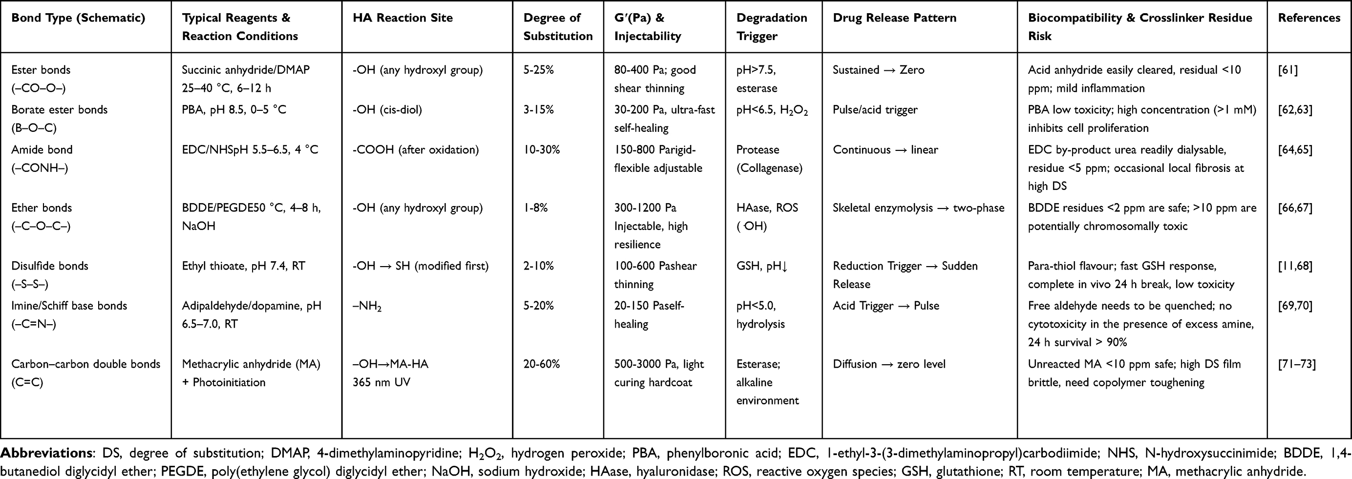 |
Table 1 Chemical Modification Strategies of Hyaluronic Acid |
Ester Bonds (–CO–O–)
HA can form ester bonds (–CO–O–) through esterification reactions with alcohols at either the carboxyl or hydroxyl group, with esterification of the carboxyl group being more prevalent. A wide range of alcohols can be used, including simple nontherapeutic ones like ethanol, propanol, and aromatic alcohols, as well as therapeutically active ones like steroids. Ester bonds significantly influence the physicochemical and biological characteristics of HA without changing its backbone, leading to numerous unique applications. For example, esterification can alter the polymer stability and water solubility of HA, as well as its cell adhesion capabilities. The water solubility of esterified HA is influenced by the specific type of alcohol involved as well as the degree of esterification. The most common ethyl and phenyl esters of HA are almost entirely insoluble in water, whereas some steroidal esters of HA are water-soluble. Esterified HA derivatives are generally biocompatible, biodegradable, and exhibit excellent processing properties. These characteristics have resulted in their extensive implementation in biomedical fields, including as drug carriers in various formulations, such as microspheres, films, and tablets, as well as in pharmaceutical dressings and implantable materials.
Many drugs are conjugated with HA via ester bonds to prepare targeted drug delivery systems. For example, HA–dexamethasone conjugates with a conjugation efficiency of over 98% have been successfully prepared for the specific delivery of dexamethasone to inflamed pulmonary tissues.61 These conjugates release active dexamethasone upon cleavage of the ester bond, thereby enhancing the effectiveness of current glucocorticoid therapies while minimizing associated adverse effects. Another research group developed a novel lubricating microneedle system to treat osteoarthritis based on a covalent conjugate formed by ester bonding between HA and the drug difluorochlorothiazide (Figure 2).74 The microneedle system consisted of a double-layered soluble microneedle where the inner layer contained the covalent conjugate and the outer layer comprised a self-adhesive lubricating copolymer. This design effectively reduced skin damage while providing sustained drug release through hydrolysis of the ester bond, physical diffusion, and breakthrough of the lubricating coating. Pang et al75 successfully prepared HA-quercetin (QT) conjugates by attaching the hydroxyl group of quercetin to hexanedihydrazide-modified HA via succinate. After a single tail vein injection of QT solution or HA-QT conjugated micelles at a dose equivalent to 8 mg/kg QT in rats, QT was detectable in plasma for up to 24 h with the HA-QT micelles, whereas free QT disappeared rapidly from circulation within one hour. This indicates that QT was continuously released from the HA-QT micelles over an extended period. The half-life of the drug was significantly increased from 0.17 h for free QT to 3.3 h for the HA-QT micelles. The mean residence time in plasma was also markedly prolonged to 4.3 h, which was 23.2 times longer than that of free QT. To simultaneously inhibit osteosarcoma recurrence and repair bone defects, Yu et al76 embedded curcumin chitosan nanoparticles (CCNPs) into a methacrylate-esterified HA/sericin hydrogel (CCNPs-SF/HAMA). The gel exhibited a flattened pH-responsive release profile over 32 days: at an acidic microenvironment (pH 5.5), the cumulative release was 77%, lower than that of bare CCNPs (92.6%), and at physiological pH 7.4, the release was only 55%, compared with 72.8% for the CCNPs alone. These results confirm that the HAMA network reduces the risk of systemic drug spikes while sustaining a low local dose in the tumor.
 |
Figure 2 Synthesis of drug compound hyaluronic acid (HA)-difluorochlorothiazide (DCF). HA-DCF is synthesized via the esterification reaction between HA-PEG-NH2 and DCF- N-hydroxysuccinimide (NHS). The first step involves reacting HA with NH2-PEG-NH2, poly(ethylene glycol) bis(amine), to form HA-PEG-NH2. The second step involves esterification of HA-PEG-NH2 with DCF-NHS—a compound derived from diclofenac (DCF, pink structure) and NHS—to synthesize HA-DCF. Adapted reprinted with permission from Ref.74 License Number: 6120810668796. Copyright © 2024 Wiley‐VCH GmbH. |
In conclusion, HA derivatives with ester bonds offer valuable and versatile functionality for clinical use. By modulating the ester bond structure, novel HA derivatives with specific functionality can be designed to meet different clinical needs. This demonstrates the broad application prospects of HA in regenerative medicine, drug delivery systems, and other biomedical applications.
Borate Ester Bonds (B–O–C)
In recent years, the borate ester bonds (B–O–C) formed between HA and boronic acid derivatives have attracted considerable attention because of their dynamic reversibility and high sensitivity to environmental factors, including pH levels, sugar molecules, and peroxides. Shi et al62 fabricated a dynamic self-healing hydrogel by grafting 3-aminomethylphenylboronic acid onto a HA backbone via borate ester bonding and combining it with polyvinyl alcohol. Owing to the sensitivity of the phenylboronic acid ester in the presence of biologically significant levels of H2O2, the hydrogel served as a proficient targeted drug delivery system that responded to H2O2 and reactive oxygen species (ROS). Furthermore, the hydrogel supported the survival of neural progenitor cells by protecting them from ROS-induced damage in the presence of H2O2. In a separate investigation, Liu et al63 synthesized an injectable cream-like hydrogel using epigallic acid and HA-based microspheres bonded to polyvinyl alcohol via dynamic borate bonding. The prepared formulation was designed to inhibit the formation of abdominal adhesions following surgical procedures. Notably, it exhibited excellent multifunctionality, with swift gelation, self-repair, antioxidant ability, anti-inflammatory properties, and inhibition of cellular adhesion. These studies highlight the promising applications of HA derivatives containing borate ester bonds in the progression of biosensors, drug delivery systems, and smart hydrogels.
Amide Bonds (–CONH–)
Amide bonds (–CONH–) can be introduced into HA through various chemical modification strategies. A common method is to react the activated carboxyl group of HA with amino groups to create stable amide linkages; for example, bipartite amino compounds such as hydrazides can be used as crosslinking agents to promote the intra- or intermolecular crosslinking of HA through amidation reactions. The free amino group generated by deacetylating the N-acetyl group (–NHCOCH3) of HA can also serve as an active site for amide bond formation. For example, it can react with activated carboxylic acids to produce amide derivatives or engage with the carboxyl group of HA to create self-crosslinked hydrogels. Deacetylation treatments, however, can degrade HA even under mild conditions;64 therefore, HA modification is generally not carried out using this method. Finally, carbon–carbon bonds containing cis-diol groups can be easily oxidized, leading to the formation of reactive aldehyde groups and amide bonds through Schiff base reactions (Figure 3A).77
 |
Figure 3 (A) Schematic diagram of AHAMA adhesive hydrogel binding to tissue and preparation process. Adapted reprinted with permission from Ref.77 based on CC BY License, Copyright © 2020 The Authors. Publishing services by Elsevier B.V. on behalf of KeAi Communications Co. Ltd. (B) Schematic of the Ce6-HA fabrication method. Adapted reprinted with permission from Ref.65 based on CC BY License, Copyright © 2024, © The Author(s) 2024. Published by Oxford University Press. |
The chemical modification of HA to form amide bonds can be used to adjust its hydrophilicity, stability, and functionality to meet specific application requirements. For example, Hong et al65 combined the photosensitizer chloride e6 with HA via amide bonding to form a photoresponsive hydrogel that produced ROS upon exposure to light, which in turn achieved an antibacterial effect (Figure 3B). Another research group synthesized biotinylated HA by grafting HA with adipic dihydrazide via amide bonding and then combining it with biotin.78 The prepared hydrogel was combined with sodium alginate and bioprinted to create a 3D hydrogel scaffold. The scaffold exhibited good biocompatibility and significantly increased the expression levels of genes associated with chondrogenesis.
The amidation of HA also improves its biocompatibility and biodegradability, increasing its safety and efficacy in clinical settings. For example, Nguyen et al79 extracted and purified HA from the eggs of Liparis tessellatus and grafted it with a variety of naturally occurring phenolic acids (eg, gallic, caffeic, and ferulic acids). They then grafted the antimicrobial peptide nisin onto HA via amide bonding to further enhance its biological activity. The experiments showed that all grafting reactions were successful. This study introduced a new avenue for the chemical modification of HA, establishing a basis for the creation of polymers characterized by extended in vivo retention times and stronger bioactivity. Another research group developed a novel nanotherapeutic diagnostic agent by conjugating the photosensitizer IR808 to HA via amide bonding and loading then complex onto the surfaces of single-walled carbon nanotubes.80 This agent generated ROS upon light activation, enabling phototherapeutic effects. Additionally, the fluorescence of IR808—initially quenched by the composite structure—was restored through the action of endogenous enzymes, facilitating the accurate detection of residual tumor cells during subsequent treatments. This method presents a promising strategy for tailored cancer treatment and demonstrates the capabilities of HA-based nanotechnology in oncological applications.
HA derivatives with amide bonds also demonstrate significant potential in the field of drug delivery systems. The bioavailability and targeting ability of drugs can be improved by linking them to the carboxyl groups of HA by amide bonding. Hou and colleagues81 developed a novel drug delivery system that utilizes HA-modified porous silica nanocarriers. The porous silica nanocarrier was embedded with Ag2S quantum dots to enhance the photothermal effect, and its surface contained a sensitive linker to enable it to be loaded with the anticancer drug doxorubicin. Finally, HA was covalently linked to the amino-functionalized carrier via amide bonding to achieve confinement and targeted drug release. Thus, this platform exhibited a photothermal chemotherapeutic effect and controlled drug release, highlighting its potential for combined oncology treatment regimens. Another group synthesized hydrogel coatings with redox-responsive properties from catechol HA and cystamine via amide bonding.82 This coating was capable of intelligently releasing drugs and hydrogen sulfide into microenvironments exhibiting inflammation and oxidative stress, while also offering better biocompatibility and drug release responsiveness compared to conventional drug coatings. The coating demonstrated not only remarkable hemocompatibility and anti-inflammatory properties but also facilitated the regeneration of endothelial cells while hindering the proliferation of smooth muscle cells and macrophages. This effectively reduced restenosis following stent placement and ensured the efficacy and safety of the interventional device.
Overall, the amide bonding of HA is of great significance for biomedical applications, as it not only enhances the mechanical robustness and stability of HA, but also broadens its range of applications, providing new possibilities for its use in the fields of drug delivery, tissue engineering, and biomaterial research. With further research, the amidation of HA is expected to hold considerable importance in the advancement of biomedicine.
Ether Bonds (–C–O–C–)
Ether bonds (–C–O–C–) constitute one of the pioneering chemical crosslinking techniques used in the modification of HA. The crosslinking of HA was first reported in 1964 when Laurent et al66 employed 1,2,3,4-diepoxybutane as a crosslinking agent in a strongly alkaline environment (pH 13–14). Several commonly used crosslinking agents, including BDDE, 1,2,7,8-diepoxyoctane, and glutaraldehyde, operate by generating ether bonds. Specifically, the epoxide groups (–CH(O)CH2) of BDDE and 1,2,7,8-diepoxyoctane undergo nucleophilic ring-opening reactions with the hydroxyl moieties of HA to create stable ether bonds. Meanwhile, glutaraldehyde can form Schiff bases (imines) with amines and hemiacetal/acetal bonds with hydroxyl groups under acid catalysis. Although both acetal and ether bonds contain C-O-C units, their properties differ significantly. Ether bonds are almost irreversible at physiological pH and require strong acids or free radicals to break, making them suitable for long-term filling applications. In contrast, acetal bonds contain ketodiethyl carbon, which is highly susceptible to acid catalysis owing to electronic effects, allowing HA scaffolds to degraded on-demand in mildly acidic environments (eg, tumors, intracellular lysosomes, and wounds) without cytotoxicity. Imine bonds, meanwhile, can reversibly break and reorganize near physiological pH, endowing hydrogels characteristics of shear-thinning and the capacity for rapid self-healing. Compared with “static” bonds such as ether and amide, imine bonds are sensitive to water and slowly hydrolyze under neutral conditions, decreasing crosslink density over time. The overall biocompatibility of the HA hydrogels is good, but potential aldehyde toxicity must be managed using strategies such as oxidation control, covalent co-cross-linking, and catalyst/coordination stabilization in practical applications.
Ether linkages exhibit a marginally higher degree of chemical stability compared to amide linkages, and both are relatively resistant to hydrolysis under physiological conditions. Ether-crosslinked HA gels are long-lasting, but their suitability for drug delivery depends on overall network properties, including crosslinking density, degradable co-crosslinkers, enzymatic/oxidative degradation, and hydrophilicity, rather than solely on bond type. Common degradation pathways of ether-crosslinked HA gels include: enzymatic cleavage, oxidative degradation, and hydrolysis. (1) Enzymatic cleavage: HA glycosidic bonds (β-1,3 vs β-1,4) are recognized by enzymes such as hyaluronidase and macrophage-secreted β-D-glucosidase. Degradation accelerates as crosslink density decreases and the backbone becomes exposed. (2) Oxidative degradation: high levels of ROS (H2O2, -OH) at lesion sites cleave both the HA backbone and cross-linker C-C/ether bonds, producing low-molecular-weight fragments.67 (3) Hydrolysis: normal ether bonds are very stable under physiological conditions of pH 7.4 and a temperature of 37 °C, with half-lives up to several years.83 To improve drug delivery efficiency, introducing cleavable motifs into ether cross-linking matrices has emerged as an effective strategy. Cleavable ether bonds, such as borate esters, break under mildly acidic or high-ROS conditions, reducing the half-life from years to tens of hours. By incorporating these motifs into reactions between HA hydroxyl groups and bis-epoxy crosslinkers, a series of stimuli-responsive gels can be obtained. For instance, boronate-HA/ polyvinyl alcohol gels released 80% of interleukin (IL)-10 in 0.5 mM H2O2 arthritic synovial fluid over 24 h, significantly suppressing local inflammation84 and offering a path toward precision drug delivery with ether-crosslinked HA hydrogels. In addition, the moderate polarity of ether bonds allows the crosslinked HA to remain hydrophilic, which helps to maintain the high water-absorption capacity of HA. This is beneficial for dermal fillers, as it enhances the filling effect. Simultaneously, ether crosslinking significantly improves the mechanical characteristics of HA hydrogels, resulting in increased elasticity and durability. Finally, ether bonds are less likely to trigger immune reactions or cytotoxicity, making them safe and suitable for long-term implantation and other biomedical uses.
Within the field of aesthetic medicine, hyaluronic acid is extensively utilized as a dermal filler owing to its excellent physicochemical properties and remarkable filling effect. Endogenous HA exhibits a brief half-life within humans, primarily because of the action of hyaluronidase; therefore, commercial HA fillers are often chemically crosslinked to enhance their anti-enzymatic ability, thus extending their longevity in the body. Ether crosslinking is widely used because of its simplicity and low reaction temperature. However, the cross-linking reaction is often incomplete, and unreacted cross-linkers may remain in the final product. These residues possess cytotoxic and sensitizing potential and must therefore be strictly controlled. According to prevailing industry standards and regulatory requirements, the acceptable upper limit of BDDE residue is 2 ppm (ie, 0.002 mg/mL). There is no uniform standard for polyethylene glycol diglycidyl ether (PEGDE), and the industry generally refers to the BDDE residue limit, which is a safety threshold established based on toxicological evaluations and animal experimental data. To achieve this standard, manufacturers typically implement a multi-step dialysis or buffer-cleaning process after the cross-linking reaction to remove free and single-ended BDDE/PEGDE molecules.85 In response to these issues, Choi et al86 developed a new dispersion process to enhance the quality of BDDE-crosslinked HA fillers. In this method, the solution is dispersed at low temperature, allowing the solvent to naturally penetrate the solute after mixing, forming a homogeneous mixture. This approach significantly improves the viscoelasticity and cohesion of the fillers in both laboratory and industrial settings, addressing the poor homogeneity and inefficient cross-linking observed in traditional BDDE cross-linking processes. Despite the generally favorable safety profile of cross-linked HA fillers, local inflammatory reactions with delayed granuloma formation are recognized potential complications. These adverse reactions are typically delayed hypersensitivity responses, appearing weeks to months after injection as redness, swelling, hardness, or nodules.87 The precise mechanisms remain inadequately elucidated; however, they may encompass the following elements: (1) activation of T-cell-mediated immune responses by residual cross-linking agents or their degradation products acting as semi antigens; (2) degradation of HA fragments triggering macrophage aggregation and granulation tissue formation; and (3) improper injection techniques leading to product aggregation and the formation of foreign body reaction foci. Kim et al88 reported in vivo that mice injected with 0.2 or 0.3 mL of HA-PEGDE filler exhibited elevated tumor necrosis factor (TNF)-α and IL-1β expression, as well as cyclooxygenase (COX)-2 protein expression, at both 1 and 4 weeks post-injection. In contrast, the HA-PEGDE filler resulted in increased mRNA expression of inflammatory cytokines solely during the first week following subcutaneous administration, with no notable differences detected at the four-week mark. This indicates that HA-PEGDE exhibits a more favorable profile than HA-BDDE concerning inflammation. Regarding cytotoxicity, Jeong et al89 investigated the impact of varying concentrations of BDDE and PEGDE (0–1000 ppm) on keratinocytes and fibroblasts within a cell culture environment. Neither cross-linker exhibited cytotoxicity at low concentrations (0–25 ppm), whereas BDDE showed cytotoxic effects at higher concentrations (50–1000 ppm). They also observed that cells exposed to HA-BDDE filler had more dead cells than those exposed to HA-PEGDE filler at equivalent concentrations. Another study reported that BDDE exhibited significant cytotoxicity above 100 ppm, whereas PEGDE maintained a safety threshold of 500 ppm, suggesting superior biocompatibility at the same residue level.90 Therefore, another group of researchers compared the toxicity and biocompatibility of HA fillers with different crosslinking agents (BDDE and PEGDE).88 They found that PEGDE-crosslinked HA had better physical properties, lower cytotoxicity, and a reduced inflammatory response.
Biomaterials based on ether-crosslinked HA are also widely used for tissue repair. For example, interpenetrating polymer networks of N-isopropylacrylamide and glutaraldehyde-crosslinked HA exhibit thermoresponsive self-shrinkage behavior and tissue adhesion properties that accelerate wound healing (Figure 4A).91 Another group of researchers synthesized a similar hydrogel intended for application as a cellular carrier, with the objective of facilitating the repair of the nucleus pulposus within intervertebral discs.92 These studies demonstrate the biocompatibility and tissue-regeneration ability of crosslinked HA hydrogels.
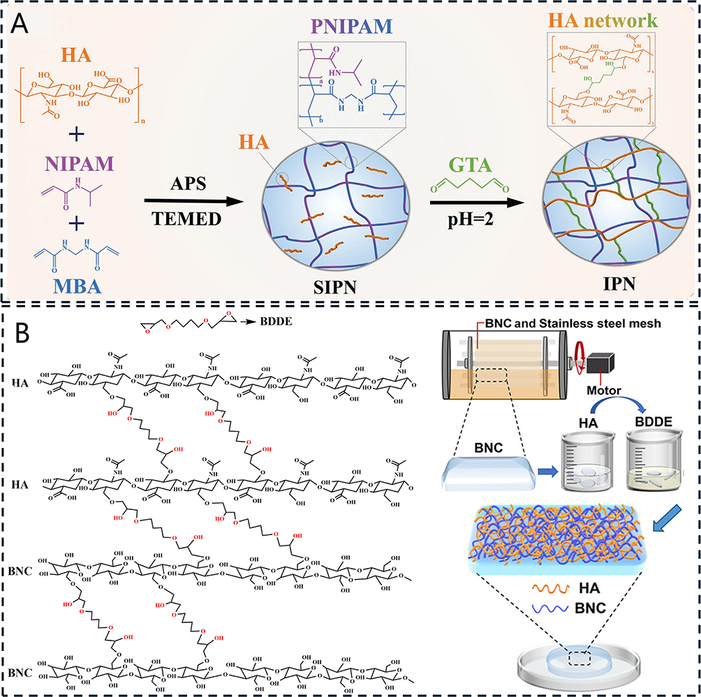 |
Figure 4 (A) Schematic synthesis of poly(N-isopropylacrylamide) (PNI)- hyaluronic acid (HA) hydrogel and its characterization. Adapted reprinted with permission from Ref.91 License Number: 6120870839490. Copyright © 2023 Wiley‐VCH GmbH. (B) Schematic diagram of the cross-linking reactions in the bacterial nanocellulose (BNC)/HA composite membrane. HA and HA acid, HA and BNC, as well as BNC and BNC, are all crosslinked via ether bonds generated by BDDE (1,4-butanediol diglycidyl ether). The red groups represent the BDDE-crosslinked structures. Adapted reprinted with permission from Ref.93 License Number: 6121400696720. Copyright © 2023, American Chemical Society. |
Ether-crosslinked HA has also been studied for use in ophthalmology. Synthetic corneas made from biogenic materials, such as human amniotic membranes, decellularized porcine corneal stroma, and collagen, suffer from poor transparency, low mechanical strength, and susceptibility to tearing. Artificial corneal materials, such as Boston keratoprosthesis and osteo-odonto-keratoprosthesis, also exhibit certain limitations. Based on this, Luo et al93 prepared a novel artificial corneal material by crosslinking HA with bionanocellulose using BDDE (Figure 4B). The composite exhibited favorable cytocompatibility, outstanding optical characteristics, resistance to sutures, and excellent moisture retention, making it a promising candidate for artificial corneal transplantation or the repair of the ocular surface.
Lai et al94 presented a pioneering method for the real-time quantitative synthesis and analysis of ester-crosslinked HA hydrogels. This approach utilized a microfluidic system in conjunction with electrospray-differential mobility analysis. By regulating the synthesis parameters in the microfluidic system, such as the pH, temperature, duration of the reaction, and molar ratio of HA to BDDE, HA hydrogels exhibiting customized particle dimensions and characteristics were effectively produced. Notably, there was a significant relationship between the synthesis conditions and particle size distribution. This research demonstrated the capabilities of microfluidic platforms for the efficient generation of homogeneous and precisely characterized hydrogel nanoparticles, thereby facilitating their utilization in tissue engineering and biomaterials.
Ether-crosslinked HA hydrogels have numerous applications across various domains including dermal fillers and soft tissue repair. Their high chemical stability, good hydrophilicity, strong mechanical properties, and low toxicity render these materials highly suitable for applications in the domains of biomedicine and tissue engineering.
Disulfide Bonds (–S–S–)
Natural HA does not contain disulfide bonds (–S–S–), but disulfide linkages can be introduced through reactions between disulfide-containing compounds and reactive groups on HA, such as hydroxyl groups, or amino groups introduced via amino modification. Two main methods are used. The first introduces an amino (–NH2) group via chemical modification, which then reacts with disulfide crosslinking agents to introduce disulfide linkages (Figure 5).95 The second uses ethyl thioesters or other sulfide reagents to introduce sulfhydryl (–SH) groups to form thiolated HA. Sulfhydryl groups can form disulfide bonds with other sulfhydryl groups in oxidizing environments. In this manner, thiolated HA can be used to form biocompatible self-crosslinked hydrogels without the use of crosslinking agents.11 Owing to the reducing nature of biological compounds such as glutathione and nicotinamide adenine dinucleotide phosphate, disulfide linkages are reduced back to sulfhydryl groups within the cellular environment. This reversible crosslinking behavior makes HA with disulfide bonds suitable for the synthesis of smart responsive hydrogels.12
 |
Figure 5 Design of doxorubicin-loaded multifunctional HA derivative-modified mesoporous silica nanoparticles (DOX/HHS-MSNs) for active targeting, endo-lysosomal escape, and multilevel drug release to reverse cancer multidrug resistance. Adapted reprinted with permission from Ref.95 License Number: 6121401266427. Copyright © 2016, American Chemical Society. |
In tissue engineering, disulfide-crosslinked HA is commonly used to create biological scaffolds with remarkable biocompatibility and robust mechanical properties. These scaffolds facilitate not only the attachment, growth, multiplication, and specialization of cells but also are essential in maintaining the cell’s phenotype. In one study, disulfide-crosslinked HA was compounded with gelatin to prepare composite hydrogels that effectively supported the growth of chondrocytes and the repair of cartilage tissue.96 Disulfide-crosslinked HA has also been used to enhance the resistance of scaffolds to degradation and prolong their retention in the body, thereby improving the effectiveness of tissue repair.
Disulfide-crosslinked HA also has promising applications in drug delivery. Hydrogels or nanocarriers prepared with disulfide-crosslinked HA offer effective loading and slow-release of anticancer drugs.97 In one study, researchers embedded the anticancer drug sulforaphane in a disulfide-crosslinked HA hydrogel and found that drug release within the tumor microenvironment via reduction reactions effectively inhibited the stem-cell-like characteristics associated with breast cancer.98 Zhang et al68 designed a nanogel composed of lactoferrin and phenylboronic acid that exhibited targeted drug release capabilities. It swiftly released encapsulated doxorubicin in environments with elevated glutathione levels, making it an excellent candidate for effective glioma-targeted therapy.
Researchers have also examined the application of disulfide-crosslinked HA hydrogels for wound repair owing to their biocompatibility and self-healing ability. These hydrogels can form a protective film over wounds, thereby preventing infection and facilitating the process of wound recovery. For example, Yang et al99 engineered a disulfide-crosslinked HA hydrogel with antimicrobial properties that effectively inhibited bacterial growth and demonstrated good self-healing ability during wound healing. The modifiable nature of these hydrogels renders them ideal for a range of applications in wound healing.
In summary, disulfide-crosslinked HA exhibits significant potential for applications within the field of biomedicine, especially in tissue engineering, drug delivery, and wound repair. It has already shown good research progress and application potential. With further research and technological development, disulfide-crosslinked HA is anticipated to have a considerable impact in these domains.
Imine/Schiff Base Bonds (–C=N–)
Imines, also referred to as Schiff bases, are characterized by the functional group –C=N–. They are typically generated through the condensation reaction involving either an aldehyde group (–CHO) or a ketone group (–CO–) in conjunction with an amine group (–NH2). Natural HA can undergo chemical modification to establish imine bonds by first introducing the necessary functional groups, followed by a condensation reaction. Three main strategies are used: (1) condensation of aldehyde-modified HA with amine-containing compounds in a weakly acidic to neutral environment (pH 4–7); (2) condensation of amino-modified HA with aldehyde-modified polymers, proteins, or small molecules (eg, glucuronide aldehydes or oxidized chitosan); and (3) condensation of aldehyde-modified hyaluronate with hydrazine groups (–NHNH2) to form an acylhydrazone bond (–C=N–NH–C(=O)–); a more stable derivative of the imine bond). Imine bonds are relatively stable in neutral or alkaline solutions (pH > 7), with a half-life of 6–12 h. Under acidic conditions (pH < 5), however, they are prone to hydrolysis, with a shortened half-life of 1–3 h, leading to degradation of the crosslinked material. Notably, this property imparts pH-responsiveness to imine-crosslinked HA (Figure 6A).100,101 In addition, imine-crosslinked HA hydrogels usually exhibit self-healing behavior, because the imine bonds can be dynamically broken and reorganized in aqueous solution.69 In comparison, acylhydrazone bonds are more stable under physiological conditions. Their stronger conjugated systems render them more resistant to hydrolysis, with a half-life of 2–5 days. This stability can be further enhanced to 1–3 weeks if the material is additionally mixed and crosslinked with BDDE, making acylhydrazone bonds more suitable for long-lasting crosslinked hydrogels (Figure 6B).102,103
 |
Figure 6 (A) Schematic illustration of HA/gelatin methacryloyl/sodium alginate–arginine carbon dots/stromal cell-derived factor-1α (HG-AA-SDF-1α) composite hydrogel, which enhances “cou-pling osteogenesis and angiogenesis” for the promotion of bone regeneration. Adapted reprinted with permission from Ref.100 based on CC BY License, Copyright © 2025 The Author(s). Advanced Science published by Wiley‐VCH GmbH. (B) Schematic illustration of pH-activatable oxidative stress amplifying dissolving microneedles for chemo-photodynamic therapy of melanoma. Adapted reprinted with permission from Ref.102 based on CC BY License, Copyright © 2022 Shenyang Pharmaceutical University. Published by Elsevier B.V. (C) Synthetic scheme of HA hydrogel modified by aldehyde groups and methacrylate (AHAMA) and its partial characterization. AHA is synthesized by reacting hyaluronic acid with sodium periodate. The blue structure indicates the highly reactive aldehyde group introduced at the hyaluronic acid chain breakpoint. Subsequently, methacrylate (red structure) is added to AHA and reacted for 12 hours at pH 8–8.5 to synthesize AHAMA. Adapted reprinted with permission from Ref.77 based on CC BY License, Copyright © 2020 The Authors. Publishing services by Elsevier B.V. on behalf of KeAi Communications Co. Ltd. |
Imine-bonded HA finds extensive application within biomedicine. In tissue engineering, the modification of HA with imine bonds provides it with superior physicochemical properties for the construction of biocompatible scaffolds. For example, Chen et al77 formulated a modified adhesive HA hydrogel incorporating aldehyde groups and methacrylate, which demonstrated improved adhesion and stability (Figure 6C). These properties were attributed to its diverse anchoring mechanisms, including amide bonding, hydrogen bonding, and physical intercalation facilitated by dynamic Schiff base reactions. The hydrogel was durable and stable for at least seven days, even in humid environments, with adhesion strengths of 43 and 52 kPa for skin and glass, respectively. Notably, these values far exceed those of commercially available fibrin glue (~10 kPa) and HAMA hydrogels (~20 kPa).
The imine modification of HA also improves its ability to respond to acidic environments and regulate cellular behaviors. Xiao et al100 conducted a study focusing on a bifunctional platform that is activated by acidic conditions. In this platform, they incorporated C-X-C (Cys-Xaa-Cys) motif chemokine ligand 12 (CXCL12), which is also known as stromal cell-derived factor (SDF-1α), alongside arginine carbon dots and calcium ions, all embedded within an oxidized hydrogel composed of HA and gelatin methacryloyl (Figure 6A). In an acidic environment, the Schiff base bonds in the hydrogel were broken, sustaining the release of CXCL12 and enhancing the movement and enlistment of intrinsic mesenchymal stem cells. In addition, the recruited cells metabolized the arginine carbon dots, resulting in the production of nitric oxide in the presence of calcium ions. This process activated the cyclic guanosine monophosphate signaling pathway, subsequently fostering angiogenesis. This composite hydrogel demonstrated good potential for coupling osteogenesis and angiogenesis and provided an effective strategy for bone regeneration.
Imine-bonded HA has also demonstrated excellent performance in drug delivery systems. HA-based carriers can link or encapsulate multiple drugs through imine bonds and achieve slow drug release in vivo, significantly improving bioavailability. For example, researchers constructed a copper–doxorubicin–anlotinib nanoconjugate bound via copper-hydrazide coordination, hydrazone linkages, and Schiff base bonds.104 The release of doxorubicin from this complex significantly enhanced copper-mediated chemodynamic therapy, whereas anlotinib effectively inhibited copper ion-induced tumor angiogenesis. This multifunctional platform enabled the multidimensional treatment of hepatocellular carcinoma with targeted synergistic chemotherapy, chemokinetic therapy, and antiangiogenesis.
Hydrogels with dynamic Schiff base crosslinks have significantly higher stability and structural integrity when subjected to external forces. For instance, Wu et al70 prepared a self-assembled herbal polysaccharide hydrogel from glycyrrhizic acid (a natural compound derived from licorice root) and a HA derivative via dynamic Schiff base crosslinking, and incorporated deferoxamine as a functional component to promote wound healing. Dynamic crosslinking significantly enhanced the stability of the hydrogel, making it less prone to disintegration under physiological conditions. Furthermore, it enabled the efficient delivery of deferoxamine to the trauma site to promote angiogenesis.
In summary, imine-bonded HA has a broad spectrum of potential uses within the domains of tissue engineering and drug delivery systems. Future studies should explore the mechanism of HA modification using imine bonds and specific applications of these materials in different biomedical fields to promote their clinical translation and use.
Carbon–Carbon Double Bonds (C=C)
HA that has been modified to incorporate carbon–carbon double bonds (C=C) presents numerous prospective applications within the biomedical sector. The main methods of C=C bond formation in HA include esterification, acrylation, and grafting via click chemistry. A prevalent technique employed in this context is methacrylation, which facilitates the formation of HAMA. In this process, methacrylic anhydride interacts with the hydroxyl groups present in HA to introduce methacryloyl groups (–CH2=C(CH3)CO–). HAMA is relatively stable under physiological conditions, but may be hydrolyzed in alkaline environments or by esterases. A similar strategy involves acrylation, whereby acryloyl chloride or acrylic acid interacts with the hydroxyl groups of HA or amino groups found in amino-modified HA when subjected to alkaline conditions, resulting in the formation of HA acrylate.105 Derivatives of HA that contain either acryloyl or methacryloyl groups can undergo crosslinking using 365 nm UV light, visible light, or free radical initiation to create biocompatible hydrogels (Figure 7).71,72 Taking advantage of this, Wang et al106 conjugated HAMA with 1,4-dihydrobenzothiazol-4-one-3-carboxylic acid via disulfide bonding to develop sequential bifunctional supramolecular hydrogels aimed at ROS scavenging and stabilizing hypoxia-inducible factor 1 subunit alpha (HIF1A), targeting the therapeutic management of myocardial infarction.
 |
Figure 7 Schematic diagram of the preparation of methacrylated hyaluronic acid (HAMA)/tetrahedral framework nucleic acid (tFNA)-GL13K hydrogel. Evaluation of HAMA/tFNA-GL13K hydrogel in vivo for skin repair and wound healing in general observation. Adapted reprinted with permission from Ref.71 based on CC BY License, Copyright © 2024, The Author(s). |
Another key method of introducing C=C bonds to HA is fumaric acid modification. In this approach, HA undergoes esterification or acrylation with a fumaric ester to introduce a conjugated C=C bond. When this double bond is conjugated to a carbonyl group (eg, fumaric acid or an acryloyl group), further modification with sulfhydryl or amine groups is possible via Michael addition. In addition, modified HA bearing fumarate groups as dienophiles (2π electron acceptors) can undergo Diels–Alder reactions with dienes (4π electron donors) to form reversible C=C bonds, enabling dynamic crosslinking in HA-based hydrogels. Finally, the use of click chemistry to introduce C=C bonds typically involves a reaction between azide-modified HA (HA-N3) and alkynyl (–C≡C)-containing compounds (eg, propargyl esters). This reaction forms a conjugated C=C bond, which can be used for subsequent functionalization or polymerization.
HA with C=C bonds shows considerable potential for the construction of drug delivery systems. The C=C bonds facilitate chemical modification and crosslinking, which are essential for the regulated release of pharmaceuticals. As an example, Qi et al107 combined HAMA with polyvinyl alcohol to form a 3D-crosslinked hydrogel using dynamic borate ester bonding and photocrosslinking techniques. The dual network structure not only improved the mechanical characteristics of the hydrogel but also facilitated the localized activation of platelet-rich plasma along with a prolonged release of growth factors. Therefore, it effectively promoted the regeneration of the endometrium and reinstated uterine functionality. In addition, photocrosslinking technologies can be employed to fabricate hydrogels based on HA with tunable network structures, enabling dynamic and sustained drug release in vivo. This method improves the bioavailability of pharmaceuticals while enabling precise drug release under particular physiological conditions.
In the domain of tissue engineering, chemically modifying HA with C=C bonds can significantly improve its mechanical characteristics and biocompatibility, rendering it more appropriate for use as a scaffold material. For example, hydrogel particles combining C=C bond-modified HA and hyperbranched poly(acrylate-capped thioketone-containing ethylene glycol) scavenge ROS and neutralize pro-inflammatory cytokines. The introduction of C=C bonds enhances the mechanical strength of HA hydrogels, making them more stable under physiological conditions.108
Although a variety of C=C bond-modified HA derivatives have been developed, the direct incorporation of C=C bonds into the HA backbone has been less studied. Buffa et al73 synthesized a novel HA derivative, 4,5-anhydro-N-acetylglucosamine hyaluronan (∆HA), which contains a double bond between the 4 and 5 positions of the N-acetylglucosamine ring. ∆HA can react with a wide range of oxidizing agents, resulting in higher chemical reactivity and biological activity. In addition, ∆HA is selectively cytotoxic to a wide range of cancer cell lines without significant effects on normal human dermal fibroblasts, which signifies its promise as a potential anticancer agent.
C=C-bond modified HA shows good prospects for application in pharmaceutical distribution and biological tissue engineering. In the future, as research in this field continues to deepen, further HA-based therapeutic solutions are expected to emerge to meet clinical demands for efficient and safe treatment.
Biomedical Applications of HA
HA has been used in a wide variety of clinical disciplines due to its unique physicochemical properties, ie, high water binding capacity, viscoelasticity, biocompatibility, and biodegradability. These areas include, but are not limited to, ophthalmic surgery, osteoarthritis treatment, aesthetic and orthopedic medicine, and advanced drug delivery systems. In the following sections, each therapeutic area will be reviewed (Table 2).
 |
Table 2 Application of Hyaluronic Acid |
Application of HA in Ophthalmology
Dry Eye Disease
Dry eye disease, referred to as keratoconjunctivitis sicca, represents a prevalent eye disorder marked by an atypical composition of the tear film and inflammation affecting the ocular surface. Patients often experience foreign body sensation, irritation, pain, and blurred vision. Studies have linked the onset of dry eye disease to factors such as being female, being over 50 years of age, connective tissue disease, contact lens use, certain medications (eg, diuretics and antihistamines), and hematopoietic stem cell transplantation.142
HA has been extensively investigated for its therapeutic potential in managing dry eye disease. As a fundamental component of the tear film, HA contributes to ocular surface hydration and protection. However, when administered in therapeutic formulations, HA is rapidly cleared and degraded in the body. Therefore, researchers have explored various approaches to ensure the long-term effectiveness of HA formulations. The main strategies involve continuous replenishment (“open source”) and metabolic regulation or degradation resistance (“throttling”).
“Open source” methods aim to counteract the rapid clearance of HA by continuously replenishing it. Long-term contact lens wearers are more prone to dry eye symptoms; however, the frequent use of eye drops can be inconvenient and may lead to poor treatment compliance. To address this problem, researchers embedded the metabolically engineered soil bacteria Corynebacterium glutamicum as a biofactory in a hydrogel-based contact lens (Figure 8A).109 The bacteria effectively regulated the rate of HA release by modulating the hydrogel’s composition and degree of crosslinking, thereby sustaining therapeutic effects for at least 3 weeks. In vivo living contact lenses are still in the early stages of transition from laboratory proof-of-concept to preclinical testing in animals and have not yet entered Phase I or any human clinical trials. The authors who proposed the concept emphasized that safety and comfort validation must be completed before subsequent clinical translation can be considered. Although in vitro cytotoxicity tests did not reveal significant issues, data from corneal epithelial models, in vivo animal studies, and immunologically abnormal populations remain lacking. Currently, bacteria are embedded only in a 1-mm annulus around the periphery of the lens, using a polyvinyl alcohol–vinyl sulfone (PVA-VS) secondary crosslinked network with a nominal aperture size of 10–30 nm, whereas the bacterial diameter is approximately 0.8 µm, theoretically preventing their passage. However, the authors did not provide quantitative data on bacterial “leakage.” In addition, no temperature-controlled, pH-induced, or antibiotic-dependent suicide genes were introduced, and the lenses were disposed of solely by rinsing with a standard care solution, with no mandatory sterilization step. From a regulatory perspective, product definition, quality control standards, and clinical endpoints are all treated as “off-the-shelf” and require a “case-by-case” De Novo/Live Biotherapeutic Product (LBP) pathway globally. Consequently, the approval timeline, costs, and uncertainty are substantially higher than those for conventional Class III medical devices. Therefore, despite the remarkable technology of living contact lenses, long-term ocular surface safety data must be established before these devices can realistically enter clinical trials.
 |
Figure 8 (A) Schematic of the fabrication process of the bacterial ring-implanted contact lens (CL) and its characterization. Research Team fabricated laboratory CL prototypes in a two-step molding process: ① Addition of PVA-VS/PVA bacterial solution Lens functional ring fabrication ② Crosslinking ③ Ring transfer and addition of PVA-VS transparent solution Lens field of view fabrication ④ Moulding and crosslinking ⑤ Contact Lens. Adapted reprinted with permission from Ref.109 based on CC BY License, Copyright © 2024 The Authors. Advanced Materials published by Wiley‐VCH GmbH. (B) Chemical structure and morphology of monoblock bottlebrush B and triblock bottlebrush ABA. Two BB polymers (Bottlebrush polymers are a type of biomimetic polymer whose structure draws inspiration from lubricin, a key protein component in synovial fluid. Featuring a bottlebrush-like molecular configuration, they effectively mitigate friction damage and demonstrate excellent lubricating properties in both in vitro and in vivo experiments) were designed and used with or without HA in different settings. The chemical structures (left) and morphology (right) of the monoblock (B) and triblock (ABA) polymers are presented. The synthesis of ABA involves attaching an A group (blue group, no corresponding scientific name) to the B base. It is synthesized from a B-type block copolymer by adding CuCl2, 4,4′Dinonyl-2,2′-Dipyridyl, 2-(dimethylamino)ethyl methacrylate, methyl methacrylate, and CuCl. Adapted reprinted with permission from Ref.143 based on CC BY License, Copyright © 2024 The Authors. Advanced Materials published by Wiley‐VCH GmbH. |
“Throttling” methods aim to reduce the clearance of HA by increasing its degradation resistance or reducing the activity of degrading enzymes. In the human body, the metabolism and degradation of HA are primarily due to the action of hyaluronidase, although metalloproteinases also contribute indirectly. Therefore, increasing the tolerance of HA to hyaluronidase or inhibiting metalloproteinases can effectively reduce its clearance and prolong its residence time in tissues. Galassi et al110 successfully synthesized a new material, HA-3, with higher resistance to hyaluronidase degradation by covalently functionalizing HA with TIMP metallopeptidase inhibitor 3 (TIMP3, previously referred to as metalloproteinase inhibitor 3 [MMPI]). HA-3 not only maintained moisture on the surface of the cornea but also inhibited the overexpression of matrix metallopeptidase (MMP-9), which has been associated with dry eye disease, thereby protecting the integrity of the corneal epithelium. In addition, the lubricating properties and biocompatibility of polymers can be enhanced by blending with HA, thereby doubling the effectiveness of dry eye disease treatments. For example, researchers have constructed triblock (ABA) bottlebrush polymers that rapidly adhere to multiple surfaces, including cartilage, the ocular surface, and contact lenses, thereby establishing a robust and biocompatible lubricating protective layer (Figure 8B).143
Ophthalmic Surgery
HA is increasingly used in cataract surgery to safeguard the corneal endothelium, improve surgical safety, and minimize the inflammatory response. HA-based eye drops can significantly improve ocular surface health after surgery. For example, in a randomized controlled trial, the Ocular Surface Disease Index (OSDI) scores of patients treated with 0.15% HA-based eye drops after cataract surgery were significantly lower than those of patients treated with 0.1% HA-based eye drops, suggesting that formulations with higher HA contents are more effective in relieving postoperative dry eye symptoms.111 In addition, the biocompatibility and slow-release properties of HA have facilitated the synthesis of various drug-loaded biomaterials to prevent or treat post-cataract surgical complications. For example, Bao et al112 prepared hydrogel films from mannose chitosan and oxidized HA and loaded them with the corticosteroid dexamethasone and antibiotic levofloxacin. These hydrogel films exhibited phased drug release, with a rapid release of levofloxacin and prolonged release of dexamethasone. This dual-drug delivery mechanism is a promising therapeutic option for postoperative endophthalmitis. Thus, HA has become an integral part of modern cataract surgery as an effective ophthalmic surgical aid.
HA has also shown importance in vitrectomy. HA mimics the biomechanical properties and functionality of the vitreous humor, leading to its widespread use as a vitreous substitute to maintain structural integrity and function within the eye. Crosslinked HA maintains postoperative intraocular pressure and promotes retinal recovery. For example, Chen et al113 developed BDDE-crosslinked HA that showed good handling, a longer degradation time, and effective drug release capabilities in vitro, effectively removing intraoperative residual trifluorochloroethylene and maintaining normal intraocular pressure during the postoperative period.113 The anti-inflammatory properties of HA also help to reduce postoperative inflammation, thereby improving recovery.11 The crosslinked HA prepared by Chen et al113 was loaded with the anti-inflammatory agent epigallocatechin gallate, further improving the anti-inflammatory effect.
Contact Lenses
Contact lenses are an increasingly popular method of vision correction; however, many wearers experience issues such as dry eye symptoms and discomfort. HA is widely used in contact lens care products because of its excellent lubricating and moisturizing properties. In addition, HA can be used as an additive in contact lenses themselves, significantly improving their moisture retention and comfort.144 For example, HA-coated contact lenses reduce ocular discomfort by enhancing moisturizing properties, wettability, and mechanical performance.114,145 They also improve lens safety by effectively reducing protein adsorption and providing antimicrobial activity against major ocular pathogens.114 Further studies revealed that HA contains approximately 4–6 hydrogen bonding donor/acceptor sites per disaccharide unit, which bind 20–30 water molecules to form a highly viscoelastic hydration shell 2–4 nm thick.146 This hydrated layer allows tear proteins (eg, lysozyme, albumin) to overcome additional resistance, thus reducing protein precipitation.147 Additionally, HA, as a negatively charged polysaccharide, can repel negatively charged bacterial surface proteins (eg, Staphylococcus aureus surface proteins) through electrostatic interactions, forming a physical barrier that inhibits bacterial adhesion to cells or material surfaces. Regarding antimicrobial activity, healthy skin and mucous membranes serve as the first line of defense against infection. HA promotes corneal epithelial wound healing by stimulating epithelial migration, adhesion, and proliferation, enhancing extracellular matrix remodeling, and activating CD44.148 An intact barrier naturally improves resistance to bacterial invasion. Furthermore, Ruppert et al149 demonstrated that HA reduces pathogenic bacterial adherence and infection by modulating the expression of lipocalin 2 (LCN2), an antimicrobial peptide component of epithelial cells, and IL-8, a pro-inflammatory factor, via regulation of the NF-κB signaling pathway, a key component of the intrinsic immune response. Although HA exhibits some antimicrobial effects, it requires combination with other antimicrobial components to enhance its effectiveness for contact lens safety. For example, Ferreres et al114 developed a biocompatible antimicrobial coating on contact lenses using cobalt, a low-toxicity antimicrobial metal, in combination with HA. This coating maintained wearer comfort and optical properties while demonstrating effective anti-protein deposition and antimicrobial activity against major ocular pathogens, including Pseudomonas aeruginosa and Staphylococcus aureus.
Additionally, HA is gradually released from the contact lens onto the ocular surface, providing a sustained lubricating effect.109 In summary, the incorporation of HA in contact lens materials and care solutions has markedly enhanced user comfort and has become a key component of modern eye care.
HA in the Treatment of Joint Diseases
Arthritis
HA, as an effective biological agent, is widely used in the treatment of osteoarthritis. It operates by improving the viscoelasticity of the synovial fluid, providing lubrication, reducing joint pain, and promoting cartilage repair and regeneration. Intraarticular injections of HA are considered a low-risk and effective solution for the nonsurgical treatment of osteoarthritis, especially for patients who do not respond well to nonsteroidal anti-inflammatory drugs.13 In a clinical trial of patients with osteoarthritis, the HA group reported significant improvements in pain and functional scores, along with more favorable postoperative recovery compared to the control group.121 However, in another study, intraarticular HA injections showed no superiority over the placebo, with all groups reporting similar pain relief scores after six months.115 In addition, patients with lower baseline pain levels showed less improvement and did not meet clinically relevant criteria for pain reduction. In summary, the therapeutic efficacy of HA in osteoarthritis requires further investigation. Currently, most HA-based treatment strategies rely on HA complexes or drug-loaded delivery systems, which show greater potential for promoting cartilage repair and regeneration.
Rheumatoid arthritis is a persistent systemic autoimmune disorder. Similarly to osteoarthritis, the potential of HA in rheumatoid arthritis treatment is gradually gaining attention. However, HA-based treatments may be particularly advantageous in rheumatoid arthritis owing to its anti-inflammatory properties and ability to bind to immune cell receptors, which render it a highly suitable vehicle for administering therapeutic agents or biologically active compounds.150 Several researches have revealed that HA effectively reduces levels of inflammatory markers and improves quality of life in patients with rheumatoid arthritis.121
In addition, HA may have a synergistic effect when combined with other treatments such as biologics to improve efficacy and reduce side effects. For example, researchers have used HA microneedle-assisted dual delivery systems to deliver both BCL2 binding component 3 (BBC3; also known as p53-upregulated modulator of apoptosis (PUMA)) and the natural product celastrol.116 This combination promoted the apoptosis of fibroblast-like synovial cells and inhibited the inflammatory response of macrophages, thereby restoring synovial membrane homeostasis and effectively treating rheumatoid arthritis. Another group designed a ROS-responsive microcapsule (denoted as HA@RH-CeOX) to deliver nanoenzymes and the anti-inflammatory compound rhein (RH) to M1 macrophages in the inflamed synovium. In a rheumatoid-arthritis rat model, the injection of HA@RH-CeOX significantly decreased the M1/M2 macrophage ratio and reduced inflammatory cytokine levels, thereby promoting cartilage regeneration and restoring joint function. Although research on HA-based treatments for rheumatoid arthritis is still in the early stages, the available findings suggest that HA has good clinical potential (Figure 9A).117
 |
Figure 9 (A) ROS-responsive microcapsule (denoted as HA@RH-CeOX) for the delivery of nanoenzymes and anti-inflammatory compounds to M1 macrophages in the inflamed synovium. Adapted reprinted with permission from Ref.117 License Number: 6122230896477. Copyright © 2023, American Chemical Society. (B) Vancomycin-loaded HA-based nanoparticle system for targeted drug delivery. Adapted reprinted with permission from Ref.120 License Number: 6122250132031. Copyright © 2025 Wiley‐VCH GmbH. (C) Preparation and characterization of dihydromyricetin (DMY)-loaded dual-responsive microspheres. Adapted reprinted with permission from Ref.125 License Number: 6122250601990. Copyright © 2023 Wiley‐VCH GmbH. |
HA-based treatments may also benefit patients with other types of arthritis, including gouty arthritis, psoriatic arthritis, and septic arthritis. For instance, Xu et al118 prepared an intelligent targeted drug delivery system to treat gouty arthritis by combining platinum-in-HA and M2 macrophage-derived exosomes. The prepared system promoted macrophage accumulation, decreased intraarticular urate levels, and promoted tissue repair through thermal induction. Another group developed a novel HA-integrated multilayer microneedle platform for the therapeutic management of cutaneous and articular manifestations in patients with psoriatic arthritis.119 By loading the anti-inflammatory drug diclofenac and immunosuppressant tacrolimus into separate layers, the system enabled targeted drug delivery to both the skin and joint cavities. In addition, Yu et al120 designed a nanoparticle system for treating septic arthritis by copolymerizing HAMA with divinylglycerol diacid ester, loading it with the antibiotic vancomycin, and encapsulating it within macrophage–platelet membranes (Figure 9B). The prepared system durably retained the loaded drugs and selectively targeted the site of infection, significantly enhancing treatment effectiveness. HA has also been studied for the treatment of synovitis151 and adhesive capsulitis (also known as frozen shoulder).152
Despite the promising clinical results of HA-based treatments for arthritis, there is a risk of complications. Common adverse reactions include pain, swelling, and localized infections.153 In a study on the injection of HA for the treatment of knee osteoarthritis, some patients reported mild local discomfort and transient joint swelling, although most did not experience serious adverse effects.121 In addition, some studies have reported allergic reactions after administering HA injections; however, this is relatively rare.13 Therefore, in clinical applications, medical personnel should adequately assess their patients to ensure the safety and effectiveness of HA treatment. Further studies should explore the optimal use of HA to minimize complications and improve therapeutic effects.
Cartilage Repair
Owing to the increasing incidence of cartilage injuries and degenerative joint diseases, effective strategies for cartilage repair are urgently needed in clinical practice. HA is a promising biomaterial in this regard because of its unique biological properties. HA occurs naturally in cartilage tissue, where it promotes cell attachment and proliferation, regulates inflammatory responses, and enhances cartilage regeneration.14 Furthermore, its biocompatibility and biodegradability provide it with broad application prospects in tissue engineering. However, HA-based hydrogels often have rapid degradation rates and poor mechanical properties, restricting their use in cartilage tissue engineering.154 To overcome these limitations, researchers have designed multifunctional hydrogels based on allyl-modified, aldehyde-modified, thiolated, and phenolized HA. The modification of HA can also help to improve the bioefficacy and therapeutic effect, thereby enhancing the clinical potential of HA-based hydrogels.
Several researchers have combined HA with other biomaterials such as collagen and gelatin to form composite hydrogels with better mechanical properties and bioactivities. For example, composite hydrogels comprising HA and gelatin can effectively support chondrocyte growth and proliferation and promote cartilage matrix formation.155 HA crosslinked with type I collagen forms lacuna-inspired HA microcarriers that inhibit chondrocyte dedifferentiation by regulating the Wnt/β-catenin signaling pathway, thus promoting cell proliferation and extracellular matrix remodeling.156 Furthermore, based on the role of cell-to-cell interactions (eg, N-cadherin) in promoting the development of tissues such as cartilage, Di Caprio et al122 developed a new strategy involving adult porcine mesenchymal stem cell spheroids. These spheroids were combined with HA hydrogel particles to form an injectable complex that promoted cellular contact and signaling to support cartilage tissue formation.
Nanomaterials have also emerged as valuable tools for enhancing the functionality of HA-based materials in the repair of cartilage. For example, Rong et al123 utilized modified HA to construct a composite hydrogel scaffold that effectively promoted the repair of lower cartilage. Notably, by integrating superparamagnetic nanoparticles into the scaffold, they achieved noninvasive monitoring of the regeneration process via magnetic resonance imaging. Han et al124 incorporated nanomaterials into modified HA to achieve a therapeutic effect. Specifically, they added piezoelectric nanomaterials that converted mechanical energy into electrical signals under ultrasound activation, thereby promoting the targeted migration and chondrogenesis of bone marrow mesenchymal stem cells.
The potential of HA-based nanogel systems for sustained drug release presents promising opportunities for developing novel therapeutic strategies in cartilage repair. These nanogels can maintain therapeutic drug levels in the osteoarthritic microenvironment for over a month, thereby improving cartilage penetration and contributing to repair and regeneration processes.157 Recent studies have also focused on integrating mitochondrial modulation into HA-based drug delivery strategies. For example, Xia et al125 developed a HA-based nanoplatform that delivered drugs in a targeted manner by responding to the osteoarthritic microenvironment (Figure 9C). This system released dihydromyricetin according to specific biochemical signals, which protected the extracellular matrix of chondrocytes by regulating the balance between mitochondrial apoptosis and mitophagy, thus facilitating precise intervention in osteoarthritis.
Another promising avenue in cartilage tissue engineering is 3D or four-dimensional (4D) bioprinting using HA-based bioinks. These techniques allow the fabrication of complex scaffolds that better mimic the microenvironment of natural cartilage.158 For example, 3D-bioprinted HAMA-based scaffolds, especially those with storage moduli above 30 kPa, have been shown to significantly enhance chondrogenesis.159 Modified HA-based 4D-bioprinted scaffolds with multilayered curved structures have also demonstrated improved efficiency in cartilage regeneration.160 This approach is expected to enable personalized treatment plans for patients with bone and joint diseases and offers new directions for cartilage repair.
In conclusion, research on the application of HA in cartilage tissue engineering is progressing. Its unique biological properties make HA an ideal material for the repair of cartilage. With continuous research, the properties of HA-based cartilage scaffolds will be further optimized, which will promote their widespread clinical use. Future research should focus on improving the degradation characteristics and mechanical properties of HA-based biomaterials and composites to satisfy the requirements of cartilage tissue engineering.
Application of HA in Aesthetic Medicine
Skin Moisturization and Anti-Aging
The largest organ of the human body is the skin. It has numerous functional roles, including protection, sensation, and temperature regulation. With age, the skin undergoes a series of physiological changes such as a decrease in collagen and HA, leading to dryness, wrinkles, and loss of elasticity.161 HA provides effective hydration owing to its unique molecular structure, which enables it to form hydrogen bonds with water molecules, thereby absorbing water and maintaining skin moisture balance. Therefore, its loss is a significant factor in dryness and manifestations of aging.
HA has been shown to significantly increase the moisture content of the skin and reduce transdermal water loss, demonstrating its broad efficacy in enhancing skin hydration.162,163 It has also been shown to upregulate the expression of proteins associated with skin barrier function, such as aquaporin 3 (AQP3) and keratin, which in turn improves the skin’s moisturizing and protective capabilities.15 HA further improves skin hydration by promoting skin cell proliferation and migration and enhancing skin repair.164 Consequently, HA is widely used in skin care products and medical aesthetic procedures to enhance the skin’s moisture content and overall appearance.165
HA also exhibits significant anti-aging effects. HA exerts an antioxidant effect that can effectively remove free radicals from the human body, thereby slowing the process of skin aging.166 For example, HA reduces collagen degradation by inhibiting the activity of matrix metalloproteinases, which protects the structural integrity of the skin. In addition, it promotes skin cell proliferation and enhances the skin’s self-repair ability, thereby improving overall skin health.167 In one study, HA was found to significantly reduce UV-induced skin damage, minimize the formation of wrinkles, and increase the skin moisture content, demonstrating its anti-aging potential.168 HA also promotes collagen synthesis by providing a favorable microenvironment.169 Moreover, HA has excellent biocompatibility and biodegradability, allowing it to be absorbed by the skin safely and further exert its anti-aging effect.
Clinical studies have shown that skincare products containing HA can significantly increase the moisture content and smoothness of the skin, improve skin texture, and achieve good anti-aging effects.164 Consequently, HA has become an important ingredient in moisturizing and anti-aging products and has received widespread attention. The relative molecular mass and degree of polymerization of HA have a significant impact on its moisturizing effect: low-molecular-weight HA better penetrates the skin, whereas high-molecular-weight HA establishes a protective film on the skin surface to prevent water loss.15,16 The lipophilic nature of the stratum corneum can impede the penetration of HA into the epidermis and dermis. Therefore, various transdermal delivery techniques have been developed, such as iontophoresis,170 fractional CO2 lasers,171 low-frequency ultrasound,172 microneedle-assisted drug delivery, and the use of ionic liquids.173 HA injections have also been widely used in aesthetic medicine to improve facial contours and reduce wrinkles through filler effects, with good clinical results.174 Therefore, HA is not only important in basic skin care, but also shows broad application prospects in modern anti-aging medicine.
Filling and Shaping
HA is an important dermal filler in aesthetic medicine, where it is used to restore the volume and shape of the face and improve wrinkles and other signs of aging. For example, HA injections can effectively fill wrinkles and hollows around the eyes, thereby restoring their natural contours.127 Furthermore, injectable HA fillers are effective at increasing the moisture content of the skin and promoting the synthesis of collagen and elastin, resulting in structural remodeling and increased skin thickness.126 HA contouring can be used to treat congenital or acquired depressions resulting from subcutaneous soft tissue deficiencies,128 including temporal depressions, tear troughs, facial asymmetry, zygomatic depressions, and soft tissue loss following facial mass resection. In addition, HA contouring is widely applied in procedures such as rhinoplasty, lip and chin augmentation, and overall facial contouring to meet contemporary aesthetic preferences. Studies have shown that soft gels with G′ values of 50–160 Pa are suitable for the superficial dermis or dynamic areas such as the lips, allowing fine lines to be smoothed while naturally deforming with facial expressions. Medium-modulus products with G′ values of 180–300 Pa are appropriate for the mid-cheeks, malar muscles, and other areas experiencing volume loss, providing support while maintaining smooth transitions. High-modulus gels with G′ values of 350–600 Pa are used for supraperiosteal or deeper scaffolds that require strong lifting power and resistance to deformation, such as the chin, jawline, and dorsal nasal contouring; these gels are less prone to postoperative displacement and have longer-lasting effects.175,176
In addition to facial contouring, HA injections are increasingly used for body and intimate area augmentation. These applications include chest and buttock enhancement, as well as penis contouring in men and vulvovaginal rejuvenation in women. HA injections into the penile shaft have been shown to significantly increase girth and improve aesthetic outcomes, while glans injections also show potential for managing premature ejaculation.177 However, owing to the risk of serious complications from improper injection, these procedures require highly skilled practitioners and are associated with limited patient uptake. In contrast, HA injections in female intimate areas are more commonly performed in clinical practice. HA-based augmentation of the vulvar skin and vaginal wall has been reported to improve vaginal laxity and enhance vulvar aesthetics.178 HA injections in certain areas (eg, the G-spot) can also enhance sexual pleasure. While this approach is appealing, caution is warranted owing to the risk of complications associated with excessive or improperly administered HA injections.179
Dermal tissue fillers based on HA or HA-based hydrogels offer limited long-term support and low tissue-inductive activity. Therefore, researchers have explored composite fillers comprising HA and other biomaterials to enhance their filling capacity and plasticizing properties. For example, the HA/lithium calcium silicate filler prepared by Huang et al129 significantly enhanced the expression of collagen- and angiogenesis-related genes, which stimulated the regeneration of mature blood vessels and promoted collagen secretion in the dermis and filler areas. In addition, when HA fillers were combined with laser treatments, skin thickness and overall quality were synergistically enhanced.180 However, the use of HA fillers poses several risks, including vascular obstruction, skin necrosis, and other complications. Consequently, physicians must remain vigilant throughout injection.181 Early diagnosis and treatment of vascular events under ultrasound guidance can also help to prevent skin necrosis.182
Wound Repair and Scar Prevention
The application of HA in wound repair and scar prevention has attracted widespread attention. HA promotes wound healing through three primary mechanisms: (1) promoting cellular growth and motility, (2) enhancing collagen synthesis and extracellular matrix formation, and (3) regulating the local microenvironment. Notably, HA enhances the movement and growth of both fibroblasts and keratinocytes by binding to cell surface receptors and activating downstream signaling pathways. Recently, the rate of cellular migration within a wound healing model was found to significantly increase when HA was present, although this effect varied based on both the molecular weight and concentration of HA utilized.183 HA also enhances cell adhesion regulating the composition of the extracellular matrix, which in turn facilitates cell migration and proliferation.184 In terms of collagen synthesis, HA promotes collagen synthesis and matrix remodeling by regulating the expression of various growth factors, including isoforms of transforming growth factor beta,185 leading to a significant increase in collagen deposition at wound sites during the remodeling stage of wound healing.186 Finally, HA facilitates wound healing by modulating the surrounding microenvironment. The initial stages of wound healing are often accompanied by an inflammatory response; HA can facilitate this process via its moisturizing and anti-inflammatory effects. HA also inhibits the secretion of proinflammatory cytokines, including interleukin 6 and tumor necrosis factor, which helps to attenuate the local inflammatory response.187 HA can further improve the microenvironment by promoting angiogenesis and local oxygenation, thus creating favorable conditions for cell migration and proliferation.188 These regulatory effects not only accelerate wound healing but also improve the strength and elasticity of the healed tissue, thereby reducing the risk of scar formation.
Clinically, HA has been shown to facilitate efficient healing processes and diminish the likelihood of scar development across a range of wound types, such as surgical incisions, burn injuries, and chronic ulcers associated with diabetes.189 For example, one study found that treating postoperative wounds with crosslinked HA gels significantly reduced the risk of scar formation and improved healing.130 The use of HA is not limited to surgical procedures but can also be used to prevent postoperative adhesions, further increasing its clinical potential in the perioperative period.190–192 HA-based wound dressings promote the healing of burns, reducing the risk of infection and improving the healed appearance. For example, Dong et al131 loaded adipose-derived stem cells into an HA-based hydrogel, which improved their survival in vivo. The hydrogel not only promoted angiogenesis but also expedited wound healing and diminished the formation of burn scars in a mouse model.131 Additionally, the antimicrobial properties of HA help to prevent infections during healing.193
Chronic diabetic ulcers pose a significant challenge for biomedicine owing to their high morbidity and difficulty in healing. To improve the therapeutic outcome, Wang et al132 formulated a novel type of self-healing dynamic hydrogel based on HA and structural color microspheres (Figure 10A). The hydrogel microparticles comprised a photothermally responsive reversible framework with a dynamically crosslinked hydrogel filler, providing them with self-healing and controlled drug release capabilities. Notably, they effectively promoted tissue regeneration, collagen deposition, and angiogenesis, while the structural color microspheres enabled the drug release mechanism to be tracked, thereby providing a new integrated solution for diabetic wound management. Another group developed composite hydrogels based on modified HA, copper-loaded polydopamine nanoparticles, and metformin (Figure 10B).133 In diabetic Sprague–Dawley rats, the prepared hydrogels significantly promoted wound healing, primarily through mechanisms such as bacterial eradication, inflammation suppression, angiogenesis stimulation, and the expedited accumulation of extracellular matrix and collagen. More importantly, this novel biomaterial effectively reduced the inflammatory response through the removal of ROS and suppression of the NF-κB signaling pathway.
Overall, the application of HA in wound repair and scar prevention provides new insights and strategies in clinical biomedicine. Further novel biomaterials based on functionalized HA hydrogels are emerging.
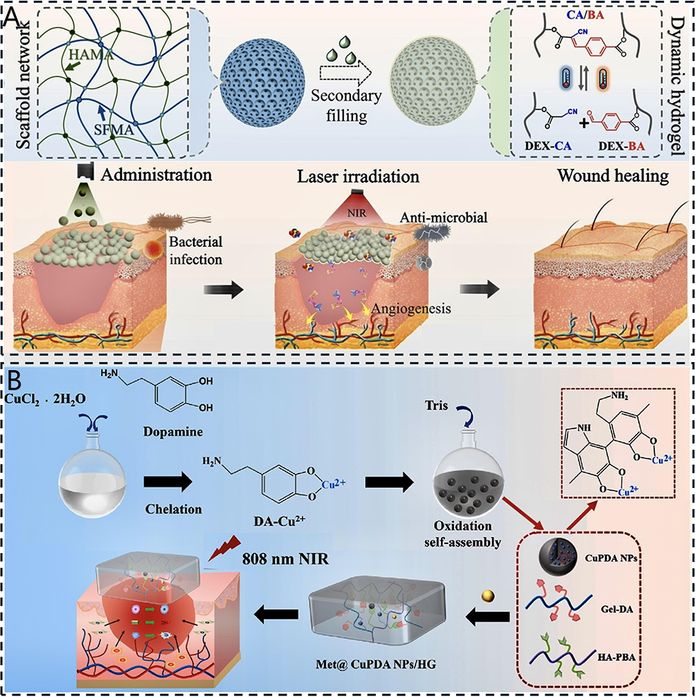 |
Figure 10 (A) Schematic representation of smart responsive structural color microspheres in trauma management. Adapted reprinted with permission from Ref.132 based on CC BY License, Copyright © 2024 The Author(s). (B) Preparation of metformin@copper-loaded polydopamine nanoparticle hydrogel (Met@CuPDA NPs/HG) dressing and its application on diabetic rat model with full-thickness skin defect and infection. Adapted reprinted with permission from Ref.133 based on CC BY License, Copyright © 2023 The Authors. Publishing services by Elsevier B.V. on behalf of KeAi Communications Co. Ltd. |
HA in Drug Delivery Systems
Advantages of HA as a Drug Carrier
HA is increasingly recognized as a promising vehicle for targeted drug delivery applications, not only because of its good biocompatibility and biodegradability, but also its ability to enhance drug targeting, increase bioavailability, and improve drug stability. HA is absorbed gradually into the body after application, eliminating the need for subsequent surgical extraction of the implant material. It also reduces the side effects of drug release, thus enhancing the safety and efficacy of their clinical application.194–197 At the same time, the molecular structure of HA allows it to interact with a variety of cell surface receptors, especially the CD44 receptor, which is highly expressed in several types of tumor cell.198,199 Through this specific binding, HA promotes drug accumulation at the tumor site, which enables targeted release and reduces toxicity to normal cells.194 This targeted delivery mechanism is used not only in tumor therapy but also in other diseases such as ophthalmic diseases and arthritis.200 HA can also improve the intestinal absorption of certain drugs by inhibiting P-glycoprotein activity, which in turn increases their oral bioavailability.201 This study demonstrates both theoretical and empirical evidence supporting the implementation of HA in targeted drug delivery platforms.
HA also shows good potential for prolonging drug release. The high viscosity of HA enables it to form a gel-like structure that can effectively control the rate of drug release. By modulating the crosslinking degree and network structure of HA-based microcapsules, sustained drug release can be achieved, thereby avoiding the phenomenon of “sudden release” that is common in traditional drug delivery systems.197,202 Additionally, HA composites with other polymers have shown good slow-release properties, offering novel approaches for developing sustained-release pharmaceutical formulations.194,200 For example, HA can form complexes with drugs through electrostatic interactions or hydrogen bonding, which can improve the drug stability and release rate.141
The ability of HA to improve drug stability in complex biological environments is of high importance for drug delivery systems. Its high hydrophilicity enables it to interact with water to form a protective film, thereby reducing drug degradation and inactivation. In one study, a complex of HA with certain antitumor drugs showed better drug stability, effectively extending the half-life of the drug in vivo and improving its bioavailability.203 Such composite systems are one of the most effective ways of enhancing drug stability. When combined with other materials, HA significantly improves both the physicochemical characteristics of pharmaceutical compounds and their biological compatibility, while simultaneously enhancing the precision of drug delivery to target sites. For example, compounding HA with synthetic polymers such as polyvinyl alcohol or polylactic acid can form composites with optimal mechanical properties and excellent biological compatibility, along with enhanced drug delivery efficiency and improved cellular attachment capabilities compared to conventional materials.204
HA has an adjustable relative molecular mass and facile chemical modification potential, making it an ideal drug carrier. Different properties can be achieved by modulating the relative molecular mass. For example, high-molecular-weight HA possesses the remarkable capability to adhere to cell surface receptors with precision via electrostatic interactions, facilitating the intracellular uptake of drugs and further enhancing their bioavailability.199 In addition, HA has been used to prepare drug delivery systems that are responsive to the tumor microenvironment, such as pH-sensitive HA–drug conjugates, that increase drug release in the tumor microenvironment, thereby enhancing antitumor effects.205 For example, the conjugation of HA with anticancer drugs can enhance their therapeutic effect by achieving targeted drug release under the acidic conditions of the tumor microenvironment.134 Furthermore, by modifying HA to introduce other functional groups, specific tumor types can be targeted. For example, conjugating HA microspheres with bone-resorbing drugs has been shown to enhance the therapeutic effect toward bone tumors.135 The successful application of HA-modified nanocarriers in drug delivery systems demonstrates their potential for tumor therapy.
HA-based drug carriers have been prepared in various formulations, including nanoparticles, micelles, microcapsules, and hydrogels, each of which has certain advantages. Micelles are only 20–50 nm in size, with a hydrophobic core that solubilizes small molecules such as adriamycin and paclitaxel. Their release is triggered by intracellular acidic or reducing environments, which induce nucleosome-shell dissociation. CD44-mediated rapid endocytosis allows the drug to escape lysosomes and enter the cytoplasm within 2–4 hours, making micelles suitable for deep tumor penetration. However, their drug-loading capacity is usually less than 10%, and hemodilution can lead to premature micelle dissociation.206 Nanoparticles, with sizes ranging from 80–200 nm, can be loaded with nucleic acids, proteins, or insoluble small molecules. Drug release occurs stepwise via backbone lysis, hyaluronidase degradation, or pH/glutathione/ROS-responsive mechanisms. Approximately 20–30% of nanoparticles are taken up by the reticuloendothelial system, while the remainder accumulates in tissues with high CD44 expression. Their half-life is extended to 6–12 hours, their surfaces are easily functionalized, and carrier residues must be cleared by the kidneys or enzymatically.207 Microcapsules, with diameters of 20–200 µm, can encapsulate vaccine antigens, growth hormones, or other active proteins within their inner core, while the outer shell is composed of HA and other biomaterials. Drugs are initially released by diffusion and subsequently through enzymatic digestion of the outer shell. Subcutaneous injection forms a localized drug reservoir, with macrophages modulating the release rate. The effect can last from 1 week to 3 months, providing low burst release and preserving protein activity; nevertheless, excessively large particles may induce foreign body reactions.208 Hydrogels form a three-dimensional network through in situ cross-linking, can be injected or 3D printed, and can carry antibodies, stem cells, or nucleic acids. Drug release occurs via a combination of network diffusion, hyaluronidase degradation, and shear thinning, with local retention exceeding 7 days. Hydrogels also provide an extracellular matrix-like microenvironment that supports cell function and tissue repair. Additionally, HA hydrogels can be combined with other biomaterials to create composites with enhanced drug-loading capacities and biocompatibility.192 Nonetheless, higher cross-linking degrees reduce drug-loading capacity, and overly rapid degradation can cause abrupt release, necessitating a balance between mechanical strength and release behavior through cross-linking density or dual-network design.209 In summary, micelles are suitable for rapid intracellular release, nanoparticles excel in multi-load co-delivery and stimulus-responsive release, microcapsules provide long-lasting sustained release, and hydrogels enable localized three-dimensional retention while supporting cells. This allows clinical selection of a single or combination of strategies depending on lesion depth, target cell type, and therapeutic duration. HA-modified nanoparticles can effectively encapsulate drugs and release them under specific conditions, such as in the highly acidic and reducing tumor microenvironment, where the degradation of HA can promote drug release.210 In addition, HA-modified nanocarriers can provide simultaneous imaging capabilities when combined with other functional materials, such as gold nanoparticles or graphene quantum dots, thus providing new strategies for integrated cancer treatment.194 HA microcapsules have also shown promise for tumor-directed drug delivery, where tumor targeting is achieved by modulating the surface properties and release mechanisms of the microcapsules.211 HA hydrogels form a physically or chemically crosslinked 3D network structure that effectively encapsulates drugs and regulates the drug release rate according to exogenous environmental factors (eg, pH and temperature). Additionally, HA hydrogels can be integrated with other biomaterials to form composites with enhanced drug-loading capacities and biocompatibility.212 These properties demonstrate their versatile therapeutic potential in oncology, tissue engineering, and regenerative medicine.
HA in Targeted Therapies
HA has good clinical prospects in several fields, especially in the delivery of antitumor drugs, anti-inflammatory drugs, and vaccines. In recent years, HA-modified antitumor drug delivery systems have made progress in clinical trials, and many studies have focused on the clinical application of HA as a drug carrier, particularly in the targeted delivery of oncology drugs. For example, PEGylated recombinant human hyaluronidase (PEGPH20) has been used to pretreat the tumor microenvironment to reduce the HA content, thereby improving the distribution and efficacy of antitumor drugs such as paclitaxel.213 Indeed, pretreatment with PEGPH20 significantly increased the concentration of paclitaxel in tumors and enhanced its antitumor activity. Although PEGPH20 significantly improved drug penetration and efficacy by degrading HA in animal models, it failed to prolong overall survival in the Phase III HALO-301 study of patients with metastatic pancreatic ductal adenocarcinoma (PDAC) exhibiting high HA expression, and was therefore not approved for marketing, with development subsequently terminated.136 This outcome serves as a reminder that the preclinical linear reasoning of “matrix degradation → improved blood flow → enhanced chemotherapy efficacy” is unreliable for remodeling the HA-rich tumor microenvironment. Changes in the microenvironment should be monitored using real-time imaging or dynamic biomarkers, and patient screening should not rely solely on static “HA-high” tissue slices but should also consider tumor heterogeneity, HA metabolism kinetics, and systemic distribution. Systemic enzymatic digestion of HA carries a risk of off-target toxicities, such as thrombosis, highlighting the need for local controlled release, nanocarriers, or precise strategies targeting the HA receptor axis. Stromal interventions must be integrated with immune, vascular, or metabolic regulation to prevent the vicious cycle of “degradation–re-fibrosis.” Moreover, combination regimens should be optimized in terms of timing rather than merely superimposed. HA-modified nanocarriers have also demonstrated promising results in the treatment of glioblastoma, with HA-modified lipid nanoparticles effectively delivering miR-181a to glioblastoma cells and inhibiting their proliferation.137,214 Although a wide range of chemotherapeutic agents is available for tumor treatment, multiple drug resistance (MDR) has become a major challenge in chemotherapy, particularly as the dose and frequency of drug administration increase, mainly owing to the overexpression of P-glycoprotein (P-gp).215 Inhibition of P-gp expression and function via redox disruption has shown considerable potential for reversing MDR. Sun et al138 constructed a nanoscale system of copper-based metal-organic frameworks modified with doxorubicin-loaded HA (HA-CuMOF@DOX) to induce mitochondrial damage, reduce intracellular adenosine triphosphate (ATP) levels, and downregulate P-gp, thereby overcoming the drug resistance of the drug-resistant human hepatocellular carcinoma cell line (HepG2-ADR) through dual regulation of aggravated redox homeostasis dysregulation. In vitro experiments demonstrated that the nanoparticles exhibited effective targeting of cancer cells while maintaining biocompatibility with normal cells, and HA-CuMOF@DOX was successfully internalized by HepG2-ADR cells. In vivo experiments showed that nude mice bearing HepG2-ADR tumors achieved 80.69% tumor growth inhibition. This study represents a significant advance in the development of effective treatments for drug-resistant tumors. These advances in clinical studies suggest that HA, as a key component of drug delivery systems, can effectively improve the therapeutic effects of antitumor drugs and provide new ideas and approaches for targeted tumor therapy.
HA has also been widely applied in anti-inflammatory drug delivery systems, especially in combination with nonsteroidal anti-inflammatory drugs, owing to its high drug-carrying capacity and targeted delivery properties. For example, HA nanocapsules containing celecoxib not only effectively encapsulate the drug but also prolong its release in the joint cavity, which significantly improves its therapeutic efficacy.139 In addition, HA can be chemically crosslinked with drugs to form hydrogel nanoparticle systems with controlled release properties, thus prolonging the intra-articular residence of the drug while simultaneously augmenting its therapeutic efficacy against inflammation (Figure 11A).216
 |
Figure 11 (A) Schematic diagram of rigid scaffold production and application. The inorganic and organic components can tightly bind with HA and become a part of the scaffold, while bioactive components are relatively freely attached and easily released. Adapted reprinted with permission from Ref.217 based on CC BY License, Copyright © 2019 The Authors. Published by Elsevier B.V. (B) Scheme of mycobacterium-loaded microneedle patches and altered frequencies of immune cells in the dermis after Mpg-microneedle patch application. Adapted reprinted with permission from Ref.140 based on CC BY License, Copyright © 2023 Lee, Seo, Lee, Kim, Kim, Lee, Oh, Shin, Jin, Jeong and Kim. (C) Schematic illustration of synthetic procedures of HA‐DOPA·Gd2+ hydrogel for the sustained in situ vaccination effect and real‐time monitoring of distribution in vivo. With the intratumoral release of DOX and PD‐1 inhibitors, synthetic hydrogels improve the suppressive immune microenvironment and enhance antitumor responses through ICD and M1 macrophage polarization in mouse STS models. Adapted reprinted with permission from Ref.218 based on CC BY License, Copyright © 2023 The Authors. Advanced Healthcare Materials published by Wiley‐VCH GmbH. |
HA offers excellent opportunities for vaccine delivery. It can bind to a wide range of biologically active molecules and form a stable system that improves the efficiency and bioavailability of vaccine delivery. HA can effectively enhance the stability of antigens and prolong their release in the body, thus improving the strength and continuity of the immune response. Additionally, the presence of HA as an endogenous component of cells and tissues helps to reduce the risk of immune rejection. The properties of HA can be optimized via molecular weight adjustments and chemical modifications to suit the requirements of different vaccines. Crosslinked HA hydrogels offer controlled vaccine release, further enhancing their immune response.217
HA-based vaccine carriers are increasingly employed in clinical trials, with promising results in terms of enhancing the immune response. In one study, the use of HA-modified microneedle patches for vaccine delivery significantly increased the activation of dermal dendritic cells and increased specific T cells (Figure 11B).140 Despite these encouraging results, more clinical trials should be conducted to verify the efficacy and long-term safety of HA in different vaccine formulations. Overall, the application of HA as a vaccine carrier is promising, especially for improving vaccine immunization efficacy and patient acceptance, which are of significant clinical importance (Figure 11C).218
The development of versatile pharmaceutical platforms is a key topic in current drug delivery research. These systems aim to achieve efficient drug delivery and targeted therapy by combining multiple functionalities. For example, researchers have developed HA-based triple-drug delivery systems that can deliver both chemotherapeutic and immunotherapeutic drugs, thereby enhancing the effectiveness of tumor therapy.141,214 In addition, the use of superparamagnetic iron oxide nanoparticles as drug carriers allows for more precise drug delivery by targeting tumor cells.219 These multifunctional drug delivery systems not only improve the bioavailability of drugs but also reduce side effects and improve the patient experience. With further development of nanotechnology and biomaterials science, future drug delivery systems will become more intelligent and able to self-adjust according to changes in the tumor microenvironment to achieve more personalized treatment.220
Summary and Outlook
As a crucial biomaterial, HA has been widely applied in several clinical disciplines. Continuous innovation in its modification and processing technologies, as well as the expansion of its clinical applications, has brought unprecedented opportunities to this field. Over the past five years, HA research has shifted from simple “chemical modification–property characterization” to more complex “focal microenvironmental response–cell fate regulation.” In 2024–2025, the focus is expected to move away from merely “discovering another new bond” toward enabling HA derivatives to release specific signals in specific spatiotemporal patterns, marking the transition from general-purpose polysaccharides to precision biomaterials. Chen et al221 developed an HA-based microgel that responds to MMP-2, which is overexpressed in OA joints, thereby achieving an enzyme-triggered intelligent drug release. In vitro experiments demonstrated that the microgel exhibits a controlled degradation rate, excellent cartilage adhesion and lubrication properties, and can effectively inhibit macrophage activation and reduce inflammatory factor production while protecting chondrocytes. In vivo experiments further confirmed that the microgel had a retention time of up to 18 days in an OA rat model, significantly alleviating OA symptoms, reducing bone spurs and ectopic mineralization, improving cartilage structure, decreasing inflammatory cell infiltration, protecting chondrocytes, and slowing disease progression. This in-depth analysis of current research trends demonstrates the diversity of HA and its potential in various medical fields, including tissue engineering, drug delivery, joint therapy, and dermatological aesthetics, which are increasingly recognized by both academia and industry. However, the clinical use of HA still faces multiple challenges.
First, the physicochemical properties of HA restrict its application in certain areas. The low mechanical strength of pure HA means it cannot withstand high mechanical stress, restricting its use to non-weight-bearing applications. HA-based hydrogels typically suffer from slow degradation and inadequate mechanical properties, which limit their use in cartilage tissue engineering.154 While crosslinking agents such as BDDE and divinyl sulfone can enhance the mechanical properties of HA hydrogels, they may compromise their biocompatibility and degradability. Additionally, the biodegradation rate of HA-based products is difficult to precisely control. HA degrades in vivo owing to the interaction between hyaluronidase and free radicals, and the degradation rate is determined by the molecular weight and crosslinking degree. Rapid degradation may reduce therapeutic efficacy, whereas overly slow degradation may affect tissue repair or trigger chronic inflammatory responses.
Second, existing HA preparation and modification techniques have many limitations. For example, physical processing technologies—most commonly heat treatment—tend to produce unstable products with limited tunability, making them unsuitable for meeting the demand for tailored biomaterials suited to different application requirements. Cutting-edge physical processing technologies such as ultrasonic treatment and electrospinning present significant technical challenges, difficulties in mass production, and high costs. Meanwhile, chemical modifications such as sulfation and acetylation may reduce the biocompatibility of HA, leading to immune reactions or inflammation. In drug delivery, HA is typically used to load small-molecule drugs, proteins, or nucleic acids; however, the loading capacity is limited, and the drug release rate is difficult to precisely regulate. Crosslinking or chemical modifications may affect the drug-binding efficiency and reduce the therapeutic efficacy. Therefore, the development and utilization of HA in the biomedical field are yet to expand further. The comparison of the efficacy of HA of different molecular weights in treating the same disease has long been a focus of research and debate. Some scholars argue that the therapeutic effect of HA may not depend solely on the physical properties associated with its molecular weight, such as viscosity, but more importantly on its pharmacological role as a signaling molecule, for example, by regulating inflammatory pathways and cellular behaviors through binding to cell surface receptors such as CD44. Currently, there is no clear evidence of a simple linear relationship between molecular weight and clinical efficacy, and the determination of the optimal molecular weight requires further high-quality clinical studies.
Looking ahead, HA research is likely to focus on developing multifunctional HA-based materials, applying HA in emerging fields, and promoting the clinical application of HA. A promising avenue is the development of intelligent crosslinking strategies, such as enzyme-sensitive crosslinking, photocrosslinking, and dynamic crosslinking, with the aim of modulating the degradation rate, mechanical properties, and controlled release ability of HA-based products. HA composites with nanomaterials, such as nanogold, graphene, carbon dots, and silica nanoparticles, also offer significant promise for drug delivery, imaging, and tissue engineering. The complexation of HA with other natural biomaterials (eg, gelatin, chitosan, and collagen) enhances its biological properties and applications.222 In addition, HA can be used in biomimetic material design to develop tissue scaffolds with stiffness gradients and adjustable porosity, thereby mimicking the architecture of the natural extracellular matrix. For instance, a “cartilage–bone integrated” layered scaffold can be constructed by dividing HA into two layers according to interfacial requirements. The upper layer (cartilage side) is cross-linked with dynamic disulfide bonds at a low substitution degree (DS ≈ 5%), imparting shear-thinning and self-healing properties, and is gradually degraded in the joint microenvironment over 6–48 h via glutathione-triggered cleavage, enabling the sustained release of TGF-β1 to induce cartilage formation. The lower layer (bone side) is cross-linked with high DS (≈20%) ether bonds (BDDE/PEGDE), providing a 1–2 MPa compressive modulus and an anti-enzymatic lifespan of 6–12 months, while supporting the slow release of BMP-2 to promote bone regeneration and repair. This design enables the outcomes typically achieved through the traditional two-step “bone followed by cartilage” grafting strategy to be realized in a single surgical procedure.
The application of HA in emerging fields, such as in the regulation of the tumor microenvironment and the construction of drug delivery systems, is another key research topic. By combining antibodies and peptides, HA-based products can be modulated to targeted tumor cells or specific tissues. The HA-based controlled drug-release system can be activated by external stimuli, including pH changes, enzymatic activity, and light exposure, thereby enhancing therapeutic precision, whereas bilayer/multilayer sustained-release systems achieve the synergistic delivery of different drugs. In addition, the development of 3D bioprinting technologies will make personalized medicine a reality, providing more precise solutions for regenerative medicine by printing synthetic cartilage, blood vessels, and skin scaffolds using HA hydrogels. The development of microneedle patch technology will also promote the application of HA in topical drug delivery, painless vaccinations, and anti-aging treatments. Finally, the clinical application of HA will be promoted, and its long-term efficacy and safety will be verified through large-scale randomized controlled trials to provide a scientific basis for clinical promotion.223 To accelerate the clinical translation of HA, it is necessary to standardize key parameters such as the upper limit of DS, control of free aldehydes, and enzyme residue assays, in addition to defining quality attributes and immunogenicity evaluation templates, thereby providing a foundation for future regulatory guidance.
In conclusion, although HA has extensive potential in the field of biomedicine, it faces many challenges for practical applications. The focus of future research should be on developing novel modification techniques and promoting clinical applications, as well as on improving the functionality and adaptability of HA, in order to promote its application in advanced biomedicine.
Abbreviations
HA, hyaluronic acid; CD44, cluster of differentiation 44; HMMR, hyaluronan mediated motility receptor; 3D, three-dimensional; ROS, reactive oxygen species; Dex, dexamethasone; DCF, difluorochlorothiazide; PBA, phenylboronic acid; Dox, doxorubicin; BDDE, 1,4-butanediol diglycidyl ether; PEGDE, polyethylene glycol diglycidyl ether; BNC, bionanocellulose; Dox/HHS-MSNs, doxorubicin-loaded multifunctional HA derivative-modified mesoporous silica nanoparticles; SFN, sulforaphane; TA, tetradecyl; HAMA, methacrylated hyaluronic acid; CXCL12, chemokine ligand 12; SDF-1α, stromal cell-derived factor; HG-AA-SDF-1α, HA/gelatin methacryloyl/sodium alginate–arginine carbon dots/stromal cell-derived factor-1α; AHAMA, HA hydrogel modified by aldehyde groups and methacrylate; Gel@DFO@GA, gelatin@deferoxamine@glycyrrhizic acid hydrogel; HIF-1α, hypoxia-inducible factor 1 subunit alpha; ∆HA, HA derivative, 4,5-anhydro-n-acetylglucosamine hyaluronan; PRP, platelet-rich plasma; PVA, polyvinyl alcohol; HMPs, hydrogel microparticles; TIMP3, tissue inhibitor of metalloproteinases 3; MMPI, metalloproteinase inhibitor 3; MMP9, matrix metallopeptidase; OSDI, ocular surface disease index; Lev, levofloxacin; BBC3, Bcl2 binding component 3; PUMA, p53-upregulated modulator of apoptosis; RH, rhein; CeOX, ceria oxide; 4D, four-dimensional; DMY, dihydromyricetin; AQP3, aquaporin 3; Met, metformin; Cu, copper; PDA, polydopamine; NPs, nanoparticles; HG, hydrogel; RHAMM, receptor for hyaluronic acid-mediated motility; LYVE-1, lymphatic vessel endothelial hyaluronan receptor 1; HARE, HA receptor for endocytosis; TLR, toll-like receptor; Siglec, sialic acid-binding immunoglobulin-like lectin; CEMIP, cell migration-inducing hyaluronidase 1; TMEM2, transmembrane protein; QT, quercetin; CCNPs, curcumin chitosan nanoparticles; SF, sericin hydrogel; TNF, tumor necrosis factor; COX, cyclooxygenase; DS, degree of substitution; DMAP, 4-dimethylaminopyridine; H2O2, hydrogen peroxide; EDC, 1-ethyl-3-(3-dimethylaminopropyl)carbodiimide; NHS, N-hydroxysuccinimide; NaOH, sodium hydroxide; HAase, hyaluronidase; ROS, reactive oxygen species; GSH, glutathione; RT, room temperature; MA, methacrylic anhydride; VS, vinyl sulfone; LBP, live biotherapeutic product; LCN2, lipocalin 2; PDAC, pancreatic ductal adenocarcinoma; MDR, multiple drug resistance; P-gp, P-glycoprotein; CuMOF, copper-based metal-organic frameworks; HepG2-ADR, HepG2 adriamycin-resistant cell line; HB-PEGDA, hyperbranched polyethylene glycol diacrylate; HA-SH, thiol-functionalised hyaluronic acid; DA, dopamine; miR-181a, microRNA-181a-5p; HARE, hyaluronan and receptor for endocytosis.
Author Contributions
All authors made a significant contribution to the work reported, whether that is in the conception, study design, execution, acquisition of data, analysis and interpretation, or in all these areas; took part in drafting, revising or critically reviewing the article; gave final approval of the version to be published; have agreed on the journal to which the article has been submitted; and agree to be accountable for all aspects of the work.
Funding
This study was funded by the Natural Science Foundation of Hunan Province, China (2024JJ5479) and Hunan Provincial Health Commission Scientific Research Project, China (C202304106744).
Disclosure
The authors report no conflicts of interest in this work.
References
1. Burdick JA, Prestwich GD. Hyaluronic acid hydrogels for biomedical applications. Adv Mater. 2011;23:H41–56. doi:10.1002/adma.201003963
2. Meyer K, Palmer JW. The polysaccharide of the vitreous humor. J Biol Chem. 1934;107:629–634. doi:10.1016/S0021-9258(18)75338-6
3. Brown MB, Jones SA. Hyaluronic acid: a unique topical vehicle for the localized delivery of drugs to the skin. J Eur Acad Dermatol Venereol. 2005;19:308–318. doi:10.1111/j.1468-3083.2004.01180.x
4. Vigetti D, Karousou E, Viola M, Deleonibus S, De Luca G, Passi A. Hyaluronan: biosynthesis and signaling. Biochim Biophys Acta. 2014;1840:2452–2459. doi:10.1016/j.bbagen.2014.02.001
5. Hintze V, Schnabelrauch M, Rother S. Chemical modification of hyaluronan and their biomedical applications. Front Chem. 2022;10:830671. doi:10.3389/fchem.2022.830671
6. Fallacara A, Baldini E, Manfredini S, Vertuani S. Hyaluronic acid in the third millennium. Polymers. 2018;11:10. doi:10.3390/polym11010010
7. Luo Z, Wang Y, Li J, Wang J, Yu Y, Zhao Y. Tailoring hyaluronic acid hydrogels for biomedical applications. Adv Funct Mater. 2023;33:2306554 doi:10.1002/adfm.202306554.
8. Rayahin JE, Buhrman JS, Zhang Y, Koh TJ, Gemeinhart RA. High and low molecular weight hyaluronic acid differentially influence macrophage activation. ACS Biomater Sci Eng. 2015;1:481–493. doi:10.1021/acsbiomaterials.5b00181
9. Trombino S, Servidio C, Curcio F, Cassano R. Strategies for hyaluronic acid-based hydrogel design in drug delivery. Pharmaceutics. 2019;11:407 doi:10.3390/pharmaceutics11080407.
10. Zhang M, Chen X, Yang K, et al. Dual-crosslinked hyaluronic acid hydrogel with self-healing capacity and enhanced mechanical properties. Carbohydr Polym. 2023;301:120372. doi:10.1016/j.carbpol.2022.120372
11. Yu S, Wang S, Xia L, et al. Injectable self-crosslinking hydrogels based on hyaluronic acid as vitreous substitutes. Int J Biol Macromol. 2022;208:159–171. doi:10.1016/j.ijbiomac.2022.03.046
12. Bermejo-Velasco D, Azémar A, Oommen OP, Hilborn J, Varghese OP. Modulating thiol p k(a) promotes disulfide formation at physiological ph: an elegant strategy to design disulfide cross-linked hyaluronic acid hydrogels. Biomacromolecules. 2019;20:1412–1420. doi:10.1021/acs.biomac.8b01830
13. Radice S, Neto MQ, Fischer A, Wimmer MA. Nickel-free high-nitrogen austenitic steel outperforms CoCrMo alloy regarding tribocorrosion in simulated inflammatory synovial fluids. J Orthop Res. 2022;40:1397–1408. doi:10.1002/jor.25174
14. Lei Y, Wang X, Liao J, et al. Shear-responsive boundary-lubricated hydrogels attenuate osteoarthritis. Bioact Mater. 2022;16:472–484. doi:10.1016/j.bioactmat.2022.02.016
15. Kim YH, Park HR, Cha SY, et al. Effect of red ginseng NaturalGEL on skin aging. J Ginseng Res. 2020;44:115–122. doi:10.1016/j.jgr.2018.09.006
16. Ni C, Zhang Z, Wang Y, Zhang Z, Guo X, Lv H. Hyaluronic acid and HA-modified cationic liposomes for promoting skin penetration and retention. J Control Release. 2023;357:432–443. doi:10.1016/j.jconrel.2023.03.049
17. López-Machado A, Díaz-Garrido N, Cano A, et al. Development of lactoferrin-loaded liposomes for the management of dry eye disease and ocular inflammation. Pharmaceutics. 2021;13 doi:10.3390/pharmaceutics13101698.
18. Fakhari A, Berkland C. Applications and emerging trends of hyaluronic acid in tissue engineering, as a dermal filler and in osteoarthritis treatment. Acta Biomater. 2013;9:7081–7092. doi:10.1016/j.actbio.2013.03.005
19. Ghatak S, Misra S, Toole BP. Hyaluronan oligosaccharides inhibit Anchorage-independent growth of tumor cells by suppressing the phosphoinositide 3-kinase/Akt cell survival pathway. J Biol Chem. 2002;277:38013–38020. doi:10.1074/jbc.M202404200
20. Oliva F, Marsilio E, Asparago G, Frizziero A, Berardi AC, Maffulli N. The Impact of hyaluronic acid on tendon physiology and its clinical application in tendinopathies. Cells. 2021;10. doi:10.3390/cells11010010
21. Shikina EV, Kovalevsky RA, Shirkovskaya AI, Toukach PV. Prospective bacterial and fungal sources of hyaluronic acid: a review. Comput Struct Biotechnol J. 2022;20:6214–6236 doi:10.1016/j.csbj.2022.11.013.
22. Kramer M, Criswell A, Marzette K, Cutcliffe E, Sewell-Loftin MK. Strain and hyaluronic acid interact to regulate ovarian cancer cell proliferation, migration, and drug resistance. Mechanobiol Med. 2024;2:100094. doi:10.1016/j.mbm.2024.100094
23. Radisky DC, Stallings-Mann M, Hirai Y, Bissell MJ. Single proteins might have dual but related functions in intracellular and extracellular microenvironments. Nat Rev Mol Cell Biol. 2009;10:228–234. doi:10.1038/nrm2633
24. Pandey MS, Harris EN, Weigel PH. Hare-mediated endocytosis of hyaluronan and heparin is targeted by different subsets of three endocytic motifs. Int J Cell Biol. 2015;2015:524707. doi:10.1155/2015/524707
25. Liu C, Liu M, Li X, et al. Unique advantages and applications of polysaccharide microneedles as drug delivery materials and in treatment of skin diseases. Nanoscale Adv. 2025;7:3631–3654. doi:10.1039/D4NA01083C
26. Gantumur MA, Jia X, Hui JH, et al. Characterization, bioactivity, and biodistribution of 35 kDa hyaluronan fragment. Life. 2024;15:14. doi:10.3390/life15010014
27. Marinho A, Nunes C, Reis S. Hyaluronic acid: a key ingredient in the therapy of inflammation. Biomolecules. 2021;11. doi:10.3390/biom12010011
28. Dovedytis M, Liu ZJ, Bartlett S. Hyaluronic acid and its biomedical applications: a review. Engineered Regeneration. 2020;1:102–113. doi:10.1016/j.engreg.2020.10.001
29. Zerbinati N, Esposito C, Cipolla G, et al. Chemical and mechanical characterization of hyaluronic acid hydrogel cross-linked with polyethylen glycol and its use in dermatology. Dermatol Ther. 2020;33:e13747. doi:10.1111/dth.13747
30. Masuko K, Murata M, Yudoh K, Kato T, Nakamura H. Anti-inflammatory effects of hyaluronan in arthritis therapy: not just for viscosity. Int J Gen Med. 2009;2:77–81. doi:10.2147/IJGM.S5495
31. Abdel-Rahman RM, Abdel-Mohsen AM. Marine biomaterials: hyaluronan. Mar Drugs. 2023;22:21. doi:10.3390/md22010021
32. Sze JH, Brownlie JC, Love CA. Biotechnological production of hyaluronic acid: a mini review. 3 Biotech. 2016;6:67. doi:10.1007/s13205-016-0379-9
33. Giji S, Arumugam M. Isolation and characterization of hyaluronic acid from marine organisms. Adv Food Nutr Res. 2014;72:61–77 doi:10.1016/b978-0-12-800269-8.00004-x.
34. Kang DY, Kim WS, Heo IS, Park YH, Lee S. Extraction of hyaluronic acid (HA) from rooster comb and characterization using flow field-flow fractionation (FlFFF) coupled with multiangle light scattering (MALS). J Sep Sci. 2010;33:3530–3536. doi:10.1002/jssc.201000478
35. Űrgeová E, Vulganová K. Comparison of enzymatic hydrolysis of polysaccharides from eggshells membranes. Nova Biotechnologica et Chimica. 2016;15:133–141. doi:10.1515/nbec-2016-0014
36. de Paula MC, Carvalho SG, Silvestre ALP, Dos Santos AM, Meneguin AB, Chorilli M. The role of hyaluronic acid in the design and functionalization of nanoparticles for the treatment of colorectal cancer. Carbohydr Polym. 2023;320(320):121257. doi:10.1016/j.carbpol.2023.121257
37. Liu L, Liu Y, Li J, Du G, Chen J. Microbial production of hyaluronic acid: current state, challenges, and perspectives. Microb Cell Fact. 2011;10:1–9. doi:10.1186/1475-2859-10-99
38. Shiedlin A, Bigelow R, Christopher W, et al. Evaluation of hyaluronan from different sources: streptococcus z ooepidemicus, rooster comb, bovine vitreous, and human umbilical cord. Biomacromolecules. 2004;5:2122–2127. doi:10.1021/bm0498427
39. Zhang X, Wang M, Li T, Fu L, Cao W, Liu H. Construction of efficient Streptococcus zooepidemicus strains for hyaluoronic acid production based on identification of key genes involved in sucrose metabolism. AMB Express. 2016;6:121. doi:10.1186/s13568-016-0296-7
40. Kim J-H, Yoo S-J, Oh D-K, et al. Selection of a Streptococcus equi mutant and optimization of culture conditions for the production of high molecular weight hyaluronic acid. Enzyme Microb Technol. 1996;19:440–445. doi:10.1016/S0141-0229(96)00019-1
41. Jia Y, Zhu J, Chen X, et al. Metabolic engineering of Bacillus subtilis for the efficient biosynthesis of uniform hyaluronic acid with controlled molecular weights. Bioresour Technol. 2013;132:427–431. doi:10.1016/j.biortech.2012.12.150
42. Sunguroğlu C, Sezgin DE, Aytar Çelik P, Çabuk A. Higher titer hyaluronic acid production in recombinant Lactococcus lactis. Prep Biochem Biotechnol. 2018;48:734–742. doi:10.1080/10826068.2018.1508036
43. Westbrook AW, Ren X, Moo-Young M, Chou CP. Engineering of cell membrane to enhance heterologous production of hyaluronic acid in Bacillus subtilis. Biotechnol Bioeng. 2018;115:216–231. doi:10.1002/bit.26459
44. Karami M, Shahraky MK, Ranjbar M, Tabandeh F, Morshedi D, Aminzade S. Preparation, purification, and characterization of low-molecular-weight hyaluronic acid. Biotechnol Lett. 2021;43:133–142. doi:10.1007/s10529-020-03035-4
45. Prasad SB, Jayaraman G, Ramachandran KB. Hyaluronic acid production is enhanced by the additional co-expression of UDP-glucose pyrophosphorylase in Lactococcus lactis. Appl Microbiol Biotechnol. 2010;86:273–283. doi:10.1007/s00253-009-2293-0
46. Jin P, Kang Z, Yuan P, Du G, Chen J. Production of specific-molecular-weight hyaluronan by metabolically engineered Bacillus subtilis 168. Metab Eng. 2016;35:21–30. doi:10.1016/j.ymben.2016.01.008
47. Schulz A, Wakili P, Januschowski K, et al. Safety and performance assessment of hyaluronic acid-based vitreous substitutes in patients with phthisis bulbi. Acta Ophthalmol. 2023;101:687–695. doi:10.1111/aos.15658
48. Zhang G, Qin M, Guo M, et al. Thiol-modified hyaluronic acid improves the physical stability of curcumin-zein nanoparticles by forming disulfide bonds with zein. Food Chem. 2023;429:136858. doi:10.1016/j.foodchem.2023.136858
49. Magdziarz S, Boguń M, Frączyk J. Coating methods of carbon nonwovens with cross-linked hyaluronic acid and its conjugates with BMP fragments. Polymers. 2023;15:1551 doi:10.3390/polym15061551.
50. Bao Y, Ge Y, Wu M, Mao Z, Ye J, Tong W. Record-high ultrasound-sensitive NO nanogenerators for cascade tumor pyroptosis and immunotherapy. Adv Sci. 2023;10:e2302278. doi:10.1002/advs.202302278
51. Di Mola A, Landi MR, Massa A, D’Amora U, Guarino V. Hyaluronic acid in biomedical fields: new trends from chemistry to biomaterial applications. Int J Mol Sci. 2022;23:14372. doi:10.3390/ijms232214372
52. Li F, Weng K, Nakamura A, et al. Preparation of waterborne silicone-modified polyurethane nanofibers and the effect of crosslinking agents on physical properties. Polymers. 2024;16:1500 doi:10.3390/polym16111500.
53. Song G, Zhou L, Zhao L, et al. Analysis of non-covalent interaction between β-lactoglobulin and hyaluronic acid under ultrasound-assisted treatment: conformational structures and interfacial properties. Int J Biol Macromol. 2024;256:128529. doi:10.1016/j.ijbiomac.2023.128529
54. Wang Y, Ji M, Xing M, et al. Effects of ultrasound and thermal treatment on the interaction between hyaluronic acid and lactoferrin: preparation, structures and functionalities. Int J Biol Macromol. 2024;272:132812. doi:10.1016/j.ijbiomac.2024.132812
55. Huang T, Zeng Y, Li C, et al. Application and development of electrospun nanofiber scaffolds for bone tissue engineering. ACS Biomater Sci Eng. 2024;10:4114–4144. doi:10.1021/acsbiomaterials.4c00028
56. Han J, Xie N, Ju J, Zhang Y, Wang Y, Kang W. Developments of electrospinning technology in membrane bioreactor: a review. Chemosphere. 2024;364:143091. doi:10.1016/j.chemosphere.2024.143091
57. Yu AC, Lian H, Kong X, Lopez Hernandez H, Qin J, Appel EA. Physical networks from entropy-driven non-covalent interactions. Nat Commun. 2021;12:746. doi:10.1038/s41467-021-21024-7
58. Debertrand L, Zhao J, Creton C, Narita T. Swelling and mechanical properties of polyacrylamide-derivative dual-crosslink hydrogels having metal-ligand coordination bonds as transient crosslinks. Gels. 2021;7 doi:10.3390/gels7020072.
59. Fallon M, Halligan S, Pezzoli R, Geever L, Higginbotham C. Synthesis and characterisation of novel temperature and pH sensitive physically cross-linked poly (N-vinylcaprolactam-co-itaconic acid) hydrogels for drug delivery. Gels. 2019;5:5. doi:10.3390/gels5010005
60. Jiang Y, Wang W, Huang Q. Impacts of crosslinking conditions on pickering emulsions stabilized by genipin-crosslinked chitosan-caseinophosphopeptides nanocomplexes. Int J Biol Macromol. 2023;242:125154. doi:10.1016/j.ijbiomac.2023.125154
61. Chen TY, Chen KC, Zhang YH, et al. Development of a dexamethasone-hyaluronic acid conjugate with selective targeting effect for acute lung injury therapy. Int J Biol Macromol. 2024;280:136149. doi:10.1016/j.ijbiomac.2024.136149
62. Shi W, Hass B, Kuss MA, et al. Fabrication of versatile dynamic hyaluronic acid-based hydrogels. Carbohydr Polym. 2020;233:115803. doi:10.1016/j.carbpol.2019.115803
63. Liu B, Kong Y, Alimi OA, et al. Multifunctional microgel-based cream hydrogels for postoperative abdominal adhesion prevention. ACS Nano. 2023;17:3847–3864. doi:10.1021/acsnano.2c12104
64. Dömling A. Recent developments in isocyanide based multicomponent reactions in applied chemistry. Chem Rev. 2006;106:17–89. doi:10.1021/cr0505728
65. Hong SH, Lee MH, Go EJ, Park JC. A promising strategy for combating bacterial infections through the use of light-triggered ROS in Ce6-immobilized hydrogels. Regen Biomater. 2024;11:rbae101. doi:10.1093/rb/rbae101
66. Laurent TC, Hellsing K, Gelotte B, Burton JS, Stevens R. Cross-linked gels of hyaluronic acid. Acta Chem Scand. 1964;18:274–275. doi:10.3891/acta.chem.scand.18-0274
67. Zheng X, Wang B, Tang X, et al. Absorption, metabolism, and functions of hyaluronic acid and its therapeutic prospects in combination with microorganisms: a review. Carbohydr Polym. 2023;299:120153. doi:10.1016/j.carbpol.2022.120153
68. Zhang M, Asghar S, Tian C, et al. Lactoferrin/phenylboronic acid-functionalized hyaluronic acid nanogels loading doxorubicin hydrochloride for targeting glioma. Carbohydr Polym. 2021;253:117194. doi:10.1016/j.carbpol.2020.117194
69. Zentner CA, Anson F, Thayumanavan S, Swager TM. Dynamic imine chemistry at complex double emulsion interfaces. J Am Chem Soc. 2019;141:18048–18055. doi:10.1021/jacs.9b06852
70. Wu H, Wang T, Liang Y, Chen L, Li Z. Self-assembled and dynamic bond crosslinked herb-polysaccharide hydrogel with anti-inflammation and pro-angiogenesis effects for burn wound healing. Colloids Surf B Biointerfaces. 2024;233:113639. doi:10.1016/j.colsurfb.2023.113639
71. Qi C, Sun Q, Xiao D, et al. Tetrahedral framework nucleic acids/hyaluronic acid-methacrylic anhydride hybrid hydrogel with antimicrobial and anti-inflammatory properties for infected wound healing. Int J Oral Sci. 2024;16:30. doi:10.1038/s41368-024-00290-3
72. Hu Z, Lv X, Zhang H, et al. An injectable gel based on photo-cross-linkable hyaluronic acid and mesoporous bioactive glass nanoparticles for periodontitis treatment. Int J Biol Macromol. 2024;257:128596. doi:10.1016/j.ijbiomac.2023.128596
73. Buffa R, Šedová P, Basarabová I, et al. A new unsaturated derivative of hyaluronic acid - synthesis, analysis and applications. Carbohydr Polym. 2017;163:247–253. doi:10.1016/j.carbpol.2017.01.071
74. Chen K, Zhao Y, Zhao W, et al. Lubricating microneedles system with multistage sustained drug delivery for the treatment of osteoarthritis. Small. 2024;20:e2307281. doi:10.1002/smll.202307281
75. Pang X, Lu Z, Du H, Yang X, Zhai G. Hyaluronic acid-quercetin conjugate micelles: synthesis, characterization, in vitro and in vivo evaluation. Colloids Surf B Biointerfaces. 2014;123:778–786. doi:10.1016/j.colsurfb.2014.10.025
76. Yu Q, Meng Z, Liu Y, Li Z, Sun X, Zhao Z. Photocuring hyaluronic acid/silk fibroin hydrogel containing curcumin loaded chitosan nanoparticles for the treatment of MG-63 cells and ME3T3-E1 cells. Polymers. 2021;14:13. doi:10.3390/polym14010013
77. Chen J, Yang J, Wang L, et al. Modified hyaluronic acid hydrogels with chemical groups that facilitate adhesion to host tissues enhance cartilage regeneration. Bioact Mater. 2021;6:1689–1698. doi:10.1016/j.bioactmat.2020.11.020
78. Nedunchezian S, Banerjee P, Lee CY, et al. Generating adipose stem cell-laden hyaluronic acid-based scaffolds using 3D bioprinting via the double crosslinked strategy for chondrogenesis. Mater Sci Eng C Mater Biol Appl. 2021;124:112072. doi:10.1016/j.msec.2021.112072
79. Nguyen TT, Neri TA, Choi BD. Characterization of hyaluronic acid extracted from Liparis tessellatus eggs grafted with phenolic acids and nisin. Int J Biol Macromol. 2020;157:45–50. doi:10.1016/j.ijbiomac.2020.04.137
80. Gao C, Jian J, Luo L, et al. Single-walled carbon nanohorns-based smart nanotheranostic: from phototherapy to enzyme-activated fluorescence imaging-guided photodynamic therapy. J Colloid Interface Sci. 2022;628:273–286. doi:10.1016/j.jcis.2022.07.168
81. Hou J, Zhao Y, Sun L, Zou X. Enzyme/GSH/pH-responsive hyaluronic acid grafted porous silica nanocarriers bearing Ag(2)S QDs for fluorescence imaging and combined therapy. Carbohydr Polym. 2023;305:120547. doi:10.1016/j.carbpol.2023.120547
82. Han X, Lu B, Zou D, et al. Allicin-loaded intelligent hydrogel coating improving vascular implant performance. ACS Appl Mater Interfaces. 2023;15:38247–38263. doi:10.1021/acsami.3c05984
83. Al-Sibani M, Al-Harrasi A, Neubert RH. Study of the effect of mixing approach on cross-linking efficiency of hyaluronic acid-based hydrogel cross-linked with 1,4-butanediol diglycidyl ether. Eur J Pharm Sci. 2016;91:131–137. doi:10.1016/j.ejps.2016.06.010
84. Luo Z, Wang Y, Xu Y, Wang J, Yu Y. Modification and crosslinking strategies for hyaluronic acid-based hydrogel biomaterials. Smart Med. 2023;2:e20230029. doi:10.1002/SMMD.20230029
85. Gholamali I, Vu TT, Jo SH, Park SH, Lim KT. Exploring the progress of hyaluronic acid hydrogels: synthesis, characteristics, and wide-ranging applications. Materials. 2024;18:17. doi:10.3390/ma18010017
86. Choi S, Cho JC, Lee S, Lee SJ. Development of dispersion process to improve quality of hyaluronic acid filler crosslinked with 1,4-butanediol diglycidyl ether. Polymers. 2024;16:3323. doi:10.3390/polym16233323
87. Park SJ, Yoo KH. One-year safety evaluation of new hyaluronic acid fillers (yys series): a prospective, multicenter, observational study. Dermatol Surg. 2024;50:731–738. doi:10.1097/DSS.0000000000004190
88. Kim DH, Jeong CH, Han JH, et al. Comparative toxicity study of hyaluronic acid fillers crosslinked with 1,4-butanediol diglycidyl ether or poly (ethylene glycol) diglycidyl ether. Int J Biol Macromol. 2025;296:139620. doi:10.1016/j.ijbiomac.2025.139620
89. Jeong CH, Kim DH, Yune JH, et al. In vitro toxicity assessment of crosslinking agents used in hyaluronic acid dermal filler. Toxicol Vitro. 2021;70:105034. doi:10.1016/j.tiv.2020.105034
90. Choi SC, Yoo MA, Lee SY, et al. Modulation of biomechanical properties of hyaluronic acid hydrogels by crosslinking agents. J Biomed Mater Res Part A. 2015;103(9):3072–3080. doi:10.1002/jbm.a.35437
91. Zhao Y, Yi B, Hu J, et al. Double cross-linked biomimetic hyaluronic acid-based hydrogels with thermo-stimulated self-contraction and tissue adhesiveness for accelerating post-wound closure and wound healing. Adv Funct Mater. 2023;33:2300710.
92. Guo W, Douma L, Hu MH, et al. Hyaluronic acid-based interpenetrating network hydrogel as a cell carrier for nucleus pulposus repair. Carbohydr Polym. 2022;277:118828. doi:10.1016/j.carbpol.2021.118828
93. Luo Y, Li G, Chen L, Hong FF. Preparation and evaluation of bacterial nanocellulose/hyaluronic acid composite artificial cornea for application of corneal transplantation. Biomacromolecules. 2023;24:201–212. doi:10.1021/acs.biomac.2c01052
94. Lai PY, Senthil Raja D, Chang JW, Huang JH, Tsai DH. Real-time quantification of microfluidic hydrogel crosslinking via gas-phase electrophoresis. J Colloid Interface Sci. 2025;684:201–212. doi:10.1016/j.jcis.2025.01.035
95. Yang D, Wang T, Su Z, Xue L, Mo R, Zhang C. Reversing cancer multidrug resistance in xenograft models via orchestrating multiple actions of functional mesoporous silica nanoparticles. ACS Appl Mater Interfaces. 2016;8:22431–22441. doi:10.1021/acsami.6b04885
96. Wang Y, Chen Y, Xu Y, et al. Effects of the bonding intensity between hyaluronan and gelatin on chondrogenic phenotypic maintenance. J Mater Chem B. 2020;8:9062–74 doi:10.1039/d0tb01816c.
97. Xu K, Yao H, Fan D, Zhou L, Wei S. Hyaluronic acid thiol modified injectable hydrogel: synthesis, characterization, drug release, cellular drug uptake and anticancer activity. Carbohydr Polym. 2021;254:117286. doi:10.1016/j.carbpol.2020.117286
98. Gu HF, Ren F, Mao XY, Du M. Mineralized and GSH-responsive hyaluronic acid based nano-carriers for potentiating repressive effects of sulforaphane on breast cancer stem cells-like properties. Carbohydr Polym. 2021;269:118294. doi:10.1016/j.carbpol.2021.118294
99. Yang K, Yang J, Chen R, et al. Antibacterial hyaluronic acid hydrogels with enhanced self-healing properties via multiple dynamic bond crosslinking. Int J Biol Macromol. 2024;256:128320. doi:10.1016/j.ijbiomac.2023.128320
100. Xiao Y, Ma J, Yuan X, et al. Acid-triggered dual-functional hydrogel platform for enhanced bone regeneration. Adv Sci;2025. e2415772. doi:10.1002/advs.202415772
101. Liu S, Zhao Y, Shen M, et al. Hyaluronic acid targeted and pH-responsive multifunctional nanoparticles for chemo-photothermal synergistic therapy of atherosclerosis. J Mater Chem B. 2022;10:562–570. doi:10.1039/D1TB02000E
102. Huang Y, Lai H, Jiang J, et al. pH-activatable oxidative stress amplifying dissolving microneedles for combined chemo-photodynamic therapy of melanoma. Asian J Pharm Sci. 2022;17:679–696 doi:10.1016/j.ajps.2022.08.003.
103. He C, Bi S, Zhang R, et al. A hyaluronic acid hydrogel as a mild photothermal antibacterial, antioxidant, and nitric oxide release platform for diabetic wound healing. J Control Release. 2024;370:543–555. doi:10.1016/j.jconrel.2024.05.011
104. Tan G, Hou G, Qian J, et al. Hyaluronan-decorated copper-doxorubicin-anlotinib nanoconjugate for targeted synergistic chemo/chemodynamic/antiangiogenic tritherapy against hepatocellular carcinoma. J Colloid Interface Sci. 2024;662:857–869. doi:10.1016/j.jcis.2024.02.085
105. Bencherif SA, Washburn NR, Matyjaszewski K. Synthesis by AGET ATRP of degradable nanogel precursors for in situ formation of nanostructured hyaluronic acid hydrogel. Biomacromolecules. 2009;10:2499–2507. doi:10.1021/bm9004639
106. Wang K, Yao SY, Wang Z, et al. A sequential dual functional supramolecular hydrogel with promoted drug release to scavenge ROS and stabilize HIF-1α for myocardial infarction treatment. Adv Healthc Mater. 2024;13:e2302940. doi:10.1002/adhm.202302940
107. Qi J, Li X, Cao Y, et al. Locationally activated PRP via an injectable dual-network hydrogel for endometrial regeneration. Biomaterials. 2024;309:122615. doi:10.1016/j.biomaterials.2024.122615
108. Song L, Zhai Z, Ouyang W, et al. Inhalation of macrophage membrane-coated hydrogel microparticles for inflammation alleviation of acute lung injury in vivo. Acta Biomater. 2025;192:409–418. doi:10.1016/j.actbio.2024.12.015
109. Puertas-Bartolomé M, Gutiérrez-Urrutia I, Teruel-Enrico LL, et al. Self-lubricating, living contact lenses. Adv Mater. 2024;36:e2313848 doi:10.1002/adma.202313848.
110. Burgalassi S, Fragai M, Francesconi O, et al. Functionalized hyaluronic acid for “in situ” matrix metalloproteinase inhibition: a bioactive material to treat the dry eye sydrome. ACS Macro Lett. 2022;11:1190–1194. doi:10.1021/acsmacrolett.2c00455
111. Yang SA, Jeong MR, Park CH, Cheon KB, Chang JH, Lee JE. Comparative efficacy of 0.1% and 0.15% sodium hyaluronate on lipid layer and meibomian glands following cataract surgery: a randomized prospective study. PLoS One. 2024;19:e0306253. doi:10.1371/journal.pone.0306253
112. Bao Z, Yu A, Shi H, et al. Glycol chitosan/oxidized hyaluronic acid hydrogel film for topical ocular delivery of dexamethasone and levofloxacin. Int J Biol Macromol. 2021;167:659–666. doi:10.1016/j.ijbiomac.2020.11.214
113. Chen HA, Tai YN, Hsieh EH, et al. Injectable cross-linked hyaluronic acid hydrogels with epigallocatechin gallate loading as vitreous substitutes. Int J Biol Macromol. 2024;275:133467. doi:10.1016/j.ijbiomac.2024.133467
114. Ferreres G, Pérez-Rafael S, Guaus E, et al. Antimicrobial and antifouling hyaluronic acid-cobalt nanogel coatings built sonochemically on contact lenses. Ultrason Sonochem. 2024;111:107131. doi:10.1016/j.ultsonch.2024.107131
115. Tschopp M, Pfirrmann CWA, Fucentese SF, et al. A randomized trial of intra-articular injection therapy for knee osteoarthritis. Invest Radiol. 2023;58:355–362. doi:10.1097/RLI.0000000000000942
116. Hua P, Liang R, Yang S, Tu Y, Chen M. Microneedle-assisted dual delivery of PUMA gene and celastrol for synergistic therapy of rheumatoid arthritis through restoring synovial homeostasis. Bioact Mater. 2024;36:83–95. doi:10.1016/j.bioactmat.2024.02.030
117. Zhou F, Li M, Chen M, et al. Redox homeostasis strategy for inflammatory macrophage reprogramming in rheumatoid arthritis based on ceria oxide nanozyme-complexed biopolymeric micelles. ACS Nano. 2023;17:4358–4372. doi:10.1021/acsnano.2c09127
118. Xu J, Wu M, Yang J, et al. Multimodal smart systems reprogramme macrophages and remove urate to treat gouty arthritis. Nat Nanotechnol. 2024;19:1544–1557. doi:10.1038/s41565-024-01715-0
119. Yu K, Yu X, Cao S, et al. Layered dissolving microneedles as a need-based delivery system to simultaneously alleviate skin and joint lesions in psoriatic arthritis. Acta Pharm Sin B. 2021;11:505–519. doi:10.1016/j.apsb.2020.08.008
120. Yu Z, Wang M, Li J, et al. A fused membrane-camouflaged biomimetic nanosystem for dual-targeted therapy of septic arthritis. Small. 2025;21:e2410710. doi:10.1002/smll.202410710
121. Costa FR, Pires L, Martins RA, Costa BR, Santos GS, Lana JF. Viscnovas: a novel classification system for hyaluronic acid-based gels in orthobiologic products and regenerative medicine. Gels. 2024;11:10. doi:10.3390/gels11010010
122. Caprio ND, Davidson MD, Daly AC, Burdick JA. Injectable MSC spheroid and microgel granular composites for engineering tissue. Adv Mater. 2024;36:e2312226. doi:10.1002/adma.202312226
123. Rong M, Liu D, Xu X, et al. A superparamagnetic composite hydrogel scaffold as in vivo dynamic monitorable theranostic platform for osteoarthritis regeneration. Adv Mater. 2024;36:e2405641. doi:10.1002/adma.202405641
124. Han Z, Wang F, Xiong W, et al. Precise cell type electrical stimulation therapy via force-electric hydrogel microspheres for cartilage healing. Adv Mater. 2024;37:e2414555 doi:10.1002/adma.202414555.
125. Xia X, Liu Y, Lu Y, et al. Retuning mitochondrial apoptosis/mitophagy balance via SIRT3-energized and microenvironment-modulated hydrogel microspheres to impede osteoarthritis. Adv Healthc Mater. 2023;12:e2302475. doi:10.1002/adhm.202302475
126. Dey N, Mohny FP, Betsy Reshma G, Rao D, Ganguli M, Santhiya D. Bioinspired synthesis of bioactive glass nanocomposites for hyaluronic acid delivery to bone and skin. Int J Biol Macromol. 2023;253:127262. doi:10.1016/j.ijbiomac.2023.127262
127. Juncan AM, Moisă DG, Santini A, et al. Advantages of hyaluronic acid and its combination with other bioactive ingredients in cosmeceuticals. Molecules. 2021;26:4429 doi:10.3390/molecules26154429.
128. Manna F, Dentini M, Desideri P, De Pità O, Mortilla E, Maras B. Comparative chemical evaluation of two commercially available derivatives of hyaluronic acid (hylaform from rooster combs and restylane from streptococcus) used for soft tissue augmentation. J Eur Acad Dermatol Venereol. 1999;13:183–192. doi:10.1111/j.1468-3083.1999.tb00881.x
129. Huang J, Xue J, Huang J, et al. An injectable hyaluronic acid/lithium calcium silicate soft tissue filler with vascularization and collagen regeneration. Bioact Mater. 2025;44:256–268.
130. Wang K, Li XL, Liu J, Sun X, Yang H, Gao X. Using cross-linked hyaluronic acid gel to prevent postoperative lumbar epidural space adhesion: in vitro and in vivo studies. Eur Spine J. 2020;29:129–140. doi:10.1007/s00586-019-06193-w
131. Dong Y, Cui M, Qu J, et al. Conformable hyaluronic acid hydrogel delivers adipose-derived stem cells and promotes regeneration of burn injury. Acta Biomater. 2020;108:56–66. doi:10.1016/j.actbio.2020.03.040
132. Wang L, Ding X, Fan L, et al. Self-healing dynamic hydrogel microparticles with structural color for wound management. Nanomicro Lett. 2024;16:232. doi:10.1007/s40820-024-01422-4
133. Zhu S, Zhao B, Li M, et al. Microenvironment responsive nanocomposite hydrogel with NIR photothermal therapy, vascularization and anti-inflammation for diabetic infected wound healing. Bioact Mater. 2023;26:306–320. doi:10.1016/j.bioactmat.2023.03.005
134. Zhang YH, Zhang YM, Yu J, Wang J, Liu Y. Boronate-crosslinked polysaccharide conjugates for pH-responsive and targeted drug delivery. Chem Commun. 2019;55:1164–1167. doi:10.1039/C8CC09956A
135. Xi Y, Jiang T, Yu Y, et al. Dual targeting curcumin loaded alendronate-hyaluronan- octadecanoic acid micelles for improving osteosarcoma therapy. Int J Nanomed. 2019;14:6425–6437. doi:10.2147/IJN.S211981
136. Van Cutsem E, Tempero MA, Sigal D, et al. Randomized phase III trial of pegvorhyaluronidase alfa with nab-paclitaxel plus gemcitabine for patients with hyaluronan-high metastatic pancreatic adenocarcinoma. J Clin Oncol. 2020;38:3185–3194. doi:10.1200/JCO.20.00590
137. Passos Gibson V, Tahiri H, Yang C, Phan QT, Banquy X, Hardy P. Hyaluronan decorated layer-by-layer assembled lipid nanoparticles for miR-181a delivery in glioblastoma treatment. Biomaterials. 2023;302:122341. doi:10.1016/j.biomaterials.2023.122341
138. Sun J, An X, Wang Y, et al. A hyaluronic acid modified copper-based metal-organic framework overcomes multidrug resistance via two-way redox dyshomeostasis under hypoxia. Int J Biol Macromol. 2025;300:140148. doi:10.1016/j.ijbiomac.2025.140148
139. El-Gogary RI, Khattab MA, Abd-Allah H. Intra-articular multifunctional celecoxib loaded hyaluronan nanocapsules for the suppression of inflammation in an osteoarthritic rat model. Int J Pharm. 2020;583:119378 doi:10.1016/j.ijpharm.2020.119378.
140. Lee MH, Seo H, Lee MS, et al. Protection against tuberculosis achieved by dissolving microneedle patches loaded with live Mycobacterium paragordonae in a BCG prime-boost strategy. Front Immunol. 2023;14:1178688. doi:10.3389/fimmu.2023.1178688
141. Ma C, Duan Y, Wu C, et al. Spatiotemporally co-delivery of triple therapeutic drugs via HA-coating nanosystems for enhanced immunotherapy. Asian J Pharm Sci. 2021;16:653–664. doi:10.1016/j.ajps.2021.07.004
142. Hakim FE, Farooq AV. Dry eye disease: an update in 2022. JAMA. 2022;327:478–479. doi:10.1001/jama.2021.19963
143. Pham DA, Wang CS, Séguy L, et al. Bioinspired bottlebrush polymers effectively alleviate frictional damage both in vitro and in vivo. Adv Mater. 2024;36:e2401689. doi:10.1002/adma.202401689
144. Chang WH, Liu PY, Lin MH, et al. Applications of hyaluronic acid in ophthalmology and contact lenses. Molecules. 2021;27:26. doi:10.3390/molecules27010026
145. Zamboni F, Okoroafor C, Ryan MP, et al. On the bacteriostatic activity of hyaluronic acid composite films. Carbohydr Polym. 2021;260:117803. doi:10.1016/j.carbpol.2021.117803
146. Gribova V, Auzely-Velty R, Picart C. Polyelectrolyte multilayer assemblies on materials surfaces: from cell adhesion to tissue engineering. Chem Mater. 2012;24:854–869. doi:10.1021/cm2032459
147. Keskin D, Tromp L, Mergel O, et al. Highly efficient antimicrobial and antifouling surface coatings with triclosan-loaded nanogels. ACS Appl Mater Interfaces. 2020;12:57721–57731. doi:10.1021/acsami.0c18172
148. Sohani Z, Jamshidi S, Koohi MK, et al. Novel ophthalmic hyaluronic acid-hydrogel with curcumin nanoparticles for enhanced healing of ulcerative keratitis in rabbit model. Sci Rep. 2024;14:23046. doi:10.1038/s41598-024-74195-w
149. Ruppert SM, Hawn TR, Arrigoni A, Wight TN, Bollyky PL. Tissue integrity signals communicated by high-molecular weight hyaluronan and the resolution of inflammation. Immunol Res. 2014;58:186–192. doi:10.1007/s12026-014-8495-2
150. Walvekar P, Lulinski P, Kumar P, Aminabhavi TM, Choonara YE. A review of hyaluronic acid-based therapeutics for the treatment and management of arthritis. Int J Biol Macromol. 2024;264:130645. doi:10.1016/j.ijbiomac.2024.130645
151. Zhou K, Yang C, Shi K, et al. Activated macrophage membrane-coated nanoparticles relieve osteoarthritis-induced synovitis and joint damage. Biomaterials. 2023;295:122036. doi:10.1016/j.biomaterials.2023.122036
152. Zhang J, Song Y, Zhu L, et al. An injectable thermosensitive hyaluronic acid/pluronic F-127 hydrogel for deep penetration and combination therapy of frozen shoulder. Int J Biol Macromol. 2024;263:130342. doi:10.1016/j.ijbiomac.2024.130342
153. Ortiz-de la Torre AM, Barrios-Moyano A, González-Perales AA, Gerbacio-Blando LA. Evaluation of the application of hyaluronic acid in postoperative arthroscopy patients with grade II-III gonarthrosis using the WOMAC scale. Acta Ortop Mex. 2023;37:36–43. doi:10.35366/112812
154. Wang M, Deng Z, Guo Y, Xu P. Designing functional hyaluronic acid-based hydrogels for cartilage tissue engineering. Mater Today Bio. 2022;17:100495. doi:10.1016/j.mtbio.2022.100495
155. Tong Z, Ma Y, Liang Q, et al. An in situ forming cartilage matrix mimetic hydrogel scavenges ROS and ameliorates osteoarthritis after superficial cartilage injury. Acta Biomater. 2024;187:82–97. doi:10.1016/j.actbio.2024.08.018
156. Ding SL, Zhao XY, Xiong W, et al. Cartilage lacuna-inspired microcarriers drive hyaline neocartilage regeneration. Adv Mater. 2023;35:e2212114. doi:10.1002/adma.202212114
157. Sun Y, Ding SL, Zhao X, et al. Self-reinforced MOF-based nanogel alleviates osteoarthritis by long-acting drug release. Adv Mater. 2024;36:e2401094. doi:10.1002/adma.202401094
158. Lan X, Ma Z, Dimitrov A, et al. Double crosslinked hyaluronic acid and collagen as a potential bioink for cartilage tissue engineering. Int J Biol Macromol. 2024;273:132819. doi:10.1016/j.ijbiomac.2024.132819
159. Martyniak K, Lokshina A, Cruz MA, Karimzadeh M, Kemp R, Kean TJ. Biomaterial composition and stiffness as decisive properties of 3D bioprinted constructs for type II collagen stimulation. Acta Biomater. 2022;152:221–234. doi:10.1016/j.actbio.2022.08.058
160. Díaz-Payno PJ, Kalogeropoulou M, Muntz I, et al. Swelling-dependent shape-based transformation of a human mesenchymal stromal cells-laden 4D bioprinted construct for cartilage tissue engineering. Adv Healthc Mater. 2023;12:e2201891. doi:10.1002/adhm.202201891
161. Shakel Z, Costa lima SA, Reis S. Strategies to make human skin models based on cellular senescence for ageing research. Ageing Res Rev. 2024;100:102430. doi:10.1016/j.arr.2024.102430
162. Lee S, Baek SW, Kim DS, et al. Injectable Microparticle-containing hydrogel with controlled release of bioactive molecules for facial rejuvenation. Mater Today Bio. 2024;24:100890. doi:10.1016/j.mtbio.2023.100890
163. Siquier-Dameto G, Boadas-Vaello P, Verdú E. Intradermal treatment with a hyaluronic acid complex supplemented with amino acids and antioxidant vitamins improves cutaneous hydration and viscoelasticity in healthy subjects. Antioxidants. 2024;14:13. doi:10.3390/antiox14010013
164. Šimek M, Turková K, Schwarzer M, et al. Molecular weight and gut microbiota determine the bioavailability of orally administered hyaluronic acid. Carbohydr Polym. 2023;313:120880. doi:10.1016/j.carbpol.2023.120880
165. Kang W, Ha Y, Jung Y, Lee H, Park T. Nerol mitigates dexamethasone-induced skin aging by activating the Nrf2 signaling pathway in human dermal fibroblasts. Life Sci. 2024;356:123034. doi:10.1016/j.lfs.2024.123034
166. Gwak MA, Hong BM, Park WH. Hyaluronic acid/tannic acid hydrogel sunscreen with excellent anti-UV, antioxidant, and cooling effects. Int J Biol Macromol. 2021;191:918–924. doi:10.1016/j.ijbiomac.2021.09.169
167. Carvalho MJ, Pedrosa SS, Mendes A, et al. Anti-aging potential of a novel ingredient derived from sugarcane straw extract (SSE). Int J Mol Sci. 2023;25:25. doi:10.3390/ijms25010025
168. Sim WJ, Lee E, Lee G, Lim W, Lim TG. Inhibition of photoaging by anthocyanin metabolites derived from rose petal extract. Mol Nutr Food Res. 2024;68:e2300611. doi:10.1002/mnfr.202300611
169. Paterska M, Czerny B, Cielecka-Piontek J. Macrofungal extracts as a source of bioactive compounds for cosmetical anti-aging therapy: a comprehensive review. Nutrients. 2024;16:2810. doi:10.3390/nu16162810
170. Pacini S, Punzi T, Gulisano M, Ruggiero M. Pulsed current iontophoresis of hyaluronic acid in living rat skin. J Dermatol Sci. 2006;44:169–171. doi:10.1016/j.jdermsci.2006.08.012
171. Zoccali G, Cinque B, La Torre C, et al. Improving the outcome of fractional CO2 laser resurfacing using a probiotic skin cream: preliminary clinical evaluation. Lasers Med Sci. 2016;31:1607–1611. doi:10.1007/s10103-016-2024-6
172. Kasetvatin C, Rujivipat S, Tiyaboonchai W. Combination of elastic liposomes and low frequency ultrasound for skin permeation enhancement of hyaluronic acid. Colloids Surf B Biointerfaces. 2015;135:458–464. doi:10.1016/j.colsurfb.2015.07.078
173. Wu X, Zhang H, He S, et al. Improving dermal delivery of hyaluronic acid by ionic liquids for attenuating skin dehydration. Int J Biol Macromol. 2020;150:528–535. doi:10.1016/j.ijbiomac.2020.02.072
174. Casiano Evans EA, Hobson DTG, Aschkenazi SO, et al. Nonestrogen therapies for treatment of genitourinary syndrome of menopause: a systematic review. Obstet Gynecol. 2023;142:555–570. doi:10.1097/AOG.0000000000005288
175. Fagien S, Bertucci V, von Grote E, Mashburn JH. Rheologic and physicochemical properties used to differentiate injectable hyaluronic acid filler products. Plast Reconstr Surg. 2019;143:707e–720e. doi:10.1097/PRS.0000000000005429
176. Heitmiller K, Ring C, Saedi N. Rheologic properties of soft tissue fillers and implications for clinical use. J Cosmet Dermatol. 2021;20:28–34. doi:10.1111/jocd.13487
177. Schifano N, Capogrosso P, Antonini G, et al. The application of hyaluronic acid injections in functional and aesthetic andrology: a narrative review. Gels. 2023;9:118 doi:10.3390/gels9020118.
178. Kolczewski P, Parafiniuk M, Zawodny P, et al. Hyaluronic acid and radiofrequency in patients with urogenital atrophy and vaginal laxity. Pharmaceuticals. 2022;16:15. doi:10.3390/ph16010015
179. Yang Y, Sheng H, Gu Q, et al. Death caused by vaginal injection of hyaluronic acid and collagen: a case report. Aesthet Surg J. 2020;40:Np263–np268. doi:10.1093/asj/sjz275
180. Huth L, Marquardt Y, Heise R, Fietkau K, Baron JM, Huth S. Biological effects of hyaluronic acid-based dermal fillers and laser therapy on human skin models. J Drugs Dermatol. 2020;19:897–899. doi:10.36849/JDD.2020.4856
181. Soares DM. Bridging a Century-Old Problem: the pathophysiology and molecular mechanisms of HA filler-induced vascular occlusion (FIVO)-implications for therapeutic interventions. Molecules. 2022;28:27. doi:10.3390/molecules28010027
182. Schelke LW, Velthuis P, Kadouch J, Swift A. Early ultrasound for diagnosis and treatment of vascular adverse events with hyaluronic acid fillers. J Am Acad Dermatol. 2023;88:79–85 doi:10.1016/j.jaad.2019.07.032.
183. Zhang M, Dong Q, Yang K, et al. Hyaluronic acid hydrogels with excellent self-healing capacity and photo-enhanced mechanical properties for wound healing. Int J Biol Macromol. 2024;267:131235. doi:10.1016/j.ijbiomac.2024.131235
184. Marin S, Popovic-Pejicic S, Radosevic-Caric B, Trtić N, Tatic Z, Selakovic S. Hyaluronic acid treatment outcome on the post-extraction wound healing in patients with poorly controlled type 2 diabetes: a randomized controlled split-mouth study. Med Oral Patol Oral Cir Bucal. 2020;25:e154–e160. doi:10.4317/medoral.23061
185. Chen RF, Wang CT, Chen YH, et al. Hyaluronic acid-povidone-iodine compound facilitates diabetic wound healing in a streptozotocin-induced diabetes rodent model. Plast Reconstr Surg. 2019;143:1371–1382. doi:10.1097/PRS.0000000000005504
186. Valachová K, El Meligy MA, Šoltés L. Hyaluronic acid and chitosan-based electrospun wound dressings: problems and solutions. Int J Biol Macromol. 2022;206:74–91. doi:10.1016/j.ijbiomac.2022.02.117
187. Wei Q, Jin Z, Zhang W, et al. Honokiol@PF127 crosslinked hyaluronate-based hydrogel for promoting wound healing by regulating macrophage polarization. Carbohydr Polym. 2023;303:120469. doi:10.1016/j.carbpol.2022.120469
188. Su X, Geng X, Zhang Y, Shi Y, Zhao L. Microenvironmental pH modulating oxygen self-boosting microalgal prodrug carboxymethyl chitosan/hyaluronic acid/puerarin hydrogel for accelerating wound healing in diabetic rats. Int J Biol Macromol. 2024;282:136669. doi:10.1016/j.ijbiomac.2024.136669
189. Zhou S, Xie M, Su J, Cai B, Li J, Zhang K. New insights into balancing wound healing and scarless skin repair. J Tissue Eng. 2023;14:20417314231185848. doi:10.1177/20417314231185848
190. Li Z, Yang L, Zhang D, et al. Mussel-inspired “plug-and-play” hydrogel glue for postoperative tumor recurrence and wound infection inhibition. J Colloid Interface Sci. 2023;650:1907–1917. doi:10.1016/j.jcis.2023.07.154
191. Tian L, Sun T, Fan M, Lu H, Sun C. Novel silk protein/hyaluronic acid hydrogel loaded with azithromycin as an immunomodulatory barrier to prevent postoperative adhesions. Int J Biol Macromol. 2023;235:123811. doi:10.1016/j.ijbiomac.2023.123811
192. Zhang T, Huang Y, Gong Y, et al. A ROS-responsive and scavenging hydrogel for postoperative abdominal adhesion prevention. Acta Biomater. 2024;184:98–113. doi:10.1016/j.actbio.2024.06.027
193. Zhang W, Zhang S, Zhao T, Zhang H. An injectable sequential dual-crosslinking catechol-functionalized hyaluronic acid hydrogel for enhanced regeneration of full-thickness burn injury. J Control Release. 2024;369:545–555. doi:10.1016/j.jconrel.2024.04.012
194. Jing Y, Wang C, Li C, et al. Development of a manganese complex hyaluronic acid hydrogel encapsulating stimuli-responsive Gambogic acid nanoparticles for targeted Intratumoral delivery. Int J Biol Macromol. 2024;270:132348. doi:10.1016/j.ijbiomac.2024.132348
195. Jin W, Shen S, Xu X, et al. All-in-one hydrogel patches with sprayed bFGF-loaded GelMA microspheres for infected wound healing studies. Int J Pharm. 2024;658:124205. doi:10.1016/j.ijpharm.2024.124205
196. Gang X, Wang L, Jia J, et al. Synthesis and biological evaluation of fluorescent hyaluronic acid modified amorphous calcium phosphate drug carriers for tumor-targeting. Int J Biol Macromol. 2021;182:1445–1454. doi:10.1016/j.ijbiomac.2021.05.068
197. Wu Z, Shi J, Song P, Li J, Cao S. Chitosan/hyaluronic acid based hollow microcapsules equipped with MXene/gold nanorods for synergistically enhanced near infrared responsive drug delivery. Int J Biol Macromol. 2021;183:870–879. doi:10.1016/j.ijbiomac.2021.04.164
198. Ouyang Q, Zhao Y, Xu K, He Y, Qin M. Hyaluronic acid receptor-mediated nanomedicines and targeted therapy. Small Methods. 2024;8:e2400513. doi:10.1002/smtd.202400513
199. Gagneja S, Capalash N, Sharma P. Hyaluronic acid as a tumor progression agent and a potential chemotherapeutic biomolecule against cancer: a review on its dual role. Int J Biol Macromol. 2024;275:133744. doi:10.1016/j.ijbiomac.2024.133744
200. Zhang X, Wei D, Xu Y, Zhu Q. Hyaluronic acid in ocular drug delivery. Carbohydr Polym. 2021;264:118006. doi:10.1016/j.carbpol.2021.118006
201. Islam N, Irfan M, Hussain T, et al. Piperine phytosomes for bioavailability enhancement of domperidone. J Liposome Res. 2022;32:172–180. doi:10.1080/08982104.2021.1918153
202. Xu M, Fan R, Fan X, Shao Y, Li X. Progress and challenges of anti-VEGF agents and their sustained-release strategies for retinal angiogenesis. Drug Des Devel Ther. 2022;16:3241–3262. doi:10.2147/DDDT.S383101
203. Fan YF, Shang WT, Lu GH, et al. Decreasing hyaluronic acid combined with drug-loaded nanoprobes improve the delivery and efficacy of chemotherapeutic drugs for pancreatic cancer. Cancer Lett. 2021;523:1–9. doi:10.1016/j.canlet.2021.09.016
204. Sepulveda AF, Kumpgdee-Vollrath M, Franco M, Yokaichiya F, de Araujo DR. Supramolecular structure organization and rheological properties modulate the performance of hyaluronic acid-loaded thermosensitive hydrogels as drug-delivery systems. J Colloid Interface Sci. 2023;630:328–340. doi:10.1016/j.jcis.2022.10.064
205. Ashrafizadeh M, Hushmandi K, Mirzaei S, et al. Chitosan-based nanoscale systems for doxorubicin delivery: exploring biomedical application in cancer therapy. Bioeng Transl Med. 2023;8:e10325. doi:10.1002/btm2.10325
206. Liu Y, Zhou C, Wei S, et al. Paclitaxel delivered by CD44 receptor-targeting and endosomal pH sensitive dual functionalized hyaluronic acid micelles for multidrug resistance reversion. Colloids Surf B Biointerfaces. 2018;170:330–340. doi:10.1016/j.colsurfb.2018.06.024
207. Mizrahy S, Raz SR, Hasgaard M, et al. Hyaluronan-coated nanoparticles: the influence of the molecular weight on CD44-hyaluronan interactions and on the immune response. J Control Release. 2011;156:231–238. doi:10.1016/j.jconrel.2011.06.031
208. Chen W, Wang J, Zhang C, Cao S, Li J, Shi J. Hyaluronic acid/chitosan microcapsules capped with hollow CuS nanoparticles for NIR/pH dual-responsive drug release. Int J Biol Macromol. 2024;280:136050. doi:10.1016/j.ijbiomac.2024.136050
209. Rajput R, Narkhede J, Naik J. Nanogels as nanocarriers for drug delivery: a review. Admet dmpk. 2020;8:1–15. doi:10.5599/admet.724
210. Jeong GW, Jeong YI, Nah JW. Triggered doxorubicin release using redox-sensitive hyaluronic acid-g-stearic acid micelles for targeted cancer therapy. Carbohydr Polym. 2019;209:161–171. doi:10.1016/j.carbpol.2019.01.018
211. Zhang F, Zhang S, Lin R, Cui S, Jing X, Coseri S. Injectable multifunctional carboxymethyl chitosan/hyaluronic acid hydrogel for drug delivery systems. Int J Biol Macromol. 2023;249:125801. doi:10.1016/j.ijbiomac.2023.125801
212. Lei C, Liu XR, Chen QB, et al. Hyaluronic acid and albumin based nanoparticles for drug delivery. J Control Release. 2021;331:416–433 doi:10.1016/j.jconrel.2021.01.033.
213. Morosi L, Meroni M, Ubezio P, et al. PEGylated recombinant human hyaluronidase (PEGPH20) pre-treatment improves intra-tumour distribution and efficacy of paclitaxel in preclinical models. J Exp Clin Cancer Res. 2021;40:286. doi:10.1186/s13046-021-02070-x
214. Li J, Chen Z, Pei Z, Pei Y. A hyaluronic acid modified advanced nanoagent activated by tumor microenvironment enables a reciprocal dual-modality therapy. Chem Eng J. 2025;507:159884. doi:10.1016/j.cej.2025.159884
215. Wan Y, Chen Z, Wang Y, et al. A hyaluronic acid modified cuprous metal-organic complex for reversing multidrug resistance via redox dyshomeostasis. Carbohydr Polym. 2023;311:120762. doi:10.1016/j.carbpol.2023.120762
216. Nabizadeh Z, Nasrollahzadeh M, Heidari F, Nasrabadi D. A drug-loaded nano chitosan/hyaluronic acid hydrogel system as a cartilage tissue engineering scaffold for drug delivery. Int J Biol Macromol. 2024;283:137314. doi:10.1016/j.ijbiomac.2024.137314
217. Zhai P, Peng X, Li B, Liu Y, Sun H, Li X. The application of hyaluronic acid in bone regeneration. Int J Biol Macromol. 2020;151:1224–1239. doi:10.1016/j.ijbiomac.2019.10.169
218. Wang C, Jing Y, Yu W, et al. Bivalent gadolinium ions forming injectable hydrogels for simultaneous in situ vaccination therapy and imaging of soft tissue sarcoma. Adv Healthc Mater. 2023;12:e2300877. doi:10.1002/adhm.202300877
219. Zhi D, Yang T, Yang J, Fu S, Zhang S. Targeting strategies for superparamagnetic iron oxide nanoparticles in cancer therapy. Acta Biomater. 2020;102:13–34. doi:10.1016/j.actbio.2019.11.027
220. Yi S, Liao R, Zhao W, Huang Y, He Y. Multifunctional co-transport carriers based on cyclodextrin assembly for cancer synergistic therapy. Theranostics. 2022;12:2560–2579. doi:10.7150/thno.70243
221. Chen K, Wang J, Cao J, et al. Enzyme-responsive microgel with controlled drug release, lubrication and adhesion capability for osteoarthritis attenuation. Acta Biomater. 2024;190:191–204. doi:10.1016/j.actbio.2024.10.026
222. Yuan H, Mears LLE, Wang Y, et al. Lubricants for osteoarthritis treatment: from natural to bioinspired and alternative strategies. Adv Colloid Interface Sci. 2023;311:102814. doi:10.1016/j.cis.2022.102814
223. Yuan N, Shao K, Huang S, Chen C. Chitosan, alginate, hyaluronic acid and other novel multifunctional hydrogel dressings for wound healing: a review. Int J Biol Macromol. 2023;240:124321. doi:10.1016/j.ijbiomac.2023.124321
 © 2025 The Author(s). This work is published and licensed by Dove Medical Press Limited. The
full terms of this license are available at https://www.dovepress.com/terms
and incorporate the Creative Commons Attribution
- Non Commercial (unported, 4.0) License.
By accessing the work you hereby accept the Terms. Non-commercial uses of the work are permitted
without any further permission from Dove Medical Press Limited, provided the work is properly
attributed. For permission for commercial use of this work, please see paragraphs 4.2 and 5 of our Terms.
© 2025 The Author(s). This work is published and licensed by Dove Medical Press Limited. The
full terms of this license are available at https://www.dovepress.com/terms
and incorporate the Creative Commons Attribution
- Non Commercial (unported, 4.0) License.
By accessing the work you hereby accept the Terms. Non-commercial uses of the work are permitted
without any further permission from Dove Medical Press Limited, provided the work is properly
attributed. For permission for commercial use of this work, please see paragraphs 4.2 and 5 of our Terms.