
Back to Journals » Drug Design, Development and Therapy » Volume 20
Bioengineering Applications of Chinese Herbal Medicine-Derived Exosomes in Cardiovascular Diseases: Mechanisms and Translational Prospects
Authors Liu Y, Wang Y  , Fang M
Received 18 December 2025
Accepted for publication 10 April 2026
Published 22 April 2026 Volume 2026:20 590015
DOI https://doi.org/10.2147/DDDT.S590015
Checked for plagiarism Yes
Review by Single anonymous peer review
Peer reviewer comments 3
Editor who approved publication: Professor Anastasios Lymperopoulos
Yixin Liu,* Yiru Wang,* Ming Fang
Department of Cardiology, Longhua Hospital, Shanghai University of Traditional Chinese Medicine, Shanghai, 200032, People’s Republic of China
*These authors contributed equally to this work
Correspondence: Ming Fang, Department of Cardiology, Longhua Hospital, Shanghai University of Traditional Chinese Medicine, Shanghai, 200032, People’s Republic of China, Tel +86 13062747415, Email [email protected]
Abstract: Cardiovascular disease remains the leading cause of death worldwide, with current therapies facing limitations in efficacy and safety. Chinese Herbal Medicine-Derived Exosomes (CHM-Exos) are nano-sized membrane vesicles secreted by herbal cells, capable of cross-species delivery of bioactive substances. While plant-derived extracellular vesicles have been extensively reviewed, analyses specifically focused on CHM-Exos in cardiovascular contexts remain limited. This review systematically examines the bioengineering applications and therapeutic mechanisms of CHM-Exos in cardiovascular diseases, addressing a critical gap in translational literature. Mechanistically, CHM-Exos show potential to alleviate oxidative stress and modulate vascular cell function, though direct cardiovascular evidence remains preliminary. Key translational barriers—including standardization challenges, scalability constraints, and regulatory uncertainties—are critically discussed, alongside strategies to advance these promising nanocarriers toward clinical application. The infographic titled ’Chinese Herbal Medicine-Derived Exosomes (CHM-Exos): From Source to Cardiovascular Therapy and Clinical Translation Barriers’ is divided into four sections. The first section, ’Source & Advanced Isolation,’ includes Astragalus, Tea and Sophora flavescens, with processes like exosome purification and optimized isolation. The second section, ’Bioengineering & Cargo Loading,’ features endogenous miRNAs, proteins, drug cargo like Simvastatin and drug loading and surface modification. The third section, ’CVD Therapeutic Mechanisms,’ covers vascular endothelium repair and protection, vascular function improvement, miR-223 increase, VSMC stabilization decrease, smooth muscle layer and vascular repair mechanisms. The final section, ’Roadmap to Progress,’ illustrates cardiovascular repair leading to clinical success and future clinical application.Infographic on CHM-Exos for cardiovascular therapy, detailing sources, bioengineering, mechanisms and progress roadmap.
Keywords: Chinese herbal medicine-derived exosomes, cardiovascular diseases, bioengineering, engineering modification, targeted delivery
Introduction
Cardiovascular diseases are among the leading causes of death worldwide, with substantial mortality and rising disease burden. Current therapies (medication and surgery) alleviate symptoms but face limitations in efficacy (restenosis after interventions) and safety. There is an urgent need for improved treatment strategies.1
Chinese Herbal Medicine-Derived Exosomes (CHM-Exos) are nano-sized extracellular vesicles (50–150 nm) derived from medicinal plant cells, distinct from general plant-derived extracellular vesicles by their origin from medicinal plants with established cardiovascular indications in traditional Chinese medicine, conferring specific phytochemical profiles and therapeutic rationales. Compared to traditional drug carriers, CHM-Exos offer advantages of low immunogenicity, cost-effective production, diverse bioactive cargo, and engineering versatility.2 Their multi-target modulation and anti-inflammatory capacity align with cardiovascular disease pathophysiology. However, current evidence predominantly derives from non-cardiac models, and the transition to clinical applications requires rigorous validation.
This review focuses specifically on CHM-Exos and their bioengineering applications in cardiovascular diseases. We systematically examine their fundamental characteristics, core isolation and engineering strategies, preclinical evidence in CVD models, underlying protective mechanisms, and critical translational challenges. By delineating the current evidence base and identifying key research gaps, this analysis aims to provide a rigorous assessment of the field’s readiness for clinical translation.
Search Strategy: A comprehensive literature search was conducted in March 2026 across PubMed, Web of Science, Embase, and China National Knowledge Infrastructure (CNKI). The search terms were combined as follows: (plant exosomes OR herbal exosomes OR plant-derived extracellular vesicles) AND (cardiovascular disease OR cardiac disorder OR atherosclerosis). The publication time was limited from January 2019 to March 2026. Studies meeting the following criteria were included: (1) original research focusing on Chinese Herbal Medicine-Derived Exosomes (CHM-Exos); (2) investigations related to cardiovascular mechanisms or therapeutic applications. Exclusion criteria were conference abstracts, studies on extracellular vesicles from non-medicinal plants, and non-cardiovascular disease applications without mechanistic relevance. This narrative review critically synthesizes findings from approximately 80 eligible studies, evaluating evidence quality and translational validity.
Basic Characteristics of Chinese Herbal Medicine-Derived Exosomes
Definition, Composition, and Functions
Chinese Herbal Medicine-Derived Exosomes (CHM-Exos) are nanoscale membrane-bound extracellular vesicles (30–200 nm) released by Chinese herbal cells through the endosomal pathway or direct plasma membrane budding. They are widely distributed in plant phloem, xylem, and intercellular spaces.3 Their molecular composition is highly complex, primarily consisting of proteins, nucleic acids, and lipids (Figure 1), which collectively determine their structure and function.4
 |
Figure 1 Isolation of Chinese Herbal Medicine-Derived Exosomes; (A) Differential centrifugation; (B) Density gradient centrifugation; (C) Size exclusion chromatography; (D) Polymer precipitation method; (E) shows the molecular structure of Chinese Herbal Medicine-Derived Exosomes; Limitations regarding scalability and reproducibility of each method are discussed in the text visually highlighted. Created with BioGDP.com.5 |
While both CHM-Exos and mammalian exosomes share fundamental characteristics as nanoscale membrane-bound extracellular vesicles, critical distinctions exist in their biogenesis, composition, and characterization that impact cardiovascular applications.6,7 Mammalian exosomes originate from the endosomal pathway and are defined by specific surface markers including CD9, CD63, CD81, and Alix, with established isolation protocols and quality control frameworks validated through clinical trials.6,8 In contrast, CHM-Exos lack universally accepted surface marker signatures; while some studies report plant-specific tetraspanins, these markers are not consistently detectable across different herbal sources and preparation methods. This compositional variability presents significant challenges for cardiovascular therapeutic development, where reproducible dosing and predictable therapeutic responses are essential.
The lipid bilayer of CHM-Exos provides structural integrity and protects encapsulated contents from enzymatic degradation.9 These vesicles integrate multidimensional functions in material transport, immune regulation, and cellular communication. They can deliver bioactive compounds—including flavonoids, glycosides, and polysaccharides—with demonstrated anti-inflammatory, anti-apoptotic, and antioxidant effects.10,11 However, critical evaluation reveals that these functional demonstrations predominantly derive from non-cardiovascular models (colitis, neuroprotection, antimicrobial applications). Direct validation of similar cargo delivery efficiency and functional potency in cardiac or vascular cell types remains limited, representing a significant evidence gap in the current literature.12
Separation and Purification Techniques
Due to their small size (30–200 nm), low density, and complex composition, efficient isolation of CHM-Exos remains a core technical challenge. Mainstream techniques must balance yield, purity, and biological activity preservation (Figure 1).
Current Methodological Approaches
Differential Ultracentrifugation (DUC), as a fundamental method, uses an ultracentrifuge for multi-step gradient centrifugation (500×g to 100,000×g) to remove impurities and precipitate Chinese herbal medicine exosomes, followed by purification via PBS washing. This method is simple, low-cost, and suitable for preliminary enrichment from plant tissues, but it tends to co-precipitate protein aggregates and has low purity.13
To address purity issues, density gradient centrifugation builds upon preliminary purification by differential centrifugation, combining with gradient media such as sucrose. After centrifugation at 100,000×g for 16 hours, the Chinese herbal medicine exosomes fraction with a density of 1.10–1.18 g/mL is collected and further purified. This method significantly improves purity and can also separate vesicle subpopulations,13 but it is time-consuming (>16 h) and complex, making it suitable for high-purity Chinese herbal medicine exosomes preparation and subpopulation analysis.
In terms of preserving biological activity, Size Exclusion Chromatography (SEC) offers advantages. It employs a chromatography system with a Sepharose CL-4B column. After sample loading, PBS is used for elution, and fractions corresponding to 30–200 nm particles are collected. This method effectively preserves the structure and activity of Chinese herbal medicine exosomes and efficiently separates free proteins.14 However, the sample requires pre-concentration, and the column has limited capacity, making it suitable for functional studies such as cell interactions.
Polymer-based precipitation methods focus on processing efficiency. Using PEG6000/8000 reagents, the sample is incubated with an 8%–10% final concentration PEG solution at 4°C for 12 hours, followed by centrifugation at 10,000×g to precipitate herbal exosomes. This method offers high throughput and simplicity, making it suitable for large-scale processing of herbal juice samples. However, residual PEG may interfere with downstream analyses, and non-exosomal particles are easily co-precipitated, resulting in low purity.4 It is thus suitable for rapid screening or diagnostic applications.
Critical Limitations for Scalability and Reproducibility
A fundamental constraint in cardiovascular therapeutic development is the lack of standardized preparation protocols. Unlike mammalian exosomes, where differential ultracentrifugation protocols have been optimized for clinical-grade production, CHM-Exos isolation remains method-dependent with significant batch-to-batch variability.15,16 This heterogeneity directly impacts cardiovascular application potential: undefined particle concentrations, variable cargo profiles, and inconsistent surface properties complicate dose-response predictions and safety assessments required for cardiac therapies.
Scalability and Standardization Challenges
For cardiovascular therapeutic applications, these methodological limitations translate to critical translational barriers. Differential ultracentrifugation, while widely employed, suffers from significant scalability constraints: the high centrifugal forces not only yield insufficient quantities for clinical dosing but also induce vesicle aggregation and membrane damage, alongside co-purification of protein aggregates that compromise purity.17 Density gradient centrifugation, despite superior purity, requires 16+ hour processing times incompatible with industrial production timelines. Size exclusion chromatography preserves biological activity but is constrained by column capacity limitations and sample dilution effects that require subsequent concentration steps.18 Polymer-based precipitation enables high-throughput processing but suffers from co-precipitation of non-exosomal proteins, generating contaminated preparations that complicate regulatory approval pathways.19 Critically, no consensus exists on optimal isolation methods for cardiovascular applications, where particle homogeneity, endotoxin removal, and preservation of cardiac-targeting surface properties remain unstandardized quality parameters.
Characterization Gaps
Current characterization approaches focus predominantly on physical properties (size, morphology) rather than functional quality metrics relevant to cardiovascular applications. Critical gaps include: (i) validated assays for cardiac cell uptake efficiency; (ii) standardized methods for cargo loading quantification; (iii) reproducible assessment of endotoxin and contaminant levels; and (iv) predictive biomarkers for in vivo therapeutic potency. These characterization deficiencies directly contribute to the reproducibility challenges noted across laboratories, where identical source materials processed via different protocols yield variable functional outcomes.
Bioengineering Applications of Chinese Herbal Medicine-Derived Exosomes
Optimization of Drug Delivery Systems
Chinese herbal medicine-derived exosomes (CHM-Exos) demonstrate significant potential as efficient drug carriers in optimizing drug delivery systems to enhance therapeutic efficacy. However, it is critical to distinguish between proof-of-concept studies and clinically relevant applications. Current evidence predominantly derives from in vitro loading optimization and small-animal disease models, with limited validation in large-animal cardiovascular systems or pharmacokinetic studies necessary for clinical translation.
Utilizing physicochemical methods such as pH gradient and electroporation, these exosomes can achieve high loading efficiency for various types of therapeutic agents (Figure 2). Thanks to their phospholipid bilayer structure, CHM-Exos can encapsulate both hydrophobic and hydrophilic drugs, substantially improving encapsulation rates and addressing issues such as poor stability and solubility of certain RNAs and chemotherapeutic drugs.20
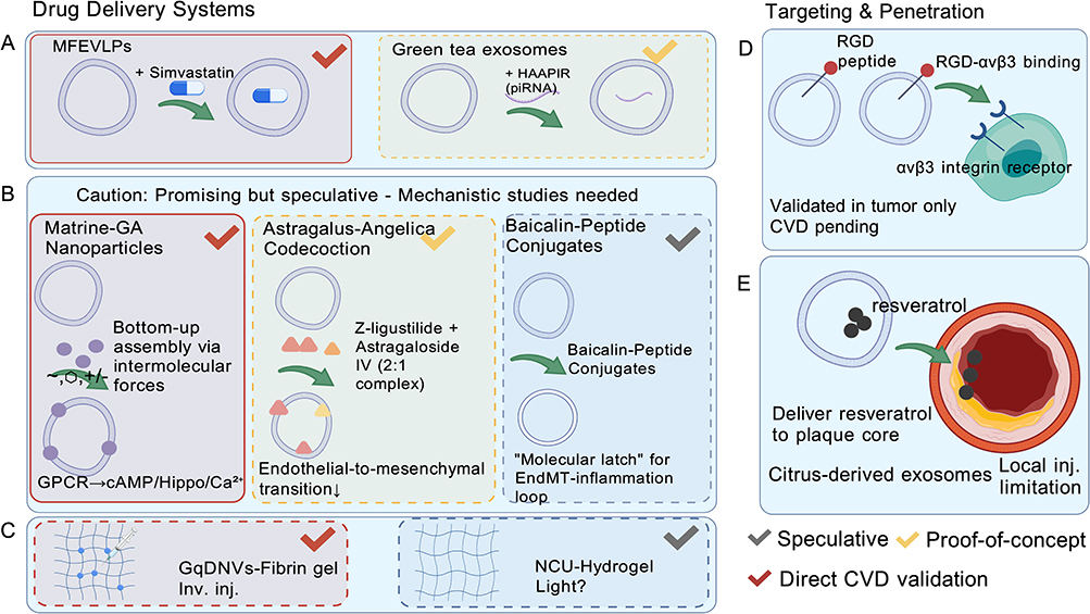 |
Figure 2 Bioengineering strategies of Chinese herbal medicine-derived exosomes (CHM-Exos). (A–C): Drug delivery systems. (A) Physicochemical loading: MFEVLPs-simvastatin (red checkmark: direct CVD validation in ApoE−/− mice) and green tea exosomes-HAAPIR (yellow: proof-of-concept). (B) Self-assembly strategies: Matrine-GA nanoparticles (red), Astragalus-Angelica complex (yellow), and Baicalin-peptide conjugates (gray: speculative; yellow banner indicates mechanistic studies needed). (C) Scaffold systems: GqDNVs-fibrin gel (red, invasive injection limitation) and NCU-hydrogel (gray, light delivery unresolved). (D and E): Targeting and penetration. (D) Active targeting: RGD-αvβ3 binding (yellow: validated in tumor models only; CVD pending). (E) Barrier penetration: Citrus-derived exosomes delivering resveratrol to plaque core (red, local injection limitation). Color key: Red checkmarks = Direct CVD validation; Yellow = Proof-of-concept/indirect evidence; Gray = Speculative. Created with BioGDP.com.5 |
In the delivery of small-molecule statins (simvastatin), CHM-Exos generally enhance drug stability and solubility. This represents a proof-of-concept demonstration; clinical-scale manufacturing, batch consistency, and regulatory-compliant quality control for statin-loaded herbal exosomes remain undeveloped. For instance, mulberry-derived exosomes, with their lipid components (phosphatidylcholine, ceramide), not only help maintain the structural stability of therapeutic agents but also target vascular endothelial cells via cross-species miRNA regulatory mechanisms, modulating the expression of lipid metabolism-related genes and indirectly improving vascular endothelial function.21 Notably, the cardiovascular effects described derive from in vitro and colitis models; direct demonstration of atheroprotective efficacy in established atherosclerosis models is lacking. Recent studies address this gap: Mori fructus-derived extracellular vesicle-like nanoparticles (MFEVLPs) demonstrate oral bioavailability in ApoE−/− mice, where plant-derived miRNAs (miR398-y, miR160-z, miR165-y) suppress hepatic HMGCR and lipogenic genes, reducing atherosclerotic plaque area and enhancing stability.21 Cardiac targeting efficiency, however, requires further investigation.
For nucleic acid drugs, CHM-Exos naturally possess the ability to load, protect, and deliver nucleic acid molecules, which can regulate gene expression and biological functions in target cells.22 While this provides important proof-of-concept for engineering applications, translation to human cardiovascular applications requires validation of oral bioavailability, plasma stability, and cardiac targeting efficiency in large-animal models. For example, green tea-derived exosomes can efficiently encapsulate small piRNA molecules (HAAPIR) and deliver them orally to aortic dissection sites in a mouse model. This complex, by regulating the mef2d and mmp9 pathways, effectively reduces the occurrence of aortic dissection, significantly lowering disease incidence and improving survival rates in mouse models.23
Beyond conventional systemic administration, scaffold-integrated systems have demonstrated enhanced local retention. Gouqi-derived nanovesicles (GqDNVs) embedded in fibrin gel inhibit p38 MAPK/NF-κB p65 signaling and reduce cardiac fibrosis in rat MI models, though local injection limits clinical translation.24 Salvia miltiorrhiza-derived exosome-like nanoparticles (SM-ELNs) provide direct cardiovascular validation in diabetic cardiomyopathy models, inhibiting NLRP3 inflammasome-mediated macrophage pyroptosis via the NEDD4/SGK1 axis.25 An innovative photosynthetic nano-unit (NCU)-hydrogel system produces ATP/NADPH under illumination to address ischemic energy depletion, but light delivery logistics remain unresolved.26
Critical limitations persist: (i) heterogeneity of isolation protocols yield variable size distributions and surface charges; (ii) pharmacokinetic profiles (systemic exposure, tissue distribution, clearance) remain uncharacterized for most CHM-Exos; (iii) scale-up from laboratory to clinical-grade manufacturing is undeveloped; (iv) regulatory classification (botanical drug vs. biological product vs. medical device) requires clarification.
Self-Assembly
Owing to the natural similarity between its lipid bilayer membrane and the cell membrane, the self-assembly characteristics of CHM-Exos provide new insights for engineering modifications (Figure 2). This section represents a promising but largely theoretical application domain; however, recent studies have provided direct cardiovascular validation of self-assembled systems.
This process is regarded as a promising biomimetic strategy, with its core lying in the utilization of the inherent physicochemical properties of CHM-Exo membranes (such as membrane fluidity, surface charge, and lipid composition). Studies have shown that through simple co-incubation, intermolecular driving forces such as hydrophobic interactions, electrostatic adsorption, and π-π stacking can be employed to spontaneously load and integrate hydrophobic drugs or functional molecules into vesicles, forming stable composite nanostructures. This approach demonstrates unique advantages in constructing carrier-free nano-platforms derived from natural plants.27 These studies demonstrate feasibility at the proof-of-concept level; however, controlled assembly efficiency, batch-to-batch reproducibility, and scalability beyond milliliter-scale preparations remain uncharacterized.
Self-assembly strategies have achieved direct cardiovascular validation through diverse mechanisms. Matrine-glycyrrhetinic acid nanoparticles ameliorate myocardial infarction via GPCR-mediated cAMP/Hippo/Ca2⁺ signaling.28 Astragalus-Angelica codecoction elucidates herb-pair self-assembly mechanisms, where Z-ligustilide and astragaloside IV form 2:1 complex that inhibit endothelial-to-mesenchymal transition in myocardial fibrosis.29 Baicalin-peptide conjugates enable ROS-responsive in situ assembly within atherosclerotic plaques, functioning as a “molecular latch” to break the EndMT-inflammation loop—though predictability in heterogeneous human plaques remains uncertain.30
For instance, super self-assemblies prepared from plant decoctions via freeze-thaw technology provide proof-of-concept for developing novel therapeutic agents using self-assembly principles.31 Clinical translation of this approach is constrained by undefined critical quality attributes, absence of standardized assembly protocols, and lack of demonstration in cardiovascular-relevant delivery scenarios.
Compared to active drug loading methods that require complex chemical conjugation or harsh physical operations, this bottom-up self-assembly modification strategy offers significant advantages: firstly, it minimizes potential damage to the natural structure and surface functional proteins of CHM-Exos under harsh conditions, better preserving their inherent biocompatibility and low immunogenicity; secondly, it enables the possibility of high-efficiency loading and programmed intelligent release of therapeutic agents. For example, through rational design, such self-assembled complexes can respond to specific microenvironmental stimuli (such as local acidic pH) to undergo triggered disassembly, precisely releasing drugs.27
Although the field holds great promise, it must be noted that key scientific questions regarding the specific mechanisms of self-assembly in CHM-Exos, precise regulation of assembly efficiency, and their stability in complex in vivo environments remain critical areas requiring focused research.27 Particularly for cardiovascular applications, critical gaps persist: (i) batch-to-batch reproducibility of AM-AS codecoction is sensitive to raw material sourcing and preparation conditions; (ii) scalability of Matrine-GA self-assembly beyond laboratory scale is uncharacterized; (iii) stability of self-assembled structures in systemic circulation, resistance to protein corona formation, and targeted cardiac delivery efficiency have not been experimentally validated; (iv) regulatory pathway for self-assembled herbal nanoparticles remains undefined. Until such validation is achieved, self-assembly applications in cardiovascular disease should be considered speculative for clinical translation, despite promising preclinical results.
Targeted Delivery Systems
CHM-Exos demonstrate significant potential in constructing targeted delivery systems for cardiovascular diseases due to their tunable targeting capabilities and innate ability to penetrate biological barriers (Figure 2). While conceptually attractive, direct experimental evidence validating cardiac-targeted delivery efficiency in vivo remains limited.
Surface modification strategies are effective means to enhance the targeted delivery of CHM-Exos. Most studies to date represent proof-of-concept demonstrations in non-cardiovascular disease models; clinical translation requires validation in relevant pathophysiological contexts.
EDC [1-ethyl-3-(3-dimethylaminopropyl) carbodiimide] and NHS (N-hydroxy succinimide) were to efficiently link molecules containing carboxyl or amino groups to nanoparticle surfaces. The αvβ3 integrin is indeed highly expressed in angiogenesis and atherosclerotic plaques, a phenomenon closely related to its critical role in cell adhesion, migration, and signal transduction.32 RGD cyclic peptides, owing to their high affinity and specificity for αvβ3 integrin,33 are extensively applied in targeted delivery systems. By employing EDC/NHS chemical conjugation technology, arginine-glycine-aspartic acid (RGD) cyclic peptides can be modified onto the surface of CHM-Exos. However, demonstration of RGD-modified herbal exosome targeting to atherosclerotic plaques in vivo, with quantified enrichment efficiency and therapeutic efficacy, is currently unavailable.
The modified CHM-Exos can specifically bind to the highly expressed αvβ3 integrin on damaged vascular endothelium, thereby significantly enhancing the enrichment efficiency of these exosomes at the lesion site and reducing potential effects on normal tissues. This mechanism has been validated primarily in tumor targeting models; cardiovascular-specific validation in atherosclerosis or ischemia models is lacking.
Additionally, studies have achieved targeted delivery of curcumin and baicalin using RGD cyclic peptide-modified nanoliposomes (RGD-Cur/Bai-Lip), markedly improving drug efficacy in liver fibrosis models. This mechanism has been validated primarily in tumor targeting models. Direct extrapolation to cardiac or vascular targeting requires experimental validation.
Engineering strategies now integrate targeting, retention, and modulation for post-infarction heart failure therapy. This framework encompasses cardiac-targeting peptide modification (WLSEAGPVVTARALRGTGSW), functional hydrogel encapsulation, and inflammatory modulation—though the cited study represents a conceptual framework rather than experimental validation in a large-animal model.34 This addresses biodistribution limitations of systemic administration, though invasive implantation restricts clinical translation.
Furthermore, due to their nanoscale particle size (generally less than 150 nm) and lipid bilayer structure, CHM-Exos inherently possess the ability to penetrate biological barriers.35,36 While this property has been demonstrated for blood-brain barrier penetration, evidence for efficient penetration of the vascular wall or fibrous cap in cardiovascular contexts is preliminary. The similarity between their lipid membrane structure (phosphatidylcholine) and vascular endothelial cells promotes fusion and penetration, allowing more effective access to deep lesion sites.36,37 This proposed mechanism is based on theoretical inference and in vitro observations; in vivo validation of deep tissue penetration efficiency in cardiac or vascular lesions is currently unavailable.
For instance, citrus-derived exosomes have demonstrated the ability to penetrate the fibrous cap in atherosclerosis models, delivering antioxidants such as resveratrol to the core region of plaques and significantly reducing oxidative stress marker.38,39 This represents one of the few direct cardiovascular validations; however, the study utilized local injection rather than systemic administration, and the translation to clinically relevant delivery routes remain uncharacterized.
Combining surface modification with innate penetration capabilities produces a synergistic effect: RGD cyclic peptide-mediated active targeting enhances the adhesion of CHM-Exos to diseased blood vessels, while their small size and lipid structure further promote penetration into deep tissues. This synergistic effect is currently theoretical; no studies have demonstrated the combination of surface modification and penetration capabilities in cardiovascular disease models. Clinical realization of this approach will require rigorous validation of targeting efficiency, off-tissue accumulation, and therapeutic index in relevant large-animal cardiovascular models.
Collectively, these engineering strategies—from native cargo utilization to scaffold-based delivery and surface modification—demonstrate varying degrees of cardiovascular validation. To provide a systematic overview of the evidence base, we have summarized the preclinical studies discussed in this section in Table 1, categorizing them by herbal source, engineering strategy, cargo composition, specific CVD model, and evidence level (direct cardiovascular validation vs. proof-of-concept). As illustrated in Table 1, while several studies now provide direct animal model validation in MI, atherosclerosis, and diabetic cardiomyopathy, critical gaps persist in large-animal pharmacokinetics, standardized dosing, and scalable manufacturing protocols necessary for clinical translation.
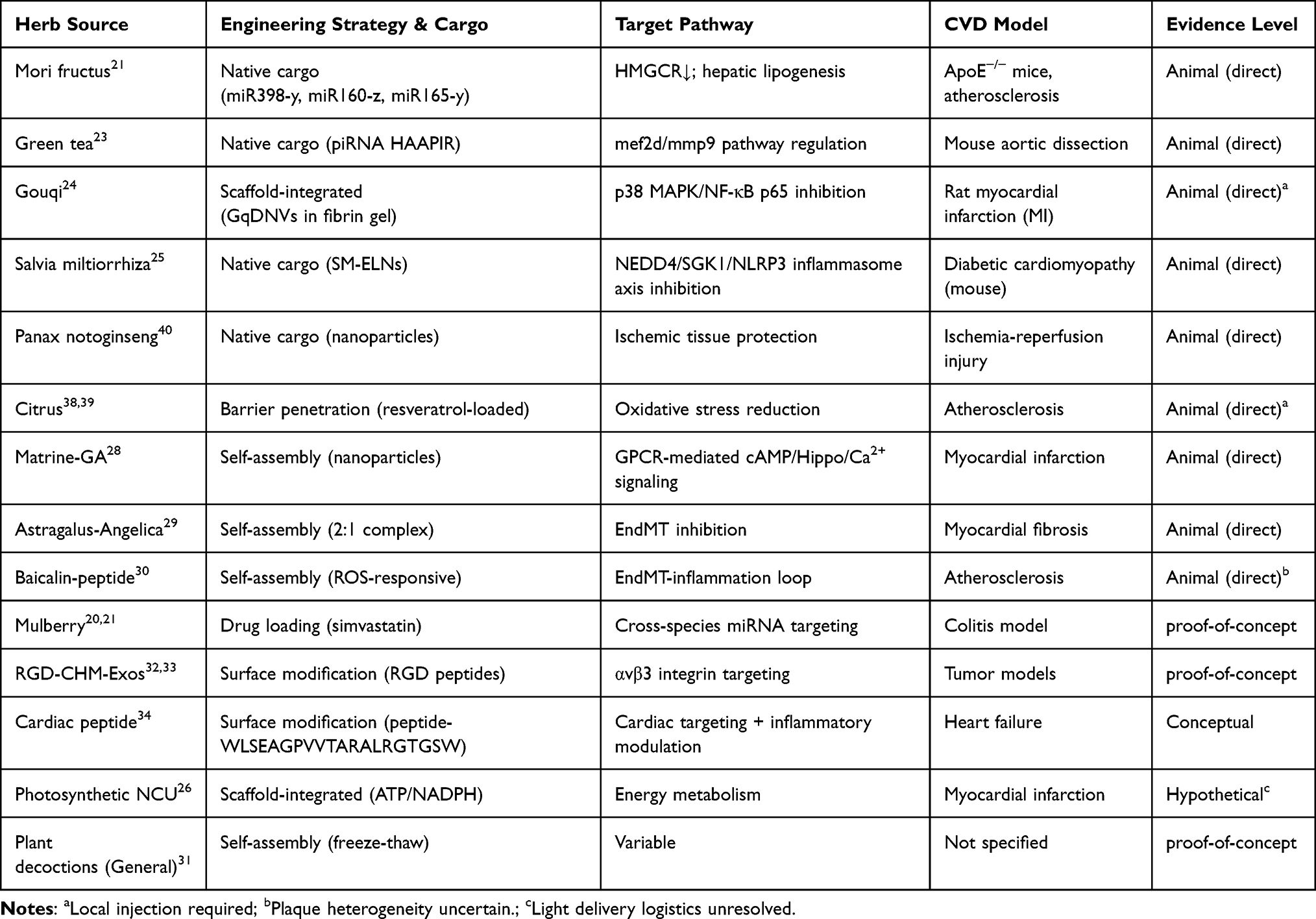 |
Table 1 Summary of Bioengineered CHM-Exosomes in Cardiovascular Diseases: Engineering Strategies and Mechanistic Pathways |
Preclinical Research: Safety, Efficacy, and Translational Barriers
The engineering strategies detailed in Optimization of Drug Delivery Systems–3.3 (native cargo delivery, scaffold-integrated systems, self-assembly, and surface modification) have been evaluated in preclinical cardiovascular models with varying degrees of validation. This section critically examines the safety profiles and therapeutic efficacy of these approaches specifically within CVD contexts, while acknowledging fundamental barriers constraining clinical translation.
Safety Profiles in Cardiovascular Models
CHM-Exos exhibit favorable safety margins in direct CVD models. In rat myocardial infarction models, fibrin gel-loaded Gouqi-derived nanovesicles (GqDNVs) showed no systemic inflammation (stable IL-6/TNF-α levels) or myocardial toxicity (normal troponin T) at therapeutic doses.24 Similarly, Salvia miltiorrhiza-derived exosome-like nanoparticles (SM-ELNs) demonstrated no hepatorenal toxicity (normal ALT/AST/Cr) in diabetic cardiomyopathy mouse models.25 The herb-derived lipid bilayer structure effectively reduces recognition by the complement system and maintains structural stability in the gastrointestinal environment after oral administration.41,42
However, these safety data derive exclusively from rodent models with distinct physiological differences from humans: (i) Immunological divergence: Plant-derived RNAs and lipids may trigger distinct pattern recognition receptor responses in humans versus rodents; (ii) Long-term safety gaps: Current studies typically monitor acute effects (≤4 weeks), whereas cardiovascular diseases require chronic administration; (iii) Batch-to-batch variability: Herbal source heterogeneity (geographical origin, harvest season) introduces composition variability that complicates safety profiling; (iv) Drug interaction uncertainty: Potential pharmacokinetic interactions with standard cardiovascular medications (statins, antiplatelet agents, antihypertensives) remain uncharacterized.
Efficacy Validation by Engineering Strategy
Efficacy validation across the engineering strategies detailed in Optimization of Drug Delivery Systems–3.3 (summarized in Table 1) reveals a spectrum from direct cardiovascular validation to proof-of-concept demonstrations. Native cargo systems have achieved the most robust preclinical validation, with Mori fructus-derived EVs demonstrating oral bioavailability and atheroprotective efficacy in ApoE−/− mice through hepatic lipogenesis suppression,21 and Panax notoginseng-derived nanoparticles attenuating ischemia-reperfusion injury.40 These studies validate systemic administration of unmodified CHM-Exos, though cardiac-specific targeting efficiency remains limited compared to engineered approaches.
Scaffold-integrated and self-assembled systems demonstrate efficacy through distinct mechanistic pathways but face delivery constraints. Fibrin gel-embedded Gouqi-derived nanovesicles reduce cardiac fibrosis in rat MI models via p38 MAPK/NF-κB inhibition,24 while photosynthetic hydrogel systems address ischemic energy depletion through ATP/NADPH production;26 however, both approaches require invasive local implantation, restricting clinical applicability to acute surgical contexts. Similarly, self-assembled Matrine-GA nanoparticles28 and ROS-responsive Baicalin-peptide conjugates30 ameliorate myocardial infarction and atherosclerosis, respectively, but utilize component-based assemblies rather than intact CHM-Exo vesicles, leaving undefined whether native membranes confer additional biodistribution advantages.
Targeted delivery strategies, while conceptually promising, lack quantitative cardiovascular validation. Surface modification approaches using cardiac-targeting peptides or RGD motifs34 have demonstrated specificity primarily in tumor models, with plaque-to-liver accumulation ratios and dose-enrichment relationships remaining unreported in atherosclerotic vasculature. This evidence gap—between sophisticated engineering design and empirical validation in large-animal cardiovascular systems—represents a critical bottleneck for translational progress.
Standardization and Extrapolation Barriers
A critical barrier to translational progress is the absence of standardized preclinical protocols. Current studies exhibit substantial heterogeneity in: (i) Isolation methods (differential ultracentrifugation vs. commercial kits yielding distinct subpopulations); (ii) Dosing parameters (particle concentrations spanning 108–1011 particles/mL without pharmacokinetic justification); (iii) Administration routes (oral, intravenous, intraperitoneal) with undefined biodistribution profiles for cardiovascular tissues; and (iv) Efficacy endpoints (inflammatory markers, plaque size, ejection fraction) lacking harmonization with clinical trial readiness criteria.
The predictive validity for human cardiovascular applications is further constrained by species-specific pathophysiology. Rodent models exhibit: (i) Atherosclerosis differences: Reliance on HDL rather than LDL cholesterol metabolism, and distinct plaque compositions (fewer fibrous caps); (ii) Cardiac regeneration: Unlike human fibrotic scarring, rodents display cardiomyocyte proliferation post-injury; (iii) Gastrointestinal processing: Herbivore-adapted rodent digestive systems alter herbal exosome bioavailability compared to human omnivore physiology. These differences necessitate validation in large-animal models (porcine or non-human primate cardiovascular systems) that approximate human anatomy before clinical translation.
Potential Role of Chinese Herbal Medicine-Derived Exosomes in Cardiovascular Diseases
Despite promising preclinical data and direct cardiovascular validation in rodent models (Figure 3, Stages A-C), the absence of Phase I clinical trials (Stage D) underscores the substantial gap between preclinical promise and clinical realization. The following sections examine the inherent biological activities of CHM-Exos that may ultimately enable clinical applications.
 |
Figure 3 Translational roadmap and critical barriers for CHM-Exos in cardiovascular medicine. Four-stage progression from standardization and sourcing (A) through bioengineering and scale-up (B), preclinical validation (C), to clinical indications and regulatory (D). Red dashed lines indicate the “Valley of Death” between stages. Red stop signs mark critical barriers: standardization gaps (geographic/seasonal variability, undefined CQAs), scalability barriers (GMP manufacturing), cross-species miRNA controversy (yellow highlight in C), and regulatory hurdles (classification unclear). Gray text in Stage D emphasizes the absence of Phase I clinical trials to date. Animal models validated in Stage C include ApoE−/− atherosclerosis, rat MI, and diabetic cardiomyopathy. Speculative technologies (Baicalin-peptide conjugates, NCU-hydrogel) are noted in Stage B; created with BioGDP.com.5 |
Chinese herbal medicine-derived exosomes (CHM-Exos) constitute a pharmacologically defined subpopulation of plant-derived extracellular vesicles isolated from medicinal species listed in traditional Chinese medicine pharmacopeias.43 While general plant EVs derive from dietary sources studied primarily for nutritional applications, CHM-Exos originate from diverse TCM herbs with established therapeutic histories spanning cardiovascular, neurological, metabolic, and immunological indications. This section specifically examines their cardiovascular therapeutic potential. Unlike conventional herbal decoctions where active components exist in free forms vulnerable to gastrointestinal degradation, CHM-Exos utilize phospholipid bilayer architectures to encapsulate labile cargo including miRNAs, ginsenosides, and flavonoids, potentially enabling enhanced oral bioavailability and systemic distribution, though direct comparative pharmacokinetic evidence against conventional extracts requires further characterization.22
While CHM-Exos demonstrate multifaceted therapeutic potential including immunomodulation, anticancer, anti-aging, and detoxification across diverse tissues, cardiovascular applications require specific validation distinct from general cytoprotection. Systematic evidence classification distinguishes direct CVD validation from indirect mechanistic precedents and extrapolation from non-cardiac models, as detailed in Table 2.
 |
Table 2 Mechanistic Insights from Non-Engineered CHM-Exosomes: Evidence from Non-CVD Models Requiring Cardiovascular Validation |
Substance Transport and Immune Regulation
CHM-Exos protect cardiovascular cells through cargo-mediated delivery mechanisms distinct from conventional extract administration.22 The phospholipid bilayer structure provides a natural biological barrier protecting active molecules such as miRNAs from enzymatic degradation.35,50 Direct cardiovascular evidence includes Salvia miltiorrhiza-derived exosome-like nanoparticles (SM-ELNs) which improve diabetic cardiomyopathy by inhibiting NLRP3 inflammasome-mediated macrophage pyroptosis via the NEDD4/SGK1 axis.25 Fibrin gel-loaded Gouqi-derived nanovesicles (GqDNVs) demonstrate cardiac repair efficacy in myocardial infarction (MI) models by inhibiting the p38 MAPK/NF-κB p65 pathway.24 These studies establish direct CVD validation for CHM-Exo therapeutic efficacy beyond general cytoprotection.
For vascular smooth muscle cell regulation, CHM-Exos demonstrate anti-inflammatory activities. The inflammatory factor LIGHT (TNFSF14) activates pro-inflammatory signaling in smooth muscle cells through receptors HVEM and LTβR, promoting cell proliferation and extracellular matrix remodeling. Perilla frutescens leaf-derived extracellular vesicle-like particles demonstrate cross-species regulatory capacity by transporting pab-miR-396a-5p to target heat shock protein 83a (HSP83A), thereby inhibiting NF-κB and JAK/STAT signaling pathways and suppressing IL-17-mediated inflammation.44 While this establishes intercellular communication potential, direct demonstration within atherosclerotic lesion smooth muscle cells—as distinct from psoriatic skin models—remains necessary for definitive cardiovascular claims. Additionally, oxidized low-density lipoprotein activates the NLRP3 inflammasome in smooth muscle cells, inducing pyroptosis and release of pro-inflammatory factors.51 While dandelion and poria-derived vesicles demonstrate NLRP3 inhibition,45,46 these effects have been characterized primarily in colitis models, requiring specific validation in atherosclerosis contexts.
Intercellular Communication: Signaling and Regulatory Mechanisms
CHM-Exos function as signal carriers maintaining cardiovascular homeostasis through intercellular communication with three principal cardiovascular cell types (Figure 4). For macrophage polarization, ginseng exosomes promote M2 polarization by inhibiting TLR4/MyD88/MAPK activation, thereby alleviating inflammation.47 Ginger-derived miRNAs (osa-mir164d) target TAB1, inhibit NF-κB signaling, and reduce pro-inflammatory cytokine secretion.48
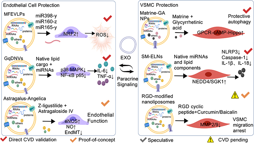 |
Figure 4 CHM-Exo-mediated cardiovascular cell protection and intercellular communication. Protective mechanisms in endothelial cells (left) and VSMCs (right). Endothelial: MFEVLPs activate NRF2; GqDNVs inhibit p38/NF-κB; Astragalus-Angelica upregulate eNOS/NO. VSMC: Matrine-GA induce autophagy; SM-ELNs suppress NLRP3; RGD inhibit MMP2/9. Central EXO hub depicts paracrine signaling. Validation: Red indicates direct CVD validation in animal models; yellow indicates proof-of-concept; warning indicates batch variability or tumor-only validation with CVD efficacy pending; created with BioGDP.com.5 |
Regarding endothelial and tissue protection, CHM-Exos mediate vascular protection through cross-species cargo delivery. Perilla frutescens particles transport pab-miR-396a-5p across species boundaries,44 while Panax notoginseng-derived extracellular-like nanoparticles deliver functional miRNAs to ischemic tissues, restoring cellular function.52 Notably, Panax notoginseng-derived exosome-like nanoparticles also attenuate ischemia-reperfusion injury specifically in cardiac contexts,40 providing direct cardiovascular validation for cross-kingdom RNA transfer mechanisms. Brucea javanica nanovesicles demonstrate miRNA delivery capabilities, though cardiovascular application remains hypothetical.49
For targeted therapeutic strategies in heart failure, recent advances demonstrate engineering plant exosomes for post-infarction therapy through targeted delivery and inflammation suppression. These systems integrate cardiac-targeting modifications with anti-inflammatory modulation, representing a convergence of inherent CHM-Exo bioactivity with bioengineering approaches discussed in Bioengineering Applications of Chinese Herbal Medicine-Derived Exosomes.
The hypothesis that CHM-Exos mediate functional gene regulation through plant-derived miRNAs remains compelling yet contentious. While Perilla EVs demonstrates functional delivery in psoriasis, and Panax notoginseng nanoparticles protect ischemic tissues in neuronal and cardiac contexts, methodological concerns persist regarding detection artifacts and Argonaute loading efficiency. Cross-kingdom miRNA delivery should be considered validated for intercellular communication potential but requiring comprehensive CVD-specific confirmation.
Disease Model Validation
The therapeutic efficacy of CHM-Exos in cardiovascular diseases requires systematic validation across diverse animal models. Mori fructus-derived extracellular vesicle-like nanoparticles regulate dyslipidemia and prevent atherosclerosis progression via functional miRNAs,21 demonstrating oral bioavailability and atheroprotective effects in ApoE−/− mice. This provides robust direct validation for CHM-Exo applications in atherosclerosis.
For myocardial infarction therapy beyond the GqDNV system,26 plant-derived hydrogels integrated with photosynthetic nano-units address ischemic energy depletion in MI models, though light delivery logistics remain unresolved. Direct cardiovascular validations detailed in Bioengineering Applications of Chinese Herbal Medicine-Derived Exosomes (Table 1) further include Matrine-GA nanoparticles28 activating GPCR-mediated signaling in MI, Astragalus-Angelica self-assembly29 inhibiting endothelial-to-mesenchymal transition in fibrosis, and ROS-responsive baicalin-peptide conjugates30 targeting atherosclerotic plaques—mechanisms that demonstrate how engineered delivery systems (Bioengineering Applications of Chinese Herbal Medicine-Derived Exosomes) translate into specific biological pathways (Potential Role of Chinese Herbal Medicine-Derived Exosomes in Cardiovascular Diseases). These self-assembled systems, alongside accessible nanomedicines from Chinese herbal medicines,53 expand the preclinical evidence landscape, though batch reproducibility and scalability require standardization.
Species-specific physiological differences constrain translational validity.22 Rodent models exhibit distinct cholesterol metabolism (HDL-dominant vs. human LDL-dominant), cardiac regeneration capacity (cardiomyocyte proliferation vs. human fibrotic scarring), and herbivore-adapted gastrointestinal processing altering CHM-Exo bioavailability. These differences necessitate large-animal validation. Inter-laboratory reproducibility barriers arise from herbal source heterogeneity affecting yield and cargo composition, while regulatory classification remains ambiguous between botanical drugs and biological products. Most critically, despite promising preclinical data summarized in Table 2, no CHM-Exo formulations have entered Phase I cardiovascular clinical trials, reflecting scalability limitations and the absence of comparative efficacy trials against conventional extracts.
The bioengineering applications in Bioengineering Applications of Chinese Herbal Medicine-Derived Exosomes emphasize surface modification, scaffold integration, and self-assembly to optimize delivery. Potential Role of Chinese Herbal Medicine-Derived Exosomes in Cardiovascular Diseases elucidates inherent CHM-Exo biological activities including cross-kingdom intercellular communication and direct CVD protective mechanisms. These approaches converge on identical translational bottlenecks: isolation heterogeneity, scalability challenges, and species extrapolation limitations. Both require harmonized preclinical protocols and comparative evidence against conventional extracts before clinical translation.
Discussion
Evidence Hierarchy and the Standardization Crisis
While Chinese herbal medicine-derived exosomes (CHM-Exos) have achieved direct cardiovascular validation in diabetic cardiomyopathy, myocardial infarction, and atherosclerosis (Table 2), these remain discrete proof-of-concept investigations rather than a cohesive evidence base. Table 1 further reveals a critical disparity: while native cargo systems demonstrate robust cardiovascular efficacy, engineered strategies (targeted delivery, self-assembly) predominantly show proof-of-concept in non-cardiac models, lacking harmonized endpoints and cross-laboratory validation necessary for clinical translation.54
This translational gap is compounded by profound methodological heterogeneity. The diverse isolation strategies catalogued across Tables 1 and 2—ranging from ultracentrifugation to microfluidics and precipitation kits—yield vesicle populations with divergent physicochemical profiles that undermine batch reproducibility.22,54,55 Herbal sourcing variability (geographic origin, harvest seasonality) introduces additional compositional uncertainty, complicating safety profiling and therapeutic standardization.22
The Cross-Species miRNA Controversy: Mechanistic Validation versus Methodological Uncertainty
A central mechanistic claim—that plant-derived miRNAs functionally regulate mammalian gene expression across species boundaries—rests on evidence from Perilla frutescens44 (pab-miR-396a-5p delivery), Brucea javanica49(cancer-targeting miRNAs), and Panax notoginseng40,52 (neuronal/cardiac protection). However, this hypothesis carries significant overinterpretation risks. Current studies demonstrate functional outcomes yet fail to rigorously distinguish direct miRNA-mediated regulation from indirect effects conferred by concurrent bioactive lipids or glycosides within the vesicular cargo.54 The stability of plant RNAs in mammalian circulation remains inadequately characterized, particularly following oral administration where gastric acid, pancreatic RNases, and intestinal mucus may compromise RNA integrity before systemic absorption.55
While Mori fructus extracellular vesicles demonstrate oral bioavailability in atherosclerosis models,21 quantitative data regarding cardiac tissue uptake versus hepatic first-pass clearance are absent, raising critical questions about systemic dosing requirements for myocardial protection versus off-target effects. The cross-kingdom RNA transfer hypothesis should therefore be considered provisionally validated pending rigorous controls—including RNase digestion experiments, miRNA-specific knockout validations, and cardiovascular-specific dose-response relationships—before clinical translation.43,54,55
Rational Comparison with Mammalian Extracellular Vesicles
Compared to mammalian extracellular vesicles—such as neonatal heart tissue-derived regenerative vesicles,56 peptide-functionalized milk-derived exosomes,57 or adipocyte-cardiomyocyte communicative particles58—CHM-Exos exhibit distinct biocompatibility and cost-effectiveness derived from their phospholipid bilayer architecture and established medicinal botanical sourcing.22,43 However, mammalian EVs possess superior cardiac homing through specific receptor-ligand interactions and characterized GMP-compliant production pathways.56–58
Pharmacokinetic disparities remain stark: while mammalian EVs have defined systemic exposure and clearance profiles,57,58 CHM-Exos face fundamental biodistribution uncertainties regarding myocardial uptake following systemic administration versus localized delivery achieved through fibrin gel scaffolds24 or photosynthetic hydrogel systems.26 Furthermore, surface modification strategies such as RGD-peptide targeting, as conceptualized in engineering frameworks,34 have demonstrated specificity primarily in oncological models; quantitative validation of plaque enrichment ratios in cardiovascular contexts remains unreported. Consequently, the cardiac targeting efficiency and systemic bioavailability of CHM-Exos require rigorous large-animal validation before clinical translation.
Systemic Barriers to Clinical Translation
Current evidence relies on laboratory-scale isolation yielding microgram quantities with undefined critical quality attributes. Transition to industrial-scale production for Phase I cardiovascular trials requires resolution of batch-to-batch variability and development of lyophilization or cryopreservation protocols that retain vesicle integrity during storage and transport.26,54,55
Regulatory ambiguity presents a formidable barrier. The dual identity of CHM-Exos—as both traditional botanical preparations and novel biological nanotherapeutics—creates classification uncertainty across FDA, EMA, and NMPA jurisdictions regarding qualification as botanical drugs, biological products, or medical devices.22,54,55 This complicates safety evaluation standards, particularly regarding cross-species RNA transfer and chronic administration immunogenicity. Furthermore, species-specific physiological differences between rodent models and human patients—including cholesterol metabolism, cardiac regeneration, and gastrointestinal processing—constrain predictive validity,55 necessitating large-animal validation before human dosing.
Strategic Roadmap: From Proof-of-Concept to Clinical Reality
Future priorities must shift from mechanistic accumulation toward resolving standardization and translational bottlenecks. Mechanistic investigations should employ RNase digestion and miRNA-specific depletion experiments to rigorously validate or refute cross-species functional RNA transfer in cardiovascular contexts.43,52,55 Technological development must establish microfluidic isolation protocols with defined critical quality attributes, alongside stimuli-responsive self-assembly strategies that enhance cardiac targeting while addressing oral delivery barriers.30,54,55
Translational validation must progress from rodent models to large-animal systems with mandatory pharmacokinetic characterization and comparative efficacy trials against conventional therapeutics. Regulatory science must clarify CHM-Exo classification within existing drug and device frameworks to establish safety evaluation standards.22,55 Only through this systematic resolution of mechanistic uncertainty, preparation heterogeneity, and regulatory ambiguity—rather than optimistic extrapolation—can these vesicles realize their potential as evidence-based cardiovascular therapeutics.
Conclusion
This systematic review demonstrates that Chinese herbal medicine-derived exosomes (CHM-Exos) possess three defining strengths warranting continued investigation: first, multi-target physiological regulation—encompassing anti-inflammatory, antioxidant, and endothelial protective mechanisms—conferred by natural phospholipid bilayer architectures with inherent biocompatibility; second, bioengineering plasticity enabling self-assembly drug loading, surface functionalization for targeted delivery, and scaffold integration; and third, cost-effective sourcing from established medicinal botanicals with potential for oral administration. However, clinical translation remains contingent upon resolving three systemic barriers. The standardization crisis presents the most immediate obstacle: methodological heterogeneity in isolation protocols coupled with herbal sourcing variability (geographic origin, harvest seasonality) yields batch-to-batch inconsistencies in physicochemical profiles that undermine reproducibility. Pharmacokinetic uncertainties constitute the second barrier: cross-species RNA stability in mammalian circulation remains inadequately characterized, particularly following oral administration where gastric acid and hepatic first-pass clearance may compromise RNA integrity before reaching cardiovascular tissues. Regulatory and manufacturing bottlenecks form the third barrier: the dual identity of CHM-Exos as both botanical preparations and biological nanotherapeutics creates classification ambiguity across FDA, EMA, and NMPA jurisdictions, while scalable GMP-compliant production and long-term storage stability protocols remain undeveloped.
A realistic translational roadmap must therefore proceed through evidence-based resolution of these bottlenecks rather than optimistic extrapolation. Immediate priorities require rigorous mechanistic validation—including RNase-resistant controls to critically assess cross-species RNA transfer hypotheses—alongside establishment of microfluidic isolation protocols with defined critical quality attributes. Medium-term objectives necessitate mandatory progression to large-animal cardiovascular models with harmonized pharmacokinetic characterization and comparative efficacy trials against conventional therapeutics. Long-term clinical realization depends upon regulatory science clarifying classification frameworks and demonstrating scalable manufacturing with batch consistency. Only through this critical, stepwise resolution of heterogeneity, biodistribution uncertainty, and regulatory ambiguity can CHM-Exos advance from proof-of-concept to evidence-based clinical reality.
Abbreviations
ALT, Alanine Aminotransferase; AM-AS, Astragalus-Angelica codecoction; ApoE−/−, Apolipoprotein E knockout; AST, Aspartate Aminotransferase; ATP, Adenosine Triphosphate; cAMP, Cyclic Adenosine Monophosphate; CHM-Exos, Chinese Herbal Medicine-Derived Exosomes; CNKI, China National Knowledge Infrastructure; Cr, Creatinine; CVD, Cardiovascular Disease; DUC, Differential Ultracentrifugation; EDC, 1-ethyl-3-(3-dimethylaminopropyl) carbodiimide; EMA, European Medicines Agency; EndMT, Endothelial-to-Mesenchymal Transition; EVs, Extracellular Vesicles; FDA, Food and Drug Administration; GA, Glycyrrhetinic acid; GMP, Good Manufacturing Practice; GPCR, G Protein-Coupled Receptor; GqDNVs, Gouqi-derived Nanovesicles; HDL, High-Density Lipoprotein; HMGCR, HMG-CoA Reductase; HVEM, Herpesvirus Entry Mediator; IL-6, Interleukin-6; JAK/STAT, Janus Kinase / Signal Transducer and Activator of Transcription; LDL, Low-Density Lipoprotein; LIGHT, Lymphotoxin-like, exhibits Inducible expression, and competes with HSV Glycoprotein D for HVEM; LTβR, Lymphotoxin Beta Receptor; MAPK, Mitogen-Activated Protein Kinase; mef2d, Myocyte Enhancer Factor 2D; MFEVLPs, Mori fructus-derived Extracellular Vesicle-Like Particles; MI, Myocardial Infarction; miRNA, microRNA; mmp9, Matrix Metallopeptidase 9; MyD88, Myeloid Differentiation Primary Response 88; NADPH, Nicotinamide Adenine Dinucleotide Phosphate; NEDD4, Neural precursor cell Expressed developmentally Downregulated 4; NF-κB, Nuclear Factor kappa-light-chain-enhancer of activated B cells; NHS, N-hydroxy succinimide; NMPA, National Medical Products Administration; NLRP3, NOD-, LRR- and pyrin domain-containing protein 3; PBS, Phosphate Buffered Saline; PEG, Polyethylene Glycol; piRNA, PIWI-interacting RNA; RGD, Arginine-Glycine-Aspartic acid; RNase, Ribonuclease; ROS, Reactive Oxygen Species; SEC, Size Exclusion Chromatography; SGK1, Serum/Glucocorticoid Regulated Kinase 1; SM-ELNs, Salvia miltiorrhiza-derived Exosome-Like Nanoparticles; TAB1, TGF-beta Activated Kinase 1 Binding Protein 1; TCM, Traditional Chinese Medicine; TLR4, Toll-Like Receptor 4; TNF-α, Tumor Necrosis Factor-alpha; TNFSF14, Tumor Necrosis Factor Superfamily Member 14.
Data Sharing Statement
Data sharing is not applicable to this article as no datasets were generated or analysed during the current study.
Acknowledgments
We thank BioGDP.com5 for assistance with the preparation of the figures.
Author Contributions
All authors made a significant contribution to the work reported, whether that is in the conception, study design, execution, acquisition of data, analysis and interpretation, or in all these areas; took part in drafting, revising or critically reviewing the article; gave final approval of the version to be published; have agreed on the journal to which the article has been submitted; and agree to be accountable for all aspects of the work.
Funding
This review was not supported by any specific grant from funding agencies in the public, commercial, or not-for-profit sectors.
Disclosure
The authors declare that they have no competing interests.
References
1. Shen H, Zhang M, Liu D, et al. Solanum lycopersicum derived exosome-like nanovesicles alleviate restenosis after vascular injury through the Keap1/Nrf2 pathway. Food Funct. 2025;16(2):539–18. doi:10.1039/d4fo03993a
2. Dhar R, Mukerjee N, Mukherjee D, Devi A, Jha SK, Gorai S. Plant-derived exosomes: a new dimension in cancer therapy. Phytother Res. 2024;38(4):1721–1723. doi:10.1002/ptr.7828
3. Chukhchin DG, Bolotova K, Sinelnikov I, Churilov D, Novozhilov E. Exosomes in the phloem and xylem of woody plants. Planta. 2019;251(1):12. doi:10.1007/s00425-019-03315-y
4. Liu X, Lou K, Zhang Y, Li C, Wei S, Feng S. Unlocking the medicinal potential of plant-derived extracellular vesicles: current progress and future perspectives. Int J Nanomedicine. 2024;19:4877–4892. doi:10.2147/ijn.S463145
5. Jiang S, Li H, Zhang L, et al. Generic Diagramming Platform (GDP): a comprehensive database of high-quality biomedical graphics. Nucleic Acids Res. 2025;53(D1):D1670–d1676. doi:10.1093/nar/gkae973
6. Kalluri R, LeBleu VS. The biology, function, and biomedical applications of exosomes. Science. 2020;367(6478). doi:10.1126/science.aau6977
7. Welsh JA, Goberdhan DCI, O’Driscoll L, et al. Minimal information for studies of extracellular vesicles (MISEV2023): from basic to advanced approaches. J Extracell Vesicles. 2024;13(2):e12404. doi:10.1002/jev2.12404
8. Xu G, Jin J, Fu Z, et al. Extracellular vesicle-based drug overview: research landscape, quality control and nonclinical evaluation strategies. Signal Transduct Target Ther. 2025;10(1):255. doi:10.1038/s41392-025-02312-w
9. Sundaram K, Miller DP, Kumar A, et al. Plant-derived exosomal nanoparticles inhibit pathogenicity of porphyromonas gingivalis. iScience. 2020;23(2):100869. doi:10.1016/j.isci.2020.100869
10. Zhang J, Tian S, Guo L, Zhao H, Mao Z, Miao M. Chinese herbal medicine-derived extracellular vesicles as novel biotherapeutic tools: present and future. J Transl Med. 2024;22(1):1059. doi:10.1186/s12967-024-05892-3
11. Pang C, Zhang J, Gu Y, Zhang Q, Zhao Y. The biological roles of exosome-encapsulated traditional Chinese medicine monomers in neuronal disorders. J Pharm Anal. 2025;15(5):101131. doi:10.1016/j.jpha.2024.101131
12. Almazroua DA, Muir KC, Abid MR. Extracellular vesicles as biomarkers and non-surgical therapeutics in cardiovascular diseases. J Clin Med. 2026;15(4):1537. doi:10.3390/jcm15041537
13. Ambrosone A, Barbulova A, Cappetta E, et al. Plant extracellular vesicles: current landscape and future directions. Plants. 2023;12(24):4141. doi:10.3390/plants12244141
14. Wang R, Zhang Y, Guo Y, et al. Plant-derived nanovesicles: promising therapeutics and drug delivery nanoplatforms for brain disorders. Fundam Res. 2025;5(2):830–850. doi:10.1016/j.fmre.2023.09.007
15. Lian MQ, Chng WH, Liang J, et al. Plant-derived extracellular vesicles: recent advancements and current challenges on their use for biomedical applications. J Extracell Vesicles. 2022;11(12):e12283. doi:10.1002/jev2.12283
16. Wang Y, Wang J, Ma J, Zhou Y, Lu R. Focusing on future applications and current challenges of plant derived extracellular vesicles. Pharmaceuticals. 2022;15(6):708. doi:10.3390/ph15060708
17. Chen M, Lin S, Zhou C, Cui D, Haick H, Tang N. From conventional to microfluidic: progress in extracellular vesicle separation and individual characterization. Adv Healthc Mater. 2023;12(8):e2202437. doi:10.1002/adhm.202202437
18. Franco C, Ghirardello A, Bertazza L, et al. Size-exclusion chromatography combined with ultrafiltration efficiently isolates extracellular vesicles from human blood samples in health and disease. Int J Mol Sci. 2023;24(4):3663. doi:10.3390/ijms24043663
19. Kimiz-Gebologlu I, Oncel SS. Exosomes: large-scale production, isolation, drug loading efficiency, and biodistribution and uptake. J Control Release. 2022;347:533–543. doi:10.1016/j.jconrel.2022.05.027
20. Zhang F, Liang X, Liu H, Anayyat U, Yang Z, Wang X. Advancements and challenges of plant-derived extracellular vesicles in anti-cancer strategies and drug delivery. Curr Drug Deliv. 2025;22(7):921–934. doi:10.2174/0115672018367056250227074828
21. Sui Y, Sun X, Liu Q, et al. Mori fructus-derived extracellular vesicle-like nanoparticles regulate dyslipidemia and prevent atherosclerosis progression via miRNAs. Phytomedicine. 2025;146:157128. doi:10.1016/j.phymed.2025.157128
22. Han X, Zheng W, Sun Z, et al. Plant-derived exosomes: unveiling the similarities and disparities between conventional extract and innovative form. Phytomedicine. 2025;145:157087. doi:10.1016/j.phymed.2025.157087
23. Liu Y, Qi H, Zong J, et al. Oral Piwi-interacting RNA delivery mediated by green tea-derived exosome-like nanovesicles for the treatment of aortic dissection. Adv Healthc Mater. 2024;13(30):e2401466. doi:10.1002/adhm.202401466
24. Zhou HH, Zhou X, Pei J, et al. A fibrin gel-loaded Gouqi-derived nanovesicle (GqDNV) repairs the heart after myocardial infarction by inhibiting p38 MAPK/NF-κB p65 pathway. J Nanobiotechnology. 2025;23(1):535. doi:10.1186/s12951-025-03615-4
25. Peng Z, Gong Z, Wang Z, Deng B, Zhang X, Lin J. Salvia miltiorrhiza-derived exosome-like nanoparticles improve diabetic cardiomyopathy by inhibiting NLRP3 inflammasome-mediated macrophage pyroptosis via targeting the NEDD4/SGK1 axis. Nanomedicine. 2025;20(12):1417–1428. doi:10.1080/17435889.2025.2506351
26. Lv Y, Zhu D, Li J, et al. Plant-derived hydrogel and photosynthetic nano-units for myocardial infarction therapy. Nat Commun. 2025;16(1):7678. doi:10.1038/s41467-025-62020-5
27. Li Z, Xu X, Wang Y, Kong L, Han C. Carrier-free nanoplatforms from natural plants for enhanced bioactivity. J Adv Res. 2023;50:159–176. doi:10.1016/j.jare.2022.09.013
28. Liu J, Xie T, Meng C, et al. Self-assembled matrine-GA supramolecular nanoparticles ameliorate myocardial infarction via GPCR-mediated regulation of cAMP/hippo/Ca2+ signaling pathways. Chem Eng J. 2026;529:173059. doi:10.1016/j.cej.2026.173059
29. Liang P, Bi T, Zhou Y, et al. Insights into the mechanism of supramolecular self-assembly in the astragalus membranaceus-angelica sinensis codecoction. ACS Appl Mater Interfaces. 2023;15(41):47939–47954. doi:10.1021/acsami.3c09494
30. Lin Z, Wu Z, Li L, et al. ROS-responsive self-assembly of baicalin-peptide as a “Molecular Latch” to break the endothelial-to-mesenchymal transition-inflammation loop in atherosclerotic plaques. J Control Release. 2026;393:114821. doi:10.1016/j.jconrel.2026.114821
31. Meng Y, Han D, Xiang J, et al. Self-assembled vectors derived from Aster tataricus L.f. mitigate bleomycin-induced pulmonary fibrosis in mice. Mater Today Bio. 2025;32:101901. doi:10.1016/j.mtbio.2025.101901
32. Yao Y, Li LB, Ji Y, et al. P1216Evaluation of alphavbeta3 integrin-targeted positron emission tomography and photoacoustic tracer for imaging of carotid plaque in apoE–/– mice. European Heart Journal. 2019;40(Supplement_1). doi:10.1093/eurheartj/ehz748.0175
33. Yamada Y, Ohki S, Matsunuma M, et al. Development and biological characterization of the cyclic peptide cRGDTFI with potent binding affinity for integrins αvβ3 and αvβ5. Materials & Design. 2025;257:114466. doi:10.1016/j.matdes.2025.114466
34. Tan X, Lu S, Qin W, et al. From targeted delivery to inflammation suppression: engineering plant exosomes for heart failure therapy post-infarction. Materials Today Bio. 2026;38:103034. doi:10.1016/j.mtbio.2026.103034
35. Shao M, Jin X, Chen S, Yang N, Feng G. Plant-derived extracellular vesicles -a novel clinical anti-inflammatory drug carrier worthy of investigation. Biomed Pharmacother. 2023;169:115904. doi:10.1016/j.biopha.2023.115904
36. Lo KJ, Wang MH, Ho CT, Pan MH. Plant-derived extracellular vesicles: a new revolutionization of modern healthy diets and biomedical applications. J Agric Food Chem. 2024;72(6):2853–2878. doi:10.1021/acs.jafc.3c06867
37. Mao X, Li T, Qi W, et al. Advances in the study of plant-derived extracellular vesicles in the skeletal muscle system. Pharmacol Res. 2024;204:107202. doi:10.1016/j.phrs.2024.107202
38. Di Raimo R, Mizzoni D, Aloi A, et al. Antioxidant effect of a plant-derived extracellular vesicles’ mix on human skin fibroblasts: induction of a reparative process. Antioxidants. 2024;13(11):1373. doi:10.3390/antiox13111373
39. Kim M, Jang H, Kim W, Kim D, Park JH. Therapeutic applications of plant-derived extracellular vesicles as antioxidants for oxidative stress-related diseases. Antioxidants. 2023;12(6). doi:10.3390/antiox12061286
40. Li S, Zhang R, Wang A, et al. Panax notoginseng: derived exosome-like nanoparticles attenuate ischemia reperfusion injury via altering microglia polarization. J Nanobiotechnology. 2023;21(1):416. doi:10.1186/s12951-023-02161-1
41. Zeng YB, Deng X, Shen LS, et al. Advances in plant-derived extracellular vesicles: isolation, composition, and biological functions. Food Funct. 2024;15(23):11319–11341. doi:10.1039/d4fo04321a
42. Fang Z, Liu K. Plant-derived extracellular vesicles as oral drug delivery carriers. J Control Release. 2022;350:389–400. doi:10.1016/j.jconrel.2022.08.046
43. Karabay AZ, Barar J, Hekmatshoar Y, Rahbar Saadat Y. Multifaceted therapeutic potential of plant-derived exosomes: immunomodulation, anticancer, anti-aging, anti-melanogenesis, detoxification, and drug delivery. Biomolecules. 2025;15(3):394. doi:10.3390/biom15030394
44. Liu Y, Tao S, Zhang Z, et al. Perilla frutescens leaf-derived extracellular vesicle-like particles carry Pab-miR-396a-5p to alleviate psoriasis by modulating IL-17 signaling. Research. 2025;8:0675. doi:10.34133/research.0675
45. Sun Y, Liu Y, Li J, et al. Taraxacum mongolicum Hand.-Mazz. derived extracellular vesicles alleviate mastitis via NLRP3 inflammasome and NF-κB/MAPK pathways. J Mater Chem B. 2025;13(34):10609–10620. doi:10.1039/d5tb00861a
46. Wang T, Zhao J, Li QY, et al. Poria cocos-derived exosome-like nanovesicles alleviate metabolic dysfunction-associated fatty liver disease by promoting mitophagy and inhibiting NLRP3 inflammasome activation. Int J Mol Sci. 2025;26(5). doi:10.3390/ijms26052253
47. Fan L, Jia X, Dong F, et al. Ginseng-derived nanoparticles accelerate diabetic wound healing by modulating macrophage polarization and restoring endothelial cell function. Mater Today Bio. 2025;34:102143. doi:10.1016/j.mtbio.2025.102143
48. Yan L, Cao Y, Hou L, et al. Ginger exosome-like nanoparticle-derived miRNA therapeutics: a strategic inhibitor of intestinal inflammation. J Adv Res. 2025;69:1–15. doi:10.1016/j.jare.2024.04.001
49. Yan G, Xiao Q, Zhao J, et al. Brucea javanica derived exosome-like nanovesicles deliver miRNAs for cancer therapy. J Control Release. 2024;367:425–440. doi:10.1016/j.jconrel.2024.01.060
50. Xu Z, Xu Y, Zhang K, et al. Plant-derived extracellular vesicles (PDEVs) in nanomedicine for human disease and therapeutic modalities. J Nanobiotechnology. 2023;21(1):114. doi:10.1186/s12951-023-01858-7
51. He X, Fan X, Bai B, Lu N, Zhang S, Zhang L. Pyroptosis is a critical immune-inflammatory response involved in atherosclerosis. Pharmacol Res. 2021;165:105447. doi:10.1016/j.phrs.2021.105447
52. Yu Y, Tan N, Xu Z, et al. Cross-kingdom miRNA delivery by Panax notoginseng-derived extracellular-like nanoparticles vesicles restores neuronal function after ischemic injury. J Nanobiotechnology. 2026;24(1). doi:10.1186/s12951-026-04103-z
53. Wang YL, Mu Y, Zhang YL, et al. Accessible and effective nanomedicines: self-assembly products from chinese herbal medicines (CHMs). Adv Funct Mater. 2025;35(9):2416151. doi:10.1002/adfm.202416151
54. Zhang J, Pang H, Tang H, et al. The pharmacodynamic and pharmacological mechanisms underlying nanovesicles of natural products: developments and challenges. Pharmacol Ther. 2025;265:108754. doi:10.1016/j.pharmthera.2024.108754
55. Guan X, Zhu M, Zhu H, et al. Oral natural extracellular vesicles for biomedical applications: advances and clinical perspectives. J Adv Res. 2025. doi:10.1016/j.jare.2025.08.003
56. Li H, Liu Y, Lin Y, et al. Cardiac repair using regenerating neonatal heart tissue-derived extracellular vesicles. Nat Commun. 2025;16(1):1292. doi:10.1038/s41467-025-56384-x
57. Tong L, Wang Q, Zhang Y, et al. Myocardial delivery of miR30d with peptide-functionalized milk-derived extracellular vesicles for targeted treatment of hypertrophic heart failure. Biomaterials. 2025;316:122976. doi:10.1016/j.biomaterials.2024.122976
58. Zhang Z, Zhu D, Liu C, et al. Small extracellular vesicle external surface adiponectin-mediated adipocytes/cardiomyocytes communication in diabetic ischemic heart failure. Circulation. 2026. doi:10.1161/circulationaha.125.076372
 © 2026 The Author(s). This work is published and licensed by Dove Medical Press Limited. The
full terms of this license are available at https://www.dovepress.com/terms
and incorporate the Creative Commons Attribution
- Non Commercial (unported, 4.0) License.
By accessing the work you hereby accept the Terms. Non-commercial uses of the work are permitted
without any further permission from Dove Medical Press Limited, provided the work is properly
attributed. For permission for commercial use of this work, please see paragraphs 4.2 and 5 of our Terms.
© 2026 The Author(s). This work is published and licensed by Dove Medical Press Limited. The
full terms of this license are available at https://www.dovepress.com/terms
and incorporate the Creative Commons Attribution
- Non Commercial (unported, 4.0) License.
By accessing the work you hereby accept the Terms. Non-commercial uses of the work are permitted
without any further permission from Dove Medical Press Limited, provided the work is properly
attributed. For permission for commercial use of this work, please see paragraphs 4.2 and 5 of our Terms.