Back to Journals » International Medical Case Reports Journal » Volume 16
Bilateral Upper Limb Complete Phocomelia: A Case Report
Authors Mesfin T , Seyoum K , Tsegaye M , Mesfin E, Tilahun G, Atlie G
Received 13 December 2022
Accepted for publication 4 March 2023
Published 14 March 2023 Volume 2023:16 Pages 167—171
DOI https://doi.org/10.2147/IMCRJ.S401298
Checked for plagiarism Yes
Review by Single anonymous peer review
Peer reviewer comments 2
Editor who approved publication: Professor Ronald Prineas
Telila Mesfin,1 Kenbon Seyoum,2 Mesfin Tsegaye,1 Eshetu Mesfin,3 Geda Tilahun,1 Gela Atlie1
1School of Medicine, Madda Walabu University Goba General Hospital, Goba, Oromia, Ethiopia; 2Department of Midwifery, Madda Walabu University Goba General Hospital, Goba, Oromia, Ethiopia; 3Department of Public Health, ICAP, Finfine, Oromia, Ethiopia
Correspondence: Telila Mesfin, Tel +251931504321, Email [email protected]
Introduction: Phocomelia is an uncommon congenital condition in which the hand or foot are normal or almost normal but the proximal section of the limb – the humerus or femur, radius or tibia, ulna or fibula –_is missing or noticeably hypoplastic. It refers to how the patient’s limbs resemble marine creatures’ flippers and its prevalence is 0.62 in 100,000 births.
Case: We present a 15-min-old male neonate born to a para-four mother who did not remember her LNMP but claimed to be amenorrheic for the past nine months. The mode of delivery was by cesarean section to extract alive neonate weighing 2.01 kg with APGAR scores of 5 and 6 at first and fifth minutes, respectively. The neonate did not cry and was resuscitated for five minutes. He was then transferred to neonatal intensive care unit for further management and investigations. His vital signs were pulse rate 160 beats per minute, respiratory rate 70 breaths per minute, temperature 33.4 degrees centigrade and saturation was 60% off oxygen. On HEENT anterior fontanelle measures 2 cm by 2 cm and has micrognathia and short neck. On the respiratory system, there were intercostal and subcostal retractions, labored breathing and grunting. On the musculoskeletal system there is bilateral upper extremity shortening, the right lower limb was normal in position and structure, the left leg rotated inward (bent in medially) at the knee joint and foot was normal in structure.
Conclusion: Phocomelia is a rare congenital anomaly in which the hand or foot are directly attached to the trunk. Ultrasonography should be done as early as possible to identify fetal anomalies in order to plan subsequent management.
Keywords: phocomelia, upper limb, ultrasonography, neonate
Introduction
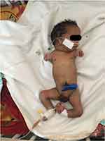
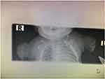
Phocomelia is an uncommon congenital condition in which the hand or foot are normal or almost normal but the proximal section of the limb – the humerus or femur, radius or tibia, ulna or fibula – is missing or noticeably hypoplastic (Figure 1). The hand or foot is directly linked to the trunk in true phocomelia, which is distinguished by the complete absence of the limb’s intermediate segments (Figure 2). The term “phocomelia” was derived from Greek words, and it refers to how the patient’s limbs resemble marine creatures’ flippers.1 It was first coined by Geoffroy Saint-Hilaire.2 Its prevalence is 0.62 in 100,000 births.3 It is also associated with Robert syndrome which is characterized by tetraphocomelia (symmetrical limb reduction) with craniofacial anomalies, growth retardation, mental retardation, cardiac and renal abnormalities.4
 |
Figure 1 A male neonate with complete upper limb phocomelia and internally rotated left lower limb. |
 |
Figure 2 X-ray showing hands directly attached to the trunk and hypoplastic scapulae. |
Case Presentation
We present a 15-min-old male neonate born to a para-four mother who did not remember her LNMP but claimed to be amenorrheic for the past nine months. His mother had antenatal care at the nearby health center twice and was uneventful. However, she had no early obstetric ultrasound. Otherwise, she has no history of chronic medical illnesses like diabetes mellitus and hypertension. During her current pregnancy, she had no fever, headache or blurring of vision. She had never used any drug other than ferrous sulfate throughout this pregnancy. Her prior children are all healthy and alive. On her arrival at our hospital, she had vaginal bleeding of two hours duration. Subsequently, she was admitted with a diagnosis of third-trimester pregnancy plus multigravida plus antepartum hemorrhage secondary to abruptio placentae plus severe oligohydramnios plus breech presentation.
On pelvic examination, she had active vaginal bleeding. Obstetric ultrasound was done on arrival. Accordingly, the gestational age was 34 weeks, the placenta was fundal with a retroplacental clot, and amniotic fluid was significantly decreased with a single deepest pocket of 1.5 cm. The estimated fetal weight was 2.4 kg and breech presentation. Other investigations of the mother are blood group AB+, VDRL=negative, RBS=120 mg/dL, on CBC, white blood cells=9000, hemoglobin=11 gm/dL, platelet=180,000 and neutrophil=60%.
After informed written consent was taken emergency cesarean section was done for already mentioned indications to extract a live neonate weighing 2.01 kg with APGAR scores of 5 and 6 at first and fifth minutes, respectively.
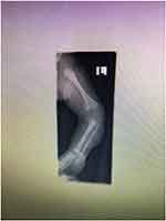
The neonate did not cry and was resuscitated for five minutes. He was then transferred to neonatal intensive care unit for further management and investigations. On arrival, the neonate was in respiratory distress. His vital signs were pulse rate 160 beats per minute, respiratory rate 70 breaths per minute, temperature 33.4 degrees centigrade and saturation was 60% off oxygen. On HEENT anterior fontanelle measures 2 cm by 2 cm and has micrognathia and short neck. On the respiratory system, there were intercostal and subcostal retractions, labored breathing and grunting. On precordial examination, s1 and s2 well heard, no murmur or gallop rhythm. On the musculoskeletal system there is bilateral upper extremity shortening, the right lower limb was normal in position and structure, the left leg rotated inward (bent in medially) at the knee joint and foot was normal in structure (Figure 3). On neurologic examination he was lethargic, the tone was normotonic on lower right extremity and the Moro reflex was difficult to assess. Laboratory investigation showed blood group=A+, CRP=reactive, WBC=30,000, neutrophil=54%, lymphocytes=21.1%, HGB=8 gm/dL and platelet=150,000. X-ray imaging was done and it shows extreme shortening of upper extremities. The left lower limb is internally rotated. Echocardiography was normal. With an assessment of late preterm (34–36 weeks by Ballard score) plus low birth weight plus appropriate for gestational age plus phocomelia plus perinatal asphyxia plus early onset neonatal sepsis he was managed with oxygen support, maintenance fluid, and intravenous antibiotics. The neonate died after three days of hospital stay.
 |
Figure 3 A limb X-ray showing internal rotation of left lower leg. |
Discussion
Phocomelia is the absence of a large portion of the upper and/or lower limbs with preservation of the affected limb’s hands and/or feet.5 Frantz and O’Rahilly defined phocomelia as a type of intercalary failure of formation. Even though their classifications are simple, it has yet to be validated in practice.6 In phocomelia, the cells stopped growing or died, obstructing the normal growth of limbs, eyes, the brain, or other tissues.7 The first instance of phocomelia was reported in Germany in 1956 in a child whose mother had taken thalidomide during pregnancy and who was born without arms and only having vestigial flipper-like hands.8 It can manifest alone as a skeletal deformity or in conjunction with other visceral abnormalities such as cleft palate, hypertelorism, microretrognathia, horseshoe kidney, and polycystic kidney.3 Our patient had micrognathia, internally rotated left leg and short neck. Otherwise, no other visceral malformations were observed. The differential diagnosis could be sporadic phocomelia, Holt–Oram syndrome, thrombocytopenia-absent radius syndrome (TAR syndrome), Roberts syndrome, and thalidomide-induced phocomelia.9
The fourth and fifth children of an Italian couple who were first cousins were born with abnormalities including “double cleft of the lip and palate, protrusion of the intermaxillary portion of the upper jaw, and imperfect development of the bones of the four extremities”, according to John B. Roberts’ description of the family in 1919. Over time, these deformities have come to be recognized as the characteristics of Roberts syndrome.10 This rare autosomal recessive disorder is characterized by tetraphocomelia (symmetrical limb reduction), craniofacial defects, growth retardation, mental impairment, and cardiac and renal abnormalities.11 SC phocomelia, which was first described in 1969 by Herrmann in a family with surname beginning with S and another with surname beginning with C has a less severe symmetric limb reduction, flexion contractures of several joints, minor facial deformities, growth retardation, and perhaps mental retardation.12 SC phocomelia is also autosomal recessive in its inheritance. The severity of the two syndromes can be thought of as falling on a scale, with SC phocomelia being on the milder end and RBS on the more severe end. While severely affected Roberts syndrome newborns may be stillborn or pass away in the postnatal period, people with SC phocomelia often live to adulthood.10
There are a number of risk factors that are believed to be associated with phocomelia. Among the many etiological factors suspected of causing this anomaly, thalidomide is a well-known teratogenic drug. In addition, it has an impact on the neurological, gastrointestinal, cardiovascular, and genitourinary systems.3 Thalidomide has anti-inflammatory, immunomodulatory, and antagonizing properties. It specifically inhibits angiogenesis in the limb, can cause cell death, and induces the formation of reactive oxygen species in limb tissue.13 However, for this particular patient his mother did not take any drugs other than the usual medications prescribed during pregnancy. Likewise, none of her previous children had this kind of malformation. Phocomelia can also be brought on by vascular disruption and ischemia, as shown in limb reduction deficits, mutant genes, chromosomal abnormalities linked to trisomy 18, environmental factors, such as teratogens, and a mix of genetic and environmental factors (multifactorial inheritance).14 Apart from this, substance abuse, such as cocaine or alcohol, gestational diabetes, and X-ray radiation exposure during pregnancy can lead to phocomelia.15,16
Antenatal care should be attended as per WHO protocol so that early in utero identification and possible termination of pregnancies with lethal congenital anomalies can be done. Prenatal diagnosis has been reported as early as 11 weeks of gestation in a high-risk pregnancy with fusion abnormalities of both upper and lower extremities and a large cystic hygroma over the lower back.17 To determine gestational age, look into possible pregnancy issues, and keep track of complex pregnancies when they happen, imaging ultrasound scans are frequently employed. The World Health Organization (WHO) updated its list of advised interventions for standard prenatal care in 2016 to include one ultrasound scan before 24 weeks of pregnancy.18 Routine antenatal ultrasound screening, frequently done in both the first and second trimesters, has long been a common procedure in the majority of high-income countries. An imaging ultrasound scan during the first trimester (up to and including 13 weeks and 6 days of gestation) aims to determine the gestational age, the location of the gestational sac, the number of fetuses, and, in the case of multiple pregnancy, the chorionicity and amnionicity. In addition, toward the end of the first trimester, nuchal translucency thickness is frequently measured in settings that allow it. Between 18 and 24 weeks, second-trimester ultrasound scans can be performed to examine the fetal anatomy in greater detail, look for fetal anomalies, count the number of fetuses present, locate the placenta, and determine the gestational age.19 Therefore, ultrasound imaging should be done as early as possible to detect the fetal anomalies and plan subsequent interventions to avoid unnecessary cesarean delivery.
Conclusion
Phocomelia is a rare congenital anomaly in which the hand or foot are directly attached to the trunk. It is less severe form of Roberts syndrome and the patients with this condition can survive up to adulthood. Even though prenatal thalidomide exposure has been extensively linked with phocomelia alcohol intake, cigarette smoking and cocaine abuse have also been reported to cause it. Ultrasonography should be done as early as possible to identify fetal anomalies in order to plan subsequent management.
Data Sharing Statement
Data on the case clinical information, informed consent form, and images are available for review from the corresponding author upon request.
Consent
Written informed consent was taken from the patient’s parent for publication of his condition and accompanying images.
Disclosure
The authors declare that there are no conflicts of interest.
References
1. Bermejo‐Sánchez E, Cuevas L, Amar E, et al. Phocomelia: a worldwide descriptive epidemiologic study in a large series of cases from the International Clearinghouse for Birth Defects Surveillance and Research, and overview of the literature. In: American Journal of Medical Genetics Part C: Seminars in Medical Genetics. Wiley Online Library; 2011.
2. Dunn P, Fisher A, Kohler H. Phocomelia. Am J Obstet Gynecol. 1962;84(3):348–355. doi:10.1016/0002-9378(62)90131-X
3. Samal SK, Rathod S, Ghose S. Tetra-phocomelia: the seal limb deformity-a case report. J Clin Diagn Res. 2015;9(2):QD01.
4. Goh ESY, Li C, Horsburgh S, et al. The Roberts syndrome/SC phocomelia spectrum—a case report of an adult with review of the literature. Am J Med Genet A. 2010;152(2):472–478. doi:10.1002/ajmg.a.33261
5. Chitayat D, Stalker HJ, Vekemans M, et al. Phocomelia, oligodactyly, and acrania: the Schinzel‐Phocomelia syndrome. Am J Med Genet. 1993;45(3):297–299. doi:10.1002/ajmg.1320450304
6. Frantz CH, O’Rahilly R. Congenital skeletal limb deficiencies. JBJS. 1961;43(8):1202–1224. doi:10.2106/00004623-196143080-00012
7. Chakre GS, Chakre S, Kulkarni P. Phocomelia syndrome-a case report. JKIMSU. 2012;1:150–151.
8. Dar IH, Dar M, Farooq O, et al. Phocomelia: case report of a rare congenital disorder. Egypt J Dermatol Venerol. 2015;35(2):82. doi:10.4103/1110-6530.162227
9. Osadsky CR. Phocomelia: case report and differential diagnosis. Radiol Case Rep. 2011;6(4):561. doi:10.2484/rcr.v6i4.561
10. Van Den Berg DJ, Francke U. Roberts syndrome: a review of 100 cases and a new rating system for severity. Am J Med Genet. 1993;47(7):1104–1123. doi:10.1002/ajmg.1320470735
11. Vega H, Waisfisz Q, Gordillo M, et al. Roberts syndrome is caused by mutations in ESCO2, a human homolog of yeast ECO1 that is essential for the establishment of sister chromatid cohesion. Nat Genet. 2005;37(5):468–470. doi:10.1038/ng1548
12. Herrmann J, Opitz JM. The SC phocomelia and the Roberts syndrome: nosologic aspects. Eur J Pediatr. 1977;125(2):117–134. doi:10.1007/BF00489985
13. Vargesson N. Thalidomide‐induced limb defects: resolving a 50‐year‐old puzzle. Bioessays. 2009;31(12):1327–1336. doi:10.1002/bies.200900103
14. Moore K, Persaud T, Torchia M. Textbook of essentials of embryology and birth defects. In: The Musculoskeletal System.
15. Hu J, Du W, Fang X, et al. A case of trisomy 18 presenting with severe facial abnormality and phocomelia. Int J Hum Genet. 2012;12(4):325–327. doi:10.1080/09723757.2012.11886187
16. Subhani M, Akangire G, Kulkarni A, et al. Al‐Awadi/Raas‐Rothschild/Schinzel (AARRS) phocomelia syndrome: case report and developmental field analysis. Am J Med Genet A. 2009;149(7):1494–1498. doi:10.1002/ajmg.a.32890
17. Naghi I, Behnam B, Behnam B. Roberts-SC phocomelia syndrome (Pseudothalidomide Syndrome): a case report. J Family Reprod Health. 2013;7:45–47.
18. World Health Organization. WHO Antenatal Care Recommendations for a Positive Pregnancy Experience: Maternal and Fetal Assessment Update: Imaging Ultrasound Before 24 Weeks of Pregnancy. World Health Organization; 2022.
19. Gabbe SG, Niebyl JR, Simpson JL, et al. Obstetrics: Normal and Problem Pregnancies e-Book. Elsevier Health Sciences; 2016.
 © 2023 The Author(s). This work is published and licensed by Dove Medical Press Limited. The full terms of this license are available at https://www.dovepress.com/terms.php and incorporate the Creative Commons Attribution - Non Commercial (unported, v3.0) License.
By accessing the work you hereby accept the Terms. Non-commercial uses of the work are permitted without any further permission from Dove Medical Press Limited, provided the work is properly attributed. For permission for commercial use of this work, please see paragraphs 4.2 and 5 of our Terms.
