Back to Journals » International Medical Case Reports Journal » Volume 15
Bilateral Corneal Melting in a Pediatric Patient with Severe Vitamin A Deficiency: A Case Report and Review of Literature
Authors Roheen ZUR, Saleh M, Mushkani TA ![]()
Received 24 February 2022
Accepted for publication 29 April 2022
Published 3 May 2022 Volume 2022:15 Pages 235—238
DOI https://doi.org/10.2147/IMCRJ.S363384
Checked for plagiarism Yes
Review by Single anonymous peer review
Peer reviewer comments 2
Editor who approved publication: Dr Scott Fraser
Zabih Ur Rahman Roheen, Mirwais Saleh, Tawfiq Ahmad Mushkani
Ophthalmology Department, Kabul University of Medical Science, Kabul, Afghanistan
Correspondence: Mirwais Saleh; Tawfiq Ahmad Mushkani, Tel +93786637018 ; +93799123501, Email [email protected]; [email protected]
Background: Keratomalacia refers to an ocular destructive dryness of the cornea that results from severe vitamin A deficiency. It is an ocular condition, usually affecting both eyes. It affects infants, children and women of reproductive age. In developing countries, vitamin A deficiency in the diet and associated keratomalacia are a major cause of childhood blindness.
Purpose: To report the clinical finding of a patient with bilateral keratomalacia arising from severe vitamin A deficiency from a malnourished diet.
Patients and Methods: Case report.
Results: A two-year-old female with severe malnutrition presented with 3 weeks of watering and tonic blepharospasm affecting the right and left eyes. She exhibited trichomegaly, severe weight loss, abnormal hairs with complete corneal melt in the right and left eyes and inflamed iris tissue in the right eye. She was treated with high dose vitamin A, but the right eye required evisceration and tarsorrhaphy was done in the left eye.
Conclusion: The visual morbidity associated with xerophthalmia secondary to vitamin A deficiency can be devastating. Diet-induced vitamin A deficiency is prevalent in developing and war-torn countries. A good history taking and review of systems are valuable in evaluating malnourished patients with corneal melt.
Keywords: keratomalacia, xerophthalmia, vitamin A deficiency
Introduction
Vitamin A deficiency is common in poorer countries, especially among children and women of reproductive age. A fat-soluble vitamin, 90% of vitamin A is stored in the liver. Vitamin A deficiency is the world’s leading cause of preventable childhood blindness. Vitamin A deficiency is estimated to affect about one-third of children under the age of five around the world.1 It is estimated to claim the lives of 670,000 children under five annually.2 About 250,000–500,000 malnourished children in the developing world go blind each year owing to vitamin A deficiency, with the highest prevalence in southeast Asia and Africa.3 Night blindness is one of the first signs of vitamin A deficiency, as the vitamin has a major role in phototransduction. Xerophthalmia, keratomalacia, and complete blindness can follow if the deficiency is more severe.4 Individuals deficient in vitamin A grow poorly, suffer more persistent or severe infections, and subsequently develop characteristic ocular manifestations termed “xerophthalmia”.5 Vitamin A deficiency can be caused by different etiologies and may lead to severe complications, when the deficiency is not treated urgently.6 We report a rare case of bilateral corneal melt arising from severe vitamin A deficiency secondary to malnutrition related to lack of appropriate diet.
Case Report
A two-year-old female child was brought to the University Eye Hospital (UEH, Kabul, Afghanistan). According to her parents’ history, in the last three weeks the patient has progressive pain, tearing and tonic blepharospasm such that she cannot open her eyes. The child weighed 10 kg and measured 60 cm in height. The patient attended the provincial hospital and due to the absence of an ophthalmologist, she was then referred to the University Eye Hospital in Kabul.
The patient’s parents were living in a remote district under insurgents’ control, where no medical facilities are present and no vaccination campaigns are allowed; it is worthwhile that during vaccination campaigns in Afghanistan, supplemental vitamin A is also given to children under the age of five years. The socio-economic situation of the parents was not good, as the child’s father was disabled with no stable payment, meanwhile family planning was not observed. At presentation the patient was cachectic and irritable, the hairs were thin and pale, and trichomegaly was found.
As slit-lamp examination was impossible, she was admitted for Exam Under Anesthesia (EUA) the next day. The EUA revealed diffusely chemotic and keratinized conjunctiva in both eyes (OU) with xerosis but no Bitot’s spots. Both corneas demonstrated total melt from limbus to limbus, with a collapsed anterior chamber. The right eye had a purulent iris that was affixed to the lens by extensive posterior synechiae(Figures 1 and 2).
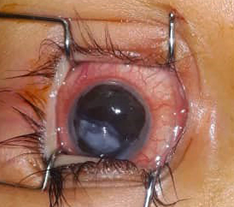 |
Figure 1 Clinical photograph of the patient shows total corneal melt in the right eye. The right eye has purulent iris attached to the lens by posterior synechiae. |
 |
Figure 2 Clinical photograph of the patient shows total corneal melt in the left eye. |
A diagnosis of X3-B keratomalacia with severe protein-energy malnutrition was made. Treatment was initiated in the form of 200.000 units of vitamin-A acetate administered by mouth in divided doses for 2 days and repeated 2 weeks later.
Because of poor visual potential and risk of sympathetic ophthalmia, the patient’s right eye was eviscerated and the patient’s left eye was treated with temporary tarsorrhaphy. The patient was then referred to a specialized eye institution for possible corneal graft.
Discussion
Keratomalacia resulting from vitamin A deficiency is an important cause of preventable corneal blindness and opacification and a major cause of pediatric ocular morbidity and severe visual impairment in developing countries.7
A major study of 162 children with nutritional keratopathy analyzed the pathology and clinical presentation of corneal xerophthalmia and keratomalacia. In this study surface changes ranged from mild haziness through generalized xerosis and formation of stromal melting.8
In a follow-up study of patients with severe xerophthalmia, of 32 children with keratomalacia, nine died because of severe protein energy malnutrition. Of those remaining, five had become bilaterally blind, nine uniocularly blind, while nine retained adequate vision.9
A study of 29 children with keratomalacia ranging from 2 months to five years of age, showed that all children belonged to families of lower socioeconomic status. 27 patients had not been immunized at all. 36 eyes (66.7%) had total corneal melting and 11 eyes (20.3%) had paracentral corneal melting.10
Our patient had extremely poor socioeconomic status with poor nutritional history, which may have contributed to development of this severe form of keratomalacia due to dietary deficiency. Based on characteristic conjunctival and corneal findings, malnourished general appearance of the patient, poor nutritional history, higher prevalence of vitamin A deficiency in Afghanistan especially in remote and war-torn areas, and absence of other local and systemic conditions to explain the ocular finding, she was diagnosed to have X3-B keratomalacia.
Beside systemic management, specific medical measures in acute keratomalacia include vitamin A supplementation. Vitamin A supplementation can help reduce morbidity and mortality in affected children.11,12
The limitation of this case report was that laboratory facilities for serum retinol level, conjunctival impression cytology and donor cornea for keratoplasty were not available.
Finally, we conclude that awareness of this condition is recommended to detect the disease at the earliest stage and manage accordingly. Health education and other preventive measures including education for families on healthy diet and importance of immunization for children, reduces the risk of vitamin A deficiency and consequent ocular morbidities in children under five years of age.
Conclusion
The visual morbidity associated with xerophthalmia secondary to vitamin A deficiency can be devastating. Diet-induced vitamin A deficiency is prevalent in developing and war-torn countries. A good history taking and review of systems are valuable in evaluating malnourished patients with corneal melt.
Abbreviations
UEH, University Eye Hospital; EUA, Exam Under Anesthesia.
Consent for Publication
The institutional approval is not required for publishing the case report. Written informed consent for publication of their details was obtained from the father of the patient.
Disclosure
The authors report no conflicts of interest in this work.
References
1. World Health Organization. Global prevalence of vitamin-A deficiency in population at risk 1999–2005. WHO global database on vitamin A deficiency; 2022.
2. Black RE, Allen LH, Bhutta ZA, et al. Maternal and child undernutrition: global and regional exposures and health consequences. Lancet. 2008;371(9608):253. doi:10.1016/S0140-6736(07)61690-0
3. World Health Organization. Micronutrient deficiencies: Vitamin A; 2013.
4. Higdone J, Delage B. “Vitamin A”. Micronutrient Information Center, Linus Pauling institute, Oregon State University, Corvallis, January 2015; 2019.
5. Sommer A. Vitamin a Deficiency and Its Consequences: A Field Guide to Detection and Control. Geneva: World Health Organization. ISBN 92-4-154478-3; 1995.
6. Susan EP, Bedrossian EH Jr, Eagle RC Jr, et al. Corneal perforation in patients with vitamin A deficiency in the United States. Arch Ophthalmol. 1990;108:350–353. doi:10.1001/archopht.1990.01070050048028
7. Rahi JS, Sripathi S, Gilbert CE, et al. Childhood blindness due to vitamin A deficiency in India: regional variations. Arch Dis Child. 1995;72:330–333. doi:10.1136/adc.72.4.330
8. Sommer A, Sugana T. Corneal Xerophthalmia and keratomalacia. Arch Ophthalmol. 1982;100:404–411. doi:10.1001/archopht.1982.01030030406003
9. Menon K, Vijayarangavan K. Sequela of sever xerophthalmia-A follow-up study. Am J Clin Nutr. 1980;33:218–220. doi:10.1093/ajcn/33.2.218
10. Vajpayee RB, Vanathi M, Tandon R, Sharma N, Titiyal JS. Keratoplasty for keratomalacia in preschool children. Br J Ophtalmol. 2003;87(5):538–542. doi:10.1136/bjo.87.5.538
11. Sommer A, Djunaedi E, Loeden AA, et al. Impact of vitamin A supplementation on childhood mortality. Lancet. 1986;327(8491):1169–1173. doi:10.1016/S0140-6736(86)91157-8
12. Vitte S, Lawani R, Bouat C, et al. Xerophthlamia; current data. Med Trop. 1995;5(4 Pt 2):434–438.
 © 2022 The Author(s). This work is published and licensed by Dove Medical Press Limited. The
full terms of this license are available at https://www.dovepress.com/terms
and incorporate the Creative Commons Attribution
- Non Commercial (unported, 3.0) License.
By accessing the work you hereby accept the Terms. Non-commercial uses of the work are permitted
without any further permission from Dove Medical Press Limited, provided the work is properly
attributed. For permission for commercial use of this work, please see paragraphs 4.2 and 5 of our Terms.
© 2022 The Author(s). This work is published and licensed by Dove Medical Press Limited. The
full terms of this license are available at https://www.dovepress.com/terms
and incorporate the Creative Commons Attribution
- Non Commercial (unported, 3.0) License.
By accessing the work you hereby accept the Terms. Non-commercial uses of the work are permitted
without any further permission from Dove Medical Press Limited, provided the work is properly
attributed. For permission for commercial use of this work, please see paragraphs 4.2 and 5 of our Terms.