
Back to Journals » International Medical Case Reports Journal » Volume 17
Bilateral Chronic Herpetic Anterior Uveitis in an Immunocompetent Patient
Authors Chu JP  , Gilhotra JS
Received 5 September 2024
Accepted for publication 31 October 2024
Published 4 November 2024 Volume 2024:17 Pages 913—918
DOI https://doi.org/10.2147/IMCRJ.S490693
Checked for plagiarism Yes
Review by Single anonymous peer review
Peer reviewer comments 2
Editor who approved publication: Dr Scott Fraser
Jan Patrick Chu, Jagjit Singh Gilhotra
Royal Adelaide Hospital, Adelaide, South Australia, Australia
Correspondence: Jan Patrick Chu, Email [email protected]
Purpose: To present a case of bilateral herpes simplex anterior uveitis in an immunocompetent patient.
Methods: Case Report.
Results: A 42-year-old Kenyan female presented with a 2-year history of intermittent painful eye redness associated with blurring of vision of both eyes. Symptoms started after childbirth. There were no associated systemic symptoms. She presented with a best corrected visual acuity (BCVA) of 6/18 and 6/30 on the right and left eyes, respectively. On examination, conjunctival hyperaemia, large keratic precipitates (KPs), posterior synechiae, poorly dilating pupil, anterior subcapsular cataract and +2 anterior chamber (AC) cells and flare were noted on both eyes. Intraocular pressures (IOP) were within normal limits. Optical coherence tomography (OCT) showed bilateral cystoid macular oedema (CMO). Serum herpes simplex virus (HSV) IgM was detected, whereas autoimmune and other infectious aetiologies were excluded. Aqueous humour samples from both eyes tested negative for HSV. Bilateral 16 mg subtenon triamcinolone injection were done. Dexamethasone 0.1% eye drops and atropine 1.0% eye drops were started. Topical anti-glaucomatous medication was started due to IOP > 30 mmHg on both eyes and Valacyclovir 1g three times a day was initiated. Final visit showed an improvement of BCVA to 6/9 on both eyes. There was complete resolution of AC cells and flare, and CMO on OCT.
Conclusion: The diagnosis of bilateral herpetic anterior uveitis was based on the criteria set by The Herpetic Eye Disease Study (HEDS). Although the patient was immunocompetent, she was in a state of transient immunodeficiency that is pregnancy, which could have led to bilateral ocular involvement.
Keywords: anterior, bilateral, chronic, herpes simplex virus, immunocompetent
Introduction
Herpes simplex virus (HSV) is a prevalent infectious agent that can cause various clinical manifestations including uveitis.1 Uveitis associated with HSV is one of the principal causes of blindness in developing countries,2 and it accounts for 5% to 10% of all uveitis cases.3
HSV associated anterior uveitis is characteristically unilateral.1,4,5 Nevertheless, bilateral cases do occur especially in the context of immunocompromise.5 However, it is rarely thought of as a differential diagnosis in bilateral anterior uveitis presentations.1 Therefore, it is imperative to support diagnosis with a positive serum HSV antibodies6 or a positive aqueous humour polymerase chain reaction (PCR) analysis to HSV antigens.5
In literature, there have been only few reported cases of bilateral anterior uveitis associated with HSV. In this article, we describe a case of bilateral chronic anterior uveitis in a serum HSV IgM positive immunocompetent patient.
Methods
This study is a case report that used the Sunrise Electronic Medical Records at the Royal Adelaide Hospital, South Australia, to acquire data that were de-identified. This study was performed according to the principles of Good Clinical Practice [Chapter 2 of the ICH Harmonized Tripartite Guideline for Good Clinical Practice (GCP)], the declaration of Helsinki, and national laws and regulations about clinical studies. Approval for research and publication was granted by the Central Adelaide Local Health Network (CALHN) Human Research Ethics Committee.
Case Report
This is a case of a 42-year-old female from Kenya who immigrated to Australia in 2011. She presented with a 2-year history of intermittent painful eye redness associated with blurring of vision of both eyes. Ocular symptoms began soon after childbirth. There were no associated systemic symptoms. Hypertension and hypothyroidism were the only known comorbidities and were well-controlled.
On examination, she presented with a best corrected visual acuity (BCVA) of 6/18 and 6/30 on the right and left eyes, respectively. Conjunctival hyperaemia, large keratic precipitates, posterior synechiae, poorly dilating pupil, anterior subcapsular cataract and +2 anterior chamber (AC) cells and flare were noted on both eyes. No iris atrophy and iris transillumination defects were seen. Intraocular pressures (IOP) were within normal limits. Fundus examinations showed no signs of vitritis, retinitis, and vasculitis; however, dull foveal reflexes were noted on both eyes. Macular optical coherence tomography (OCT) showed bilateral cystoid macular oedema (CMO) (Figure 1A and B).
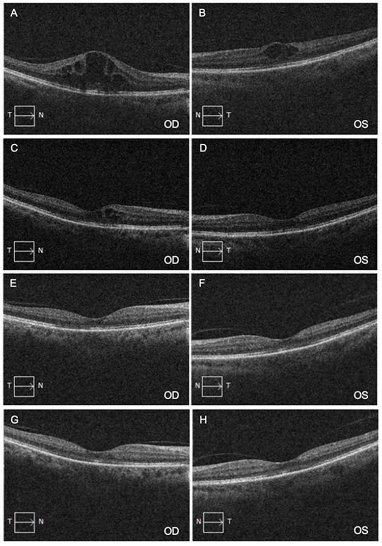 |
Figure 1 (A and B) Macular OCT showing uveitic macular oedema in both eyes; (C and D) Macular OCT 2 weeks post-subtenon triamcinolone injections; (E and F) Macular OCT 4 weeks post-subtenon triamcinolone injections; (G and H) Macular OCT 2 months post-subtenon triamcinolone injections. |
Dexamethasone 0.1% eye drop 6 times a day and atropine 1.0% eye drop 3 times a day to both eyes were initiated at initial visit. A week later, CMOs were still present in both eyes, which led to a decision to perform bilateral subtenon injections of 16 mg triamcinolone.
Two weeks post-subtenon injections, the BCVA improved to 6/12 on the right eye and 6/9 on the left eye. In the anterior chamber of both eyes, only 0.5 cells with resolution of flare and KPs were noted. On macular OCT, the CMO in the right eye partially resolved while it completely resolved in the left eye (Figure 1C and D). IOP measured at 32 mmHg and 34 mmHg on the right and left eye, respectively, were attributed as a steroid response, which were treated with topical anti-glaucomatous medication. The patient was monitored every 2 weeks.
Subsequent visits showed further improvements. BCVA was 6/9 on both eyes and IOP of both eyes were back to normal range. No AC cells were appreciated in both eyes. However, the patient still presented with poorly dilating pupils due to posterior synechiae, and an anterior subcapsular cataract in both eyes (Figure 2). Macular OCT showed complete resolution of CMO in both eyes (Figure 1E and F).
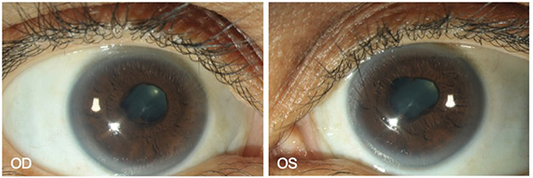 |
Figure 2 Anterior segment photos of both eyes 2 months into treatment. Quiet anterior chamber, presence of posterior synechiae and anterior subcapsular cataract in both eyes were observed. |
The patient tested positive for serum HSV IgM, which resulted in initiation of Valacyclovir 1 g three times a day. Autoimmune and other infectious aetiologies were excluded. HSV DNA were not detected on aqueous humour PCR analyses of both eyes.
On the last visit, there were no recurrence of eye redness, pain and signs of active intraocular inflammation. Although the patient reported cloudy blurring of vision, this was attributable to the cataract formation in both eyes. Dexamethasone 0.1% eye drop was reduced to 4 times a day and atropine 1.0% eye drop to once at night, and oral valacyclovir 1 g three times a day was continued.
Discussion
Herpes simplex virus associated anterior uveitis typically presents unilaterally accompanied by ocular hypertension, keratic precipitates (KPs) and sectoral iris atrophy with or without transillumination defects.1 Concomitant keratitis can occur in 30%–40%,5 but its absence does not exclude herpetic uveitis origin.4
It mainly occurs in middle-aged inidividuals,4,7 and also has a female preponderance.4,8 A case series in Italy reported that 59.8% of their patients diagnosed with HSV uveitis were female.8 These are consistent with our patient’s demographics. However, the case presented has several deviations from the typical presentation of HSV anterior uveitis.
To date, only few studies have described bilateral HSV anterior uveitis. The incidence of bilateral ocular involvement varies among literatures. One study stated that the frequency of bilateral involvement ranges from 3% to 11.9% and is more common in younger individuals,8 while another study reported bilateral occurrence in 18% of their patients.9
It has been reported that bilateral ocular involvement is associated with compromised immunity.5 Although our patient does not have comorbidities that would significantly impact her immune system, it is important to note that the ocular symptoms began after childbirth. Kump et al reported that high levels of progesterone during pregnancy have an immunosuppressive effect similar to corticosteroids. Similarly, alpha fetoprotein, which is the major fetal serum protein that protects the fetus from the maternal immune system also has immunosuppressive effects especially on T-helper cells.10 These could explain the patient’s bilateral ocular involvement since she was just recovering from a transient immunocompromised state that is pregnancy.
In 1969, a clinical report described a 34-year-old Caucasian woman who developed bilateral herpes simplex panuveitis 10 days postpartum. Her past ocular history revealed a previous bout of anterior uveitis in the right eye in 1963 that also started in the second week postpartum.11
Our patient also presented with normal IOP in both eyes despite active ocular inflammation and posterior synechiae formation. Ocular hypertension is frequently observed in 46% to 90% of patients.4 However, the IOP elevation seems to be transient as only few patients progress to secondary glaucoma.4 However, raised IOP in HSV anterior uveitis could also be permanent in some cases. A case series in Turkey reported that 68% of their patients exhibited transient rise in IOP, while only 31.8% had permanent IOP elevation.12
KPs in HSV anterior uveitis can appear as fine stellate or granulomatous KPs,1 but the typical herpetic KPs are large.13 These usually disappear with treatment13 as in our patient whose KPs in both eyes resolved within two weeks of treatment.
Sectorial iris atrophy is considered pathognomonic for HSV anterior uveitis. It is known to be not present at disease onset, but eventually develops as the course of the disease progresses.4 It is also thought to be more frequently seen in eyes without corneal involvement.13 Our patient did not show signs of it is atrophy despite the chronic presentation; however, not all patients would present with such finding. Neumann et al reported that only 50.9% of the patients in their case series presented with iris atrophy,7 which is almost similar to the finding of Tugal-Tutkun et al in their case series at 48%.13
Although macular oedema secondary to uveitis is more commonly observed in intermediate, posterior and panuveitis, it could still present in 11% of patients with isolated anterior segment inflammation.14 A case series of 1417 uveitis patients in Italy reported that macular oedema was present in 20–26% of anterior uveitis,15 while another case series of 1510 eyes in USA observed macular oedema in 28% of anterior uveitis.16
CMO is the most common form of macular oedema and is mostly observed in patients with chronic uveitis.14 Our patient presented with bilateral CMO confirmed by OCT for which she received bilateral subtenon triamcinolone injections for. Corticosteroids continue to be the mainstay of treatment of uveitic macular oedema.17 Although systemic corticosteroids are more commonly utilized for bilateral CMO,17 the decision to perform bilateral subtenon injections was based on pending negative tuberculosis result at the time, and patient compliance concern.
Subtenon triamcinolone injection is said to be comparable to intravitreal injection as both are able to reach similar vitreous concentrations.18 It still has the ability to cause secondary ocular hypertension, but anti-glaucomatous medications are highly effective in controlling IOP17 as in our patient.
The patient was diagnosed to have bilateral HSV anterior uveitis based on the HEDS criteria supported by the presence of serum antibodies to HSV.6 In most centres, the diagnosis of herpetic anterior uveitis remains to be clinical and intraocular fluid analysis is only done to confirm diagnosis in some cases.9
In the advent of PCR techniques, it is important to confirm diagnosis with such procedures especially in bilateral anterior uveitis.5 PCR analysis of intraocular fluid has a high sensitivity and specificity,7 and it is reported that its sensitivity ranges from 25% to 100% in different studies.19 HSV DNA was undetected in the aqueous humour samples of our patient. However, this does not rule out the disease because viral DNA may reach below the detection limit in the chronic stages whereas PCR tests tend to be positive at acute onset disease or during early reactivation.20 It is important to mention that the PCR analyses results of our patient were expressed as “not detected” rather than “negative” because the results may be interpreted as false negative especially that the patient tested positive for serum HSV IgM that is indicative of a recent infection.21
Topical corticosteroids and oral antiviral agents remain to be the mainstay of treatment in patients with herpetic anterior uveitis.13,19 The HEDS demonstrated that prophylactic use of acyclovir reduced the probability of recurrence of ocular HSV in a 12-month treatment period by nearly half.22 Moreover, a retrospective study reported that long-term prophylactic use of acyclovir was found to be effective in reducing recurrences beyond 12 months.23 Two studies recommended the use of prophylactic dose of acyclovir at 800 mg daily for at least 2 years after the episode of uveitis, with the possibility of life-long treatment.9,24
Conclusion
The diagnosis of herpetic anterior uveitis is still a clinical diagnosis in most centres and intraocular fluid analysis is only done in a few cases where confirmatory diagnosis is needed.9 In cases of bilateral presentations, it is important to rule out other possible autoimmune and infectious aetiologies and have a high index of suspicion. A proper thorough history taking is also imperative as it could uncover clinical scenarios that could potentially be associated with the clinical presentation as in our patient. As in the patient’s case, the bilateral presentation could be due to the transient state of immunodeficiency that is pregnancy. Prompt treatment should be given to limit secondary complications, and continuous monitoring is important as recurrences are common.
Consent
The patient was consented to the publication of this study. This study does not contain patient identifiers or personal information.
Funding
No funding was received for this manuscript.
Disclosure
The authors declare no conflicts of interest.
References
1. de-la-Torre A, Valdes-Camacho J, Foster CS. Bilateral herpes simplex uveitis: review of the literature and own reports. Ocul Immunol Inflammation. 2017;25(4):497–502. doi:10.3109/09273948.2016.1142572
2. Liesegang TJ. Herpes simplex virus epidemiology and ocular importance. Cornea. 2001;20(1):1–13. doi:10.1097/00003226-200101000-00001
3. Rathinam SR, Namperumalsamy P. Global variation and pattern changes in epidemiology of uveitis. Indian J Ophthalmol. 2007;55(3):173–183. doi:10.4103/0301-4738.31936
4. Wensing B, Mochizuki M, De Boer JH. Clinical characteristics of herpes simplex virus associated anterior uveitis. Ocul Immunol Inflammation. 2018;26(3):333–337. doi:10.1080/09273948.2017.1420806
5. Standardization of Uveitis Nomenclature (SUN) Working Group, Classification criteria for herpes simplex virus anterior uveitis. Am J Ophthalmol. 2021;(228):231–236. doi:10.1016/j.ajo.2021.03.053
6. The Herpetic Eye Disease Study Group. A controlled trial of oral Acyclovir for iridocyclitis caused by herpes simplex virus. Archiv Ophthalmol. 1996;114(9):1065–1072. doi:10.1001/archopht.1996.01100140267002
7. Neumann R, Barequet D, Rosenblatt A, et al. Herpetic anterior uveitis - analysis of presumed and PCR proven cases. Ocul Immunol Inflammation. 2019;27(2):211–218. doi:10.1080/09273948.2018.1483521
8. Miserocchi E, Fogliato G, Bianchi I, Bandello F, Modorati G. Clinical features of ocular herpetic infection in an Italian referral center. Cornea. 2014;33(6):565–570. doi:10.1097/ICO.0000000000000129
9. Miserocchi E, Waheed NK, Dios E, et al. Visual outcome in herpes simplex virus and varicella zoster virus uveitis: a clinical evaluation and comparison. Ophthalmology. 2002;109(8):1532–1537. doi:10.1016/s0161-6420(02)01113-2
10. Kump LI, Cervantes-Castañeda RA, Androudi SN, Foster CS, Christen WG. Patterns of exacerbations of chronic non-infectious uveitis in pregnancy and puerperium. Ocul Immunol Inflammation. 2006;14(2):99–104. doi:10.1080/09273940500557027
11. Pavan-Langston D, Brockhurst RJ. Herpes simplex panuveitis. A clinical report. Archiv Ophthalmol. 1969;81(6):783–787. doi:10.1001/archopht.1969.00990010785005
12. Nalcacioglu-Yüksekkaya P, Ozdal PC, Teke MY, Kara C, Ozturk F. Presumed herpetic anterior uveitis: a study with retrospective analysis of 79 cases. European J Ophthalmol. 2014;24(1):14–20. doi:10.5301/ejo.5000331
13. Tugal-Tutkun I, Otük-Yasar B, Altinkurt E. Clinical features and prognosis of herpetic anterior uveitis: a retrospective study of 111 cases. Intl Ophthalmol. 2010;30(5):559–565. doi:10.1007/s10792-010-9394-8
14. Accorinti M, Okada AA, Smith JR, Gilardi M. Epidemiology of macular edema in uveitis. Ocul Immunol Inflammation. 2019;27(2):169–180. doi:10.1080/09273948.2019.1576910
15. Pivetti-Pezzi P, Accorinti M, La Cava M, Colabelli Gisoldi RA, Abdulaziz MA. Endogenous uveitis: an analysis of 1417 cases. Ophthalmologica. 1996;210(4):234–238. doi:10.1159/000310715
16. Levin MH, Pistilli M, Daniel E, et al.; Systemic Immunosuppressive Therapy for Eye Diseases Cohort Study. Incidence of visual improvement in uveitis cases with visual impairment caused by macular edema. Ophthalmology. 2014;121(2):588–95.e1. doi:10.1016/j.ophtha.2013.09.023
17. Koronis S, Stavrakas P, Balidis M, Kozeis N, Tranos PG. Update in treatment of uveitic macular edema. Drug Des Devel Ther. 2019;13:667–680. doi:10.2147/DDDT.S166092
18. Fardeau C, Champion E, Massamba N, LeHoang P. Uveitic macular edema. Eye. 2016;30(10):1277–1292. doi:10.1038/eye.2016.115
19. Siverio Júnior CD, Imai Y, Cunningham ET. Diagnosis and management of herpetic anterior uveitis. Int Ophthalmol Clin. 2002;42(1):43–48. doi:10.1097/00004397-200201000-00007
20. Khan L, Kashyap H, Mantur MA. Diagnostic challenges in herpetic anterior uveitis: a case report. Indian J Ophthalmol. 2023;3(3):721–723. doi:10.4103/ijo.ijo_3102_22
21. Sugita S, Takase H, Nakano S. Role of recent PCR tests for infectious ocular diseases: from laboratory-based studies to the clinic. Int J Mol Sci. 2023;24(9):8146. doi:10.3390/ijms24098146
22. Wilhelmus KR, Beck RW, Moke PS; Herpetic Eye Disease Study Group. Acyclovir for the prevention of recurrent herpes simplex virus eye disease. New Engl J Med. 1998;339(5):300–306. doi:10.1056/NEJM199807303390503
23. Uchoa UB, Rezende RA, Carrasco MA, Rapuano CJ, Laibson PR, Cohen EJ. Long-term Acyclovir use to prevent recurrent ocular herpes simplex virus infection. Archiv Ophthalmol. 2003;121(12):1702–1704. doi:10.1001/archopht.121.12.1702
24. Sudesh S, Laibson PR. The impact of the herpetic eye disease studies on the management of herpes simplex virus ocular infections. Curr Opinion Ophthalmol. 1999;10(4):230–233. doi:10.1097/00055735-199908000-00002
 © 2024 The Author(s). This work is published and licensed by Dove Medical Press Limited. The
full terms of this license are available at https://www.dovepress.com/terms
and incorporate the Creative Commons Attribution
- Non Commercial (unported, 3.0) License.
By accessing the work you hereby accept the Terms. Non-commercial uses of the work are permitted
without any further permission from Dove Medical Press Limited, provided the work is properly
attributed. For permission for commercial use of this work, please see paragraphs 4.2 and 5 of our Terms.
© 2024 The Author(s). This work is published and licensed by Dove Medical Press Limited. The
full terms of this license are available at https://www.dovepress.com/terms
and incorporate the Creative Commons Attribution
- Non Commercial (unported, 3.0) License.
By accessing the work you hereby accept the Terms. Non-commercial uses of the work are permitted
without any further permission from Dove Medical Press Limited, provided the work is properly
attributed. For permission for commercial use of this work, please see paragraphs 4.2 and 5 of our Terms.