Back to Journals » Clinical Interventions in Aging » Volume 18
Association Between Oral Health and Swallowing Function in the Elderly
Authors Yamano T ![]() , Nishi K, Omori F, Wada K, Naito T
, Nishi K, Omori F, Wada K, Naito T
Received 2 December 2022
Accepted for publication 17 February 2023
Published 6 March 2023 Volume 2023:18 Pages 343—351
DOI https://doi.org/10.2147/CIA.S400032
Checked for plagiarism Yes
Review by Single anonymous peer review
Peer reviewer comments 2
Editor who approved publication: Dr Maddalena Illario
Takafumi Yamano,1 Kensuke Nishi,1 Fumitaka Omori,2 Kaori Wada,2 Toru Naito3
1Section of Otorhinolaryngology, Department of Medicine, Fukuoka Dental College, Fukuoka, 814-0193, Japan; 2Department of Otorhinolaryngology, Fukuoka Dental College Hospital, Fukuoka, 814-0193, Japan; 3Section of Gerodontology, Department of General Dentistry Fukuoka Dental College, Fukuoka, 814-0193, Japan
Correspondence: Takafumi Yamano, Section of Otorhinolaryngology, Department of Medicine, Fukuoka Dental College, 2-15-1 Tamura,Sawara-ku, Fukuoka, 814-0193, Japan, Fax +81 928013678, Email [email protected]
Objective: Although the oral environment significantly affects the risk of pneumonia, there have been few studies regarding its relation with swallowing. There is no doubt that there is a significant link between the oral environment and the development of pneumonia; however, there have been few comparative studies of swallowing using video endoscopy (VE) and video fluorography (VF) as indicators to determine the actual availability of oral intake and the choice of food form. This study was performed to examine whether the oral environment or swallowing function contributes more to the development of pneumonia in the elderly.
Methods: The study population consisted of 24 patients (7 men and 17 women; age range: 64– 97 years; average age: 86 years) assessed using the Oral Health Assessment Tool (OHAT), VE and VF at Fukuoka Dental College Hospital. The most common disease was pneumonia (17 patients), followed by cerebral infarction (5 patients), pyelonephritis (4 patients), bronchitis (2 patients), Parkinson’s disease (2 patients), scleroderma (1 patient), diabetes (1 patient), eosophageal cancer (1 patient) and Parkinson’s syndrome> (1 patient). Some patients had multiple diseases. Oral intake was possible in 20 patients (80%), whereas tube feeding and gastric banding were required in 4 patients.
Results: The OHAT score was not correlated with either the VE or VF score. Furthermore, the OHAT score was not significantly different between the multiple- and no/single-pneumonia episode groups. The group with multiple episodes of pneumonia had lower VE and VF scores than those with no or only a single episode of pneumonia.
Conclusion: Oral assessment, VE and VF are necessary to evaluate swallowing in patients with suspected dysphagia. Swallowing function, especially as assessed by VE and VF, is more important than examination of the oral environment for evaluating risk of recurrent aspiration pneumonia in the elderly. In addition, multiple factors contribute to recurrent pneumonia in patients with a good oral environment, including subclinical aspiration, pharyngeal clearance and delayed activation of the gag reflex.
Keywords: dysphagia, Oral Health Assessment Tool, video endoscopy, video fluorography
Introduction
Several studies have reported that oral care can prevent pneumonia,1–3 but few have evaluated the relation between the oral environment and swallowing function. Anecdotal evidence suggests that oral care has limited efficacy for preventing aspiration and gastroeosophageal reflux, especially in older patients.
Although a good oral environment provides conditions less conducive to the development of pneumonia in older adults, a proportion of patients show repeated pneumonia even with the provision of frequent oral care. Therefore, the oral environment is not the sole risk factor for the onset of pneumonia.1–3 On the other hand, the actual risk of pneumonia during oral intake, and whether oral intake is possible, as well as the choice of food form are often determined using video endoscopy (VE) or video fluorography (VF). However, there have been few reports comparing oral health and pneumonia risk, and the relation between the oral environment and swallowing function is unclear in many cases. Furthermore, it is difficult to share information as the oral environment is assessed mainly by dentists, while swallowing function is evaluated mainly by doctors.
This study was performed to determine whether the oral environment or swallowing function contributes more to the risk of pneumonia in the elderly.
In the present study, relations between the Oral Health Assessment Tool (OHAT) score, an assessment tool reflecting the oral environment, and VE and VF, which reflect swallowing function, were investigated.
Materials and Methods
Patients
All patients admitted to our hospital received a basic oral health check, including OHAT, by a dental hygienist to identify those requiring dental treatment and oral care. OHAT was developed for regular oral assessment and protocolisation of oral care, in an attempt to provide equitable, high-quality oral care, regardless of the personnel administering care4 and for objective assessment of the oral environment in clinical practice.
Patients with swallowing dysfunction are commonly referred to the Ear, Nose and Throat department for VE and VF, followed by treatment and rehabilitation.
This study included 24 patients (7 men and 17 women; age range: 64–97 years; average age: 86 years) who were examined by OHAT, VE and VF at Fukuoka Dental College Hospital between April 2014 and October 2019. Patients with head and neck cancers were excluded from the study, as these conditions may affect the oral environment.
A flow chart of patient selection is shown in Figure 1.
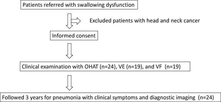 |
Figure 1 Flow chart of patient selection. |
A diagnosis of pneumonia was made in cases fulfilling the following criteria: chest X-ray or chest computed tomography (CT) showing an alveolar infiltration shadow, with a fever of 37.5°C or higher and an abnormally high C-reactive protein level, a peripheral white blood cell count of more than 9000/µL and/or the presence of any two or more airway symptoms, such as sputum. We evaluated the associations of the OHAT score with VE and VF and compared the associations between patients with no or only a single episode of pneumonia (no/single-pneumonia episode group) and patients with multiple pneumonia episodes (multiple-pneumonia episode group).
Evaluation Method
OHAT was performed to evaluate the oral environment.5 Scores for items pertaining to the lips, tongue, gingival mucosa, saliva, remaining teeth, dentures, oral cleaning and toothache were recorded using a 3-point scale (0–2). Higher OHAT scores indicate a worse oral environment.
VE was evaluated using the Hyodo scoring system6 based on four parameters: salivary pooling at the vallecula and piriform sinuses; induction of glottal closure reflex on touching the epiglottis or arytenoid with the endoscope; swallowing reflex initiation, assessed based on the “whiteout” timing (where whiteout was defined as the period during which the endoscopic image is obscured due to pharyngeal closure); and pharyngeal clearance after swallowing 3 cc of blue-dyed water. These parameters were scored on a 4-point scale (0: normal; 1: mildly impaired; 2: moderately impaired; 3: severely impaired), with higher scores indicating greater swallowing dysfunction.
VF was evaluated using the method described previously.7 Briefly, the patient in a sitting or semi-sitting position was asked to swallow 10 cc of non-ionic iodinated contrast agent (Omnipaque 300®) for a single examination. Impaired feeding was defined as 0 if 10 cc of contrast medium (Omnipaque 300®) could be delivered in one full swallow, 1 if less than 50% remained in the oral cavity and 2 if more than 50% remained. Premature pharyngeal inflow was scored 0 for no and 1 for yes. Laryngeal entry was rated on a 3-point scale (0: no laryngeal entry; 1: laryngeal entry only; 2: aspiration). The elicitation of the swallowing reflex was assessed with the laryngeal elevation delay time (LEDT), which is considered to a useful visual marker during pharyngeal swallowing, with normal defined as a value of 0.35 s or less;8 prolongation was defined according to two levels: 0 for no and 1 for yes. Residuals in the glottis valley were defined as 0 for none, 1 for very little, 2 for not overflowing the glottis valley and 3 for overflowing the pharynx in four stages. Residuals in the pear-shaped depression were defined as 0 for none, 1 for very little, 2 for not overflowing into the laryngeal valley and 3 for overflowing into the pharynx. The score was 0 for normal swallowing function, with higher scores indicating poorer swallowing function (Table 1).
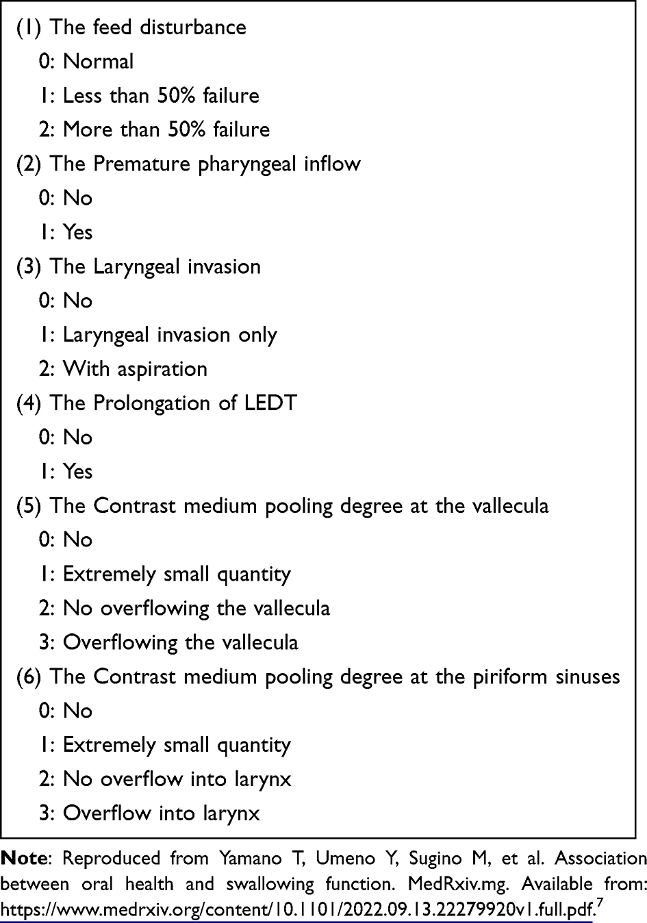 |
Table 1 Video Fluorography Scoring System Proposed by Yamano et al in 2019 |
Data Analysis
Spearman’s rank correlation coefficient was used to determine the correlations of the OHAT score with the results of swallowing endoscopy and swallowing angiography. Welch’s t-test was applied for comparisons between the no/single-pneumonia episode group and the multiple-pneumonia episode group.
The study was approved by the Fukuoka Gakuen Research Ethics Committee (permission no. 314).
Results
Primary Diseases and Oral Intake
The most common disease was pneumonia (17 patients), followed by cerebral infarction 5 patients), pyelonephritis (4 patients), bronchitis (2 patients), Parkinson’s disease (2 patients), scleroderma (1 patient), diabetes (1 patient), eosophageal cancer (1 patient) and Parkinson’s syndrome (1 patient). Some patients had multiple diseases. Oral intake was possible in 20 patients (80%), whereas tube feeding and gastric banding were required in four patients.
Correlation Between OHAT and VE Scores
There was no correlation between OHAT and VE scores (r = 0.3153) (Figure 2A).
 |
Figure 2 Comparison of oral environment and swallowing function. (A) There was no correlation between the Oral Health Assessment Tool (OHAT) and video endoscopy (VE) scores (r = 0.3153). (B) There was no correlation between the OHAT and video fluorography (VF) scores (r = 0.2211). |
Correlation Between OHAT and VF Scores
There was no correlation between OHAT and VF scores (r = 0.2211) (Figure 1B).
Comparison Between the No/Single- and Multiple-Pneumonia Episode Groups
There was no significant difference in OHAT score between the no/single-pneumonia episode group and the multiple-pneumonia episode group (Figure 3).
 |
Figure 3 Comparison of OHAT scores between the no/single- and multiple-pneumonia episode groups. |
The VE score was significantly higher in the multiple-pneumonia episode group than the no/single-pneumonia episode group (p < 0.01) (Figure 4A). Analysis of the OHAT items showed that scores for salivary retention in the laryngeal trough and pisiform depression; the elicitation of glottic closure, cough and gag reflexes, and pharyngeal clearance by coloured water swallow, were higher in the multiple-pneumonia episode group than the no/single-pneumonia episode group (Figure 4B).
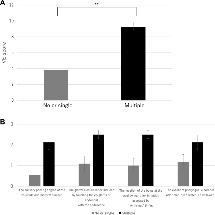 |
Figure 4 Comparison of VE between the no/single- and multiple-pneumonia episode groups. (A) Comparison of VE scores between the no/single- and multiple-pneumonia episode groups. (B) Comparison of VE item scores between the no/single- and multiple-pneumonia episode groups. |
Similar to the VF score, the swallowing angiography score was significantly higher in the multiple-pneumonia episode group than in the no/single-pneumonia episode group (p < 0.01) (Figure 5A). Patients with multiple episodes of pneumonia had higher scores for all items compared to those with no or only a single episode of pneumonia (Figure 5B).
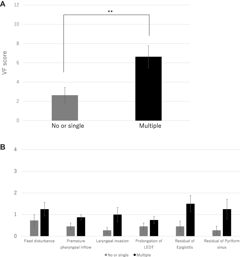 |
Figure 5 Comparison of VF between the no/single- and multiple-pneumonia episode groups. (A) Comparison of VF scores between the no/single- and multiple-pneumonia episode groups. (B) Comparison of VF item scores between the no/single- and multiple-pneumonia episode groups. |
Discussion
Some previous studies have suggested that oral care can prevent pneumonia in older patients, with relative risks of pneumonia of 19% and 11% in older patients with poor and good oral health, respectively In addition, older patients with poor oral health were reported to have a 1.7-fold higher relative risk of pneumonia compared to those with good oral health.1 It has also been suggested that the cough reflex threshold was significantly reduced after 1 month of meticulous oral care in a nursing home for older adults compared to before commencement of care.2 Furthermore, individuals receiving oral care from a dental hygienist twice a week for 24 months in nursing homes had significantly lower likelihoods of fever and death from aspiration pneumonia compared to individuals who did not receive oral care.3 Therefore, oral care is recommended as part of the treatment of dysphagia, especially in older residents of nursing homes.
Although the oral environment significantly affects the risk of pneumonia, few studies have evaluated its relation with swallowing. There is no doubt that there is a significant link between the oral environment and the development of pneumonia. However, there have been few studies comparing VE and VF scores to determine the actual availability of oral intake and the choice of food form.
In our previous study,9 the Functional Oral Intake Scale (FOIS) score was shown to be significantly associated with saliva and denture scores in patients admitted to a convalescent hospital. However, after excluding patients on non-oral nutrition, no significant association between FOIS and OHAT scores was observed. Importantly, the FOIS score reflects oral intake status rather than swallowing function.
VE is a comparable to VF,10 and recent studies using the Hyodo scoring system have shown that VE is simple to perform and effective for predicting aspiration, with a score of ≥ 6 corresponding to the highest risk of aspiration.11 The Hyodo score was shown to be significantly correlated with handgrip strength and peak expiratory flow rate, making it useful for examining the effects of strength training on dysphagia.12
VF is the most reliable swallowing assessment method because it can evaluate the oral, pharyngeal and eosophageal phases.13 In addition, VF is extremely useful when making policy decisions, and whiteout due to pharyngeal contraction does not occur. At our institution, VF is also used to determine the timing of a return to oral intake after prolonged fasting and the optimal treatment for swallowing rehabilitation or surgery to improve swallowing.
In the present study, we found no correlations between the OHAT score for oral assessment and VE and VF scores for swallowing. In addition, the OHAT score was not significantly different between the no/single-pneumonia episode and multiple-pneumonia episode groups, while the rates of swallowing endoscopy and angiography were significantly different between the two groups. In our previous study,9 we identified significant differences in the oral and pharyngeal phases on VE between older patients with pneumonia and those without a history of pneumonia; notably, we observed a significant group difference in elicitation of the gag reflex. These findings suggest that VE or VF is necessary for the evaluation of swallowing, in addition to oral assessment in patients with fever, difficulty in oral intake or with other findings suggestive of dysphagia. Pneumonia is more likely in older adults with both poor oral health and poor pharyngeal swallowing function. However, we speculated that poor pharyngeal swallowing function is the dominant factor related to the risk of pneumonia in these patients.
This study had some limitations in that we did not perform a simple comparison of patients with and without a history of aspiration pneumonia. In addition, there were no cases with extremely poor oral health, as the participants were inpatients and some nursing care was provided. Therefore, the number of cases was small and the underlying disease background was heterogeneous.
Age-related swallowing dysfunction is multifactorial, with 63% of older patients (mean age: 83 years) having oral abnormalities, such as difficulty ingesting, controlling or delivering a bolus to initiate swallowing; this prevalence rate was higher than that in young patients without dysphagia. In addition, 25% of older patients have pharyngeal dysfunction, such as bolus retention and tongue propulsion or pharyngeal muscle paralysis, and 36% have eosophageal abnormalities due to cricopharyngeal muscle dysfunction.14 Age-related swallowing dysfunctions in older adults without organic diseases, such as stroke, cancer or dementia, include prolonged oral transit time and aspiration.15
In the present study, the multiple-pneumonia episode group had higher VE and VF scores for all items than the no/single-pneumonia episode group. Therefore, the group with repeated pneumonia, despite having a good oral environment, was likely to have several issues, including those associated with clearing of the pharynx and delayed induction of the swallowing reflex, as indicated by VE and VF scores. These observations indicated that the multiple items of swallowing function assessed by VE and VF are more important than the oral environment for evaluating the risk of recurrent aspiration pneumonia in the elderly.
Conclusions
Oral assessment, VE and VF are necessary to evaluate swallowing in patients with suspected dysphagia. In addition, multiple factors contribute to recurrent pneumonia among patients with a good oral environment, including subclinical aspiration, pharyngeal clearance and delayed activation of the gag reflex. Determination of swallowing function, especially by VE and VF, is more important than examination of the oral environment for evaluating risk of recurrent aspiration pneumonia in the elderly.
Data Sharing Statement
The data that support the findings of this study are available from the Fukuoka Gakuen Research Ethics Committee. But restrictions apply to the availability of these data, which were used under license for the current study, and so are not publicly available. Data are however available from the authors upon reasonable request and with permission of the Fukuoka Gakuen Research Ethics Committee.
Ethics Approval and Consent to Participate
Paper consent is obtained from all study patients. The study was conducted on human subjects and in accordance with the Declaration of Helsinki. A full explanation of the research and other details will be given in accordance with the attached explanatory document. The decision to participate in the study is of the individual’s own free will. Informed consent was obtained from all study patients. The study was approved by the Fukuoka Gakuen Research Ethics Committee (permission no. 314).
Author Contributions
All authors made a significant contribution to the work reported, whether that is in the conception, study design, execution, acquisition of data, analysis and interpretation, or in all these areas; took part in drafting, revising or critically reviewing the article; gave final approval of the version to be published; have agreed on the journal to which the article has been submitted; and agree to be accountable for all aspects of the work.
Funding
There is no funding to report.
Disclosure
The authors declare no conflicts of interest associated with this manuscript.
References
1. Yanehara T, Yoshida M, Matsui T, Sasaki H. Oral care and pneumonia. Oral Care Working Group. Lancet. 1999;354:515. doi:10.1016/s0140-6736(05)75550-1
2. Watando A, Ebihara S, Okazaki T, Takahashi H, Asada M, Sasaki H. Daily oral care and cough reflex sensitivity in elderly nursing home patients. Chest. 2004;126:1066–1070. doi:10.1378/chest.126.4.1066
3. Adachi M, Ishihara K, Abe S, Okuda K, Ishikawa T. Effect of professional oral health care on the elderly living in nursing homes. Oral Surg Oral Med Oral Pathol Oral Radiol Endod. 2002;94:191–195. doi:10.1067/moe.2002.123493
4. Ames NJ, Sulima P, Yates JM, et al. Effects of systematic oral care in critically ill patients: a multicenter study. Am J Crit Care. 2011;20:e103–14. doi:10.4037/ajcc2011359
5. Chalmers JM, King PL, Spencer AJ, Wright AC, Carter KD. The oral health assessment tool--validity and reliability. Aust Dent J. 2005;50:191–199. doi:10.1111/j.1834-7819.2005.tb00360.x
6. Hyodo M, Nishikubo K, Hirose K. New scoring proposed for endoscopic swallowing evaluation and clinical significance. Nippon Jibi-inkoka Gakkai Kaiho. 2010;113:670–678. doi:10.3950/jibiinkoka.113.670
7. Yamano T, Umeno Y, Sugino M, et al. medRxiv.org. Association between oral health and swallowing function, 2019. https://www.medrxiv.org/content/10.1101/2022.09.13.22279920v1
8. Miyaji H, Umezaki T, Adachi K, et al. Videofluoroscopic assessment of pharyngeal stage delay reflects pathophysiology after brain infarction. Laryngoscope. 2012;122:2793–2799. doi:10.1002/lary.23588
9. Nakayama E, Tohara H, Sato M, et al. Relationship between oral intake level and oral health assessment tool scores in the convalescent ward. J Oral Sci. 2020;63:79–82. doi:10.2334/josnusd.20-0414
10. Langmore SE. Evaluation of oropharyngeal dysphagia: which diagnostic tool is superior? Curr Opin Otolaryngol Head Neck Surg. 2003;11:485–489. doi:10.1097/00020840-200312000-00014
11. Chiba Y, Sano D, Ikui Y, et al. Predictive value of the Hyodo score in endoscopic evaluation of aspiration during swallowing. Auris Nasus Larynx. 2018;45:1214–1220. doi:10.1016/j.anl.2018.03.005
12. Sugaya N, Goto F, Okami K, Nishiyama K. Association between swallowing function and muscle strength in elderly individuals with dysphagia. Auris Nasus Larynx. 2021;48:261–264. doi:10.1016/j.anl.2020.09.007
13. Langmore SE, Schatz K, Olson N. Endoscopic and videofluoroscopic evaluations of swallowing and aspiration. Ann Otol Rhinol Laryngol. 1991;100:678–681. doi:10.1177/000348949110000815
14. Ekberg O, Feinberg MJ. Altered swallowing function in elderly patients without dysphagia: radiologic findings in 56 cases. AJR Am J Rentgenol. 1991;156:1181–1184. doi:10.2214/ajr.156.6.2028863
15. Mehraban-Far S, Alrassi J, Patel R, et al. Dysphagia in the elderly population: a videofluoroscopic study. Am J Otolaryngol. 2021;42:102854. doi:10.1016/j.amjoto.2020.102854
 © 2023 The Author(s). This work is published and licensed by Dove Medical Press Limited. The
full terms of this license are available at https://www.dovepress.com/terms
and incorporate the Creative Commons Attribution
- Non Commercial (unported, 3.0) License.
By accessing the work you hereby accept the Terms. Non-commercial uses of the work are permitted
without any further permission from Dove Medical Press Limited, provided the work is properly
attributed. For permission for commercial use of this work, please see paragraphs 4.2 and 5 of our Terms.
© 2023 The Author(s). This work is published and licensed by Dove Medical Press Limited. The
full terms of this license are available at https://www.dovepress.com/terms
and incorporate the Creative Commons Attribution
- Non Commercial (unported, 3.0) License.
By accessing the work you hereby accept the Terms. Non-commercial uses of the work are permitted
without any further permission from Dove Medical Press Limited, provided the work is properly
attributed. For permission for commercial use of this work, please see paragraphs 4.2 and 5 of our Terms.