
Back to Journals » International Journal of Women's Health » Volume 18
Association Between Fetal Nuchal Translucency Measurements and Pregnancy Outcomes
Authors Zhao XN, Chang J, Sun HG, Meng H, Yao Y, Deng C, Gu YJ
Received 13 October 2025
Accepted for publication 23 January 2026
Published 12 February 2026 Volume 2026:18 574073
DOI https://doi.org/10.2147/IJWH.S574073
Checked for plagiarism Yes
Review by Single anonymous peer review
Peer reviewer comments 5
Editor who approved publication: Dr Matteo Frigerio
Xiang-Na Zhao,1,* Jing Chang,2,* Hao-Gang Sun,1 Hua Meng,3 Yuan Yao,1 Chao Deng,4 Ya-juan Gu5
1First Affiliated Hospital, Shihezi University, Shihezi, 832008, People’s Republic of China; 2Shihezi University School of Medicine, Shihezi, 832008, People’s Republic of China; 3Department of Pharmacy, First Affiliated Hospital, Shihezi University, Shihezi, 832008, People’s Republic of China; 4Department of Anesthesiology, First Affiliated Hospital, Shihezi University, Shihezi, 832008, People’s Republic of China; 5Department of Gynecology and Obstetrics, First Affiliated Hospital,Shihezi University, Shihezi, 832008, People’s Republic of China
*These authors contributed equally to this work
Correspondence: Ya-juan Gu, Department of Gynecology and Obstetrics, First Affiliated Hospital, Shihezi University, No. 107, North 2nd Road, Hongshan Sub-District, Shihezi, 832008, Xinjiang Uygur Autonomous Region, People’s Republic of China, Tel +8618703096536, Email [email protected] Chao Deng, Department of Anesthesiology, First Affiliated Hospital, Shihezi University, No. 107, North 2nd Road, Hongshan Sub-District, Shihezi, 832008, Xinjiang Uygur Autonomous Region, People’s Republic of China, Tel +8613565558780, Email [email protected]
Objective: To explore the optimal cutoff value of fetal nuchal translucency (NT) in the Shihezi region and analyze the predictive value of NT and high-risk factors for pregnancy outcomes.
Methods: This retrospective study included pregnant women who underwent NT screening at the First Affiliated Hospital of Shihezi University between January 2021 and December 2023. Prenatal examination results and pregnancy outcomes were collected and analyzed. The optimal cutoff value of NT for predicting pregnancy outcomes was calculated.
Results: Using NT=2.5mm as the cutoff value, three groups were defined (< 2.5mm, 2.5– 3.5mm, ≥ 3.5mm). For NT≥ 2.0mm, the AUC for predicting outcomes was 0.652 (P< 0.001). NT thickening was significantly associated with ≥ 2 abnormalities in soft ultrasound indicators, structural abnormalities (P< 0.001), chromosomal abnormalities (P=0.008), and adverse outcomes (P< 0.001). Compared with the NT< 2.5 mm group, the risk of adverse outcomes increased with NT thickness (OR=2.009, 95% CI: 1.472– 2.743, for 2.5– 3.5mm; OR=13.090, 95% CI: 6.571– 26.074, for ≥ 3.5mm). The top three adverse pregnancy outcomes were: 210 cases of macrosomia (7%), 158 cases of preterm birth (5.3%), and 109 cases of structural abnormalities (3.6%). As NT thickened, structural abnormalities, chromosomal abnormalities, and miscarriage were correlated with NT (P< 0.05). HDP was associated with fetal growth retardation and preterm birth (P< 0.001). Maternal age ≥ 35 was linked to structural abnormalities (P=0.017) and gestational diabetes mellitus (GDM) (P< 0.001).
Conclusion: NT=2.5mm is the cutoff value of NT thickening in Shihezi area; structural and chromosomal abnormalities and miscarriage are related to the degree of NT thickening; NT, age and HDP are independent risk factors affecting pregnancy outcome, and age is associated with GDM. NT cannot serve as an independent risk factor for adverse pregnancy outcomes and should be evaluated in conjunction with prenatal screening ultrasound and maternal high-risk factors.
Keywords: nuchal translucency thickness, adverse pregnancy outcomes, correlation, cutoff value, fetus
Introduction
Fetal nuchal translucency (NT) is a key ultrasonographic marker used in the prenatal assessment of chromosomal abnormalities, congenital heart defects, other structural anomalies, and overall pregnancy outcomes.1 NT refers to the subcutaneous fluid accumulation between the fetal nuchal skin and soft tissue, measured between 11 and 13+6 weeks of gestation. This transient accumulation is generally attributed to temporary lymphatic drainage obstruction.2,3 Due to its non-invasive nature and capacity for early risk detection, NT measurement has become a widely adopted initial screening method of fetal malformations. However, the effectiveness of this approach is heavily reliant on strict adherence to standardized diagnostic protocols. A central point of ongoing debate is the determination of an appropriate cutoff value for defining NT thickening.
Emerging evidence suggests that regional, ethnic, and methodological variations may significantly influence NT measurements, thereby limiting the universal applicability of a single cutoff threshold. Some researchers support the use of gestational age-specific percentiles (such as the 99th percentile) for dynamic risk assessment,4 whereas others advocate for a fixed cutoff value of 2.5 mm to enhance screening sensitivity, despite the associated rise in false positive rates.5 Currently, there is a lack of large-scale, authoritative data from Chinese or broader Asian populations to establish a definitive NT threshold.6 If the NT cut-off value is set too low, it may lead to over-diagnosis and unnecessary interventions. Conversely, setting it too high may result in missed diagnoses. Therefore, healthcare institutions across different regions have adopted varying cutoff values for defining NT thickening.
Most existing studies classify NT measurements < 2.5 mm as low risk, recommending follow-up with standard prenatal screening protocols such as maternal serum screening for trisomy 21 and expanded non-invasive prenatal testing (NIPT-plus). Based on the associated risk profiles identified by these screenings, decisions are subsequently made regarding the need for invasive diagnostic procedures. An NT measurement ≥ 3.5 mm is typically regarded as indicative of significant abnormality, warranting direct referral for invasive prenatal diagnostic procedures and chromosomal karyotyping.7,8 However, the intermediate NT range of 2.5–3.5 mm, often referred to as the “borderline thickening” zone, lacks standardized criteria for cutoff value selection and clinical management. Limited data specific to this range has contributed to inconsistency in clinical decision-making, with approaches varying considerably among individual physicians. This variability may result in cases of unnecessary invasive intervention, resulting in iatrogenic pregnancy termination, or in delayed diagnosis and intervention due to missed abnormalities.
ROC (Receiver Operating Characteristic) analysis, which plots the false positive rate (1 - specificity) on the x-axis against the true positive rate (sensitivity) on the y-axis, provides a visual representation of a diagnostic method’s performance across different thresholds. Its overall diagnostic accuracy can be quantified using the area under the curve (AUC), aiding researchers or clinicians in quickly assessing the method’s efficacy. This analysis is widely applicable in various diagnostic scenarios, such as disease screening, imaging evaluation, and biomarker testing. In the context of prenatal screening, prioritizing sensitivity aims to maximize the protection of fetal and maternal health by minimizing missed diagnoses.9 This approach enables earlier intervention to reduce disease burden, while also taking into account the psychological and social needs of the family.
The Shihezi region is a relatively underdeveloped area in Western China, where the proficiency of sonographers performing prenatal screening varies significantly. Adopting a universal high-risk threshold of NT ≥ 3.0 mm, as used in most hospitals, may lead to missed diagnoses in this context. Furthermore, there is no uniformity in the NT cut-off values used for recommending invasive prenatal diagnosis across local hospitals in this region. This study aims to investigate the optimal NT cut-off value for predicting adverse pregnancy outcomes specific to the Shihezi region. It seeks to assess and quantify the risk of fetal structural and chromosomal abnormalities associated with NT measurements between 2.5 mm and 3.5 mm, and to analyze the predictive performance of NT for pregnancy outcomes. The findings are intended to provide a reference for developing personalized prenatal screening strategies for the local population and to fill a gap in regional data on this subject.
Based on the principles of ROC analysis and a sensitivity-first screening strategy, we hypothesized that an NT cut-off value lower than the commonly used 3.0 mm threshold would demonstrate better predictive performance for adverse pregnancy outcomes in the local context of the Shihezi region. Specifically, we anticipated that a cut-off of 2.5 mm would optimize the balance between detection sensitivity and specificity, and that the degree of NT thickening would be positively correlated with the risks of fetal structural abnormalities, chromosomal anomalies, and other adverse outcomes.
Materials and Methods
Study Participants
This retrospective cohort study included pregnant women who underwent NT screening at the First Affiliated Hospital of Shihezi University between January 2021 and December 2023 (n = 2993). Data collected comprised general demographic and clinical information (eg, maternal age at expected delivery, gestational age at NT examination, and contact information), pregnancy complications (including gestational diabetes mellitus (GDM), pregnancy complicated by hypothyroidism, and hypertensive disorders of pregnancy (HDP), the three most frequently observed complications at this institution during the study period, NT measurement results, and findings from other prenatal assessments. These included results from prenatal diagnostic procedures and prenatal ultrasound examinations.
Inclusion criteria were as follows: (1) Gestational age between 11 and 13+6 weeks (2), Singleton pregnancy; (3) No contraindications to examination; (4) Satisfactory compliance throughout the study.
Exclusion criteria were: (1) Multiple gestation; (2) Incomplete medical records or loss to follow-up; (3) Use of medications known to potentially affect fetal development, including those classified as FDA pregnancy category D or X; (4) Elective pregnancy termination without medical indications; (5) Refusal to participate in the study; (6) Poor compliance; (7) Fetal conditions that prevented completion of the NT assessment.
This study was approved by the Medical Ethics Committee of the First Affiliated Hospital of Shihezi University (Approval number: KJ2023-435-02).
Research Methods
Ultrasound Examination
1) Standardized Measurement Techniques
(1) Fetal Position: Ensure the fetus was in a neutral position, with the head neither hyperflexed nor hyperextended. (2) Image Acquisition: Obtain a midsagittal plane image, clearly delineating the fetal head and upper thoracic outline. (3) Measurement Criteria: Measure the maximum thickness at the junction of the fetal head and neck, avoiding the measurement of amniotic fluid beyond the skin. (4) Image Clarity: Ensure the image was sufficiently sharp to distinguish fetal anatomical structures. (5) Contrast and Brightness Adjustment: Adjusted the image contrast and brightness as needed to achieve optimal measurement conditions.
2) Detailed recording of measurement results, operator information, equipment model, and calibration status was required.
3) Measurement results and images were periodically reviewed by senior experts to ensure consistency and accuracy. Operator Qualifications: All NT measurements must be performed by senior sonographers certified in prenatal screening and prenatal diagnosis, with at least two sonographers completing the same measurement.
The equipment used for prenatal screening was the Samsung W10 system. The dedicated probes for this purpose were: (1) Single Crystal Abdominal Convex Array Probe: Frequency range 3–10 MHz. (2) Single Crystal Convex Array Probe: Frequency range 1–7 MHz. (3) Single Crystal Volume Convex Array Probe: Frequency range 1–8 MHz. (4) Intracavitary Volume Probe: Frequency range 2–10 MHz. Examinations were conducted between 11 and 13+6 weeks of gestation, corresponding to a fetal crown-rump length (CRL) of 45–84 mm. The procedure was performed in accordance with the Standardized Scanning Technique and Diagnostic Criteria for 11–13+6 Week Early Pregnancy Fetal Ultrasound published in the Chinese Journal of Medical Ultrasound. A multi-plane ultrasound screening method was adopted, with NT measurements repeated multiple times. The highest value obtained was recorded for analysis.10 The screening equipment undergoes annual calibration by qualified engineers. Image acquisition quality control was performed manually by senior physicians in accordance with the regulations stipulated in the “Guidelines for Ultrasound Prenatal Screening” and the “Quality Control Indicators for Prenatal Screening and Diagnosis” established by the Maternal and Fetal Ultrasound Group of the Chinese Society of Ultrasound in Medicine in 2022. The protocol of performing three NT measurements and recording the highest value was established as a standardized procedure, balancing measurement precision, fetal physiological characteristics and clinical diagnostic needs, with the aim of providing more reliable data for fetal health assessment.
Ultrasound soft markers were defined as non-specific, potentially transient sonographic findings, typically reflecting minor structural variations in the fetus.11
Ultrasound structural abnormalities refer to clinically significant congenital anomalies, and include defects of the anterior abdominal wall, central nervous system, cardiovascular system, skeletal system, limbs, and cases of increased NT thickness, among others.12
Follow-Up
Participants were followed up to 42 days postpartum, with outcome data collected from hospital medical records and supplemented by telephone contact when necessary. Collected follow-up data included pregnancy outcomes, gestational age at delivery, mode of delivery, and neonatal condition within the first 42 days postpartum.
Pregnancy outcomes were categorized as either normal or adverse. A normal pregnancy outcome was defined as delivery occurring between 37 and 42 weeks of gestation with no adverse maternal or neonatal health events. Adverse pregnancy outcomes:
Miscarriage (Spontaneous Abortion): Termination of pregnancy before 28 completed weeks of gestation or when the fetal weight was less than 1000 grams.
Preterm Birth: Delivery occurring from 28 weeks to less than 37 completed weeks of gestation.
Stillbirth (Fetal Death): Death of a fetus in utero at or after 20 completed weeks of gestation.
Fetal Growth Restriction (FGR)/Intrauterine Growth Restriction (IUGR): A condition where the fetus failed to achieve its inherent growth potential due to pathological maternal, fetal, or placental factors. It was commonly defined as an estimated fetal weight below the 10th percentile for gestational age on prenatal ultrasound.
Macrosomia: A condition where a newborn’s birth weight was 4000 grams or greater.
Structural Abnormality (Fetal Malformation): Refers to morphological abnormalities in the organs or body structures of the fetus that occurred during development.
Chromosomal Abnormality (Aneuploidy/Chromosomal Abnormality): Refers to changed in the number or structure of chromosomes within cells, leading to an imbalance or error in genetic information.
The diagnosis of structural abnormalities in live-born neonates was primarily based on physical examinations conducted within 42 days postpartum, ICD diagnosis codes from pediatric outpatient visits or inpatient discharge summaries, and necessary imaging reports. For cases involving induced abortion or miscarriage, the diagnosis relied on records from fetal ultrasound examinations, post-delivery medical records, or ICD diagnosis codes from the corresponding inpatient discharge summaries. Cases where pregnancy was terminated due to major fetal structural or chromosomal abnormalities were classified as “adverse pregnancy outcomes” in the outcome analysis.
Data Statistics and Analysis
The sample size for this study was based on the retrospective data available from a single center over a three-year period and represents an observational exploratory analysis. The current sample size provided sufficient statistical power to detect associations between NT thickness and the primary pregnancy outcomes. However, it should be noted that an a priori sample size calculation was not performed for this study. For cases with missing data on key variables, this study employed listwise deletion and performed no data imputation. All cases included in the analysis possessed complete baseline NT measurements, essential maternal characteristic data, and definitive pregnancy outcome information. All statistical analyses were conducted using SPSS version 22.0. Continuous variables following a normal distribution are expressed as mean ± standard deviation (Mean ± SD), and those not conforming to a normal distribution are expressed as median and interquartile range [M(IQR)]. Comparisons between groups were conducted using the independent-samples t-test or Z test. Categorical variables are expressed as frequencies and percentages [n (%)], and comparisons were analyzed using the chi-squared (χ2) test, continuity correction χ2 test, or Fisher’s exact test, as applicable. Receiver operating characteristic (ROC) curve analysis was performed to calculate the area under the curve (AUC) for evaluating the predictive value of NT measurements in relation to adverse pregnancy outcomes, with the determination of the optimal cutoff value for NT thickening. The odds ratio (OR) was used to estimate the relative risk associated with different exposures. Spearman correlation analysis was applied to evaluate the relationship between NT value ranges and pregnancy outcomes. Using postnatal surface structural findings from terminated fetuses as the diagnostic reference standard, kappa (κ) statistics were employed to assess the consistency between prenatal ultrasound alone, NT examination alone, and their combined application in detecting structural abnormalities. Logistic regression analysis was used to identify risk factors for adverse pregnancy outcomes. A p value < 0.05 was considered statistically significant.
Results
Baseline Characteristics of the Study Population and Determination of the NT Thickening Cutoff Value
Among the 2,993 pregnant women included in the study, 2,384 (79.7%) experienced normal pregnancy outcomes, while 609 (20.3%) experienced adverse outcomes. In the normal outcome group, the mean maternal age was 30.49 ± 3.91 years, the mean gestational age at delivery was 38.87 ± 1.03 weeks, and the median NT measurement was 1.40 (IQR): 1.20–1.80 mm. Within this group, 487 cases (20.4%) were complicated by GDM, 192 cases (8.1%) by HDP, and 398 cases (16.7%) by hypothyroidism during pregnancy. In contrast, the group with adverse pregnancy outcomes had a mean maternal age of 30.95 ± 4.35 years, mean gestational age at delivery of 34.37 ± 7.42 weeks, and a median NT measurement of 1.60 (IQR: 1.20–2.00 mm). This group included 145 cases (23.8%) with GDM, 77 cases (12.6%) with HDP, and 85 cases (14.0%) with hypothyroidism during pregnancy.
Statistical analysis indicated that maternal age (Z = 10.916, p < 0.001), gestational age at delivery (Z = −6.584, p < 0.001), NT measurement (Z = −5.397, p < 0.001), and HDP (χ2 = 12.494, p < 0.001) were significantly associated with pregnancy outcomes. No statistically significant associations were found for GDM (χ2 = 3.330, p = 0.068) or hypothyroidism during pregnancy (χ2 = 2.686, p = 0.101), as presented in Table 1.
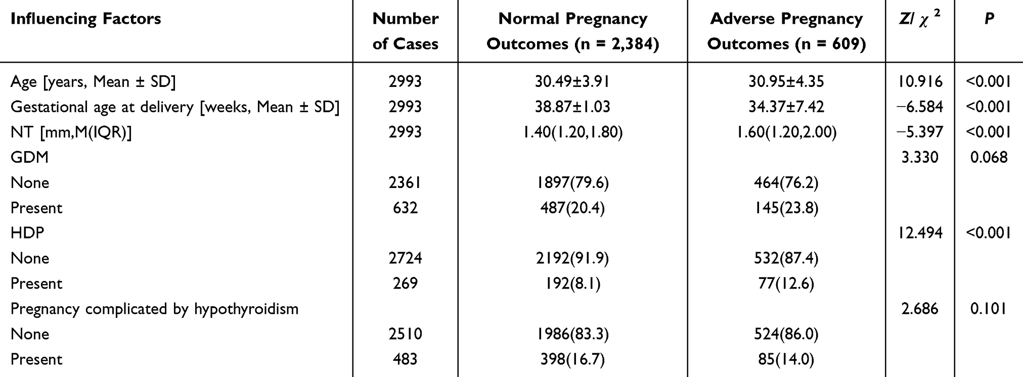 |
Table 1 Comparison of Baseline Characteristics Between Groups with Normal and Adverse Pregnancy Outcomes [n (%)] |
ROC curve analysis based on pregnancy outcomes revealed that an NT threshold of ≥ 2.0 mm yielded an AUC of 0.652 (95% CI: 0.597–0.707, p < 0.001). The optimal cut-off value was an NT = 2.6 mm, yielding a sensitivity, specificity, and Youden index of 48%, 79%, and 0.27, respectively. When the NT cut-off values were set at 2.5 mm, 3.0 mm, and 3.5 mm, the corresponding sensitivity, specificity, and Youden index were as follows: 2.5 mm: 54%, 72%, and 0.26. 3.0 mm: 28%, 92%, and 0.20. 3.5 mm: 21%, 98%, and 0.19.
Given the clinical implications of missed diagnoses and the importance of early risk identification, an NT threshold of 2.5 mm, demonstrating the highest sensitivity, was selected as the cutoff value for defining NT thickening in this study. Based on this cutoff, participants were stratified into three groups: Group A (NT < 2.5 mm), 2,746 cases (91.7%); Group B (2.5 mm ≤ NT < 3.5 mm), 203 cases (6.8%); and Group C (NT ≥ 3.5 mm), 44 cases (1.5%). These group distributions are illustrated in Figure 1 and detailed in Table 2.
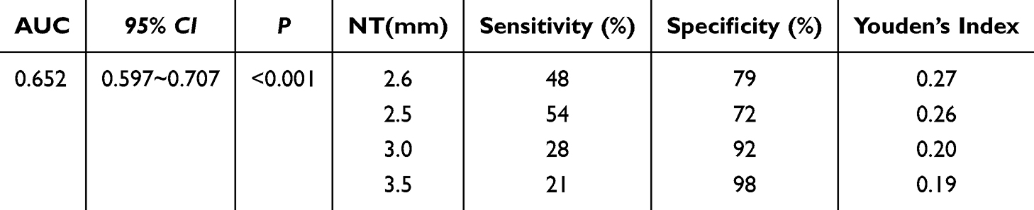 |
Table 2 ROC Curve Analysis for Predictive Value of NT ≥ 2.0 mm |
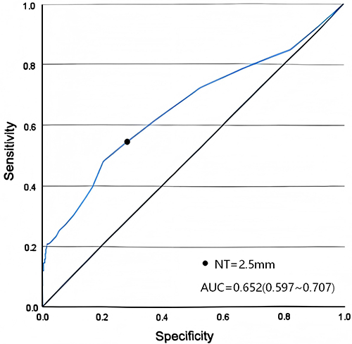 |
Figure 1 ROC Curve for predictive value of NT ≥ 2.0 mm in adverse pregnancy outcomes. |
Correlation Between NT Thickening and Fetal Structural and Chromosomal Abnormalities
Correlation analysis between NT measurement groups and prenatal ultrasound findings demonstrated the following distributions: In the NT < 2.5 mm group, 2,601 cases (95.8%) exhibited fewer than two ultrasound soft marker abnormalities, 60 cases (2.2%) had 2 or more soft marker abnormalities, and 54 cases (2.0%) demonstrated structural abnormalities. In the 2.5 mm ≤ NT < 3.5 mm group, 169 cases (88.5%) had fewer than 2 soft marker abnormalities, 14 cases (7.3%) had 2 or more soft marker abnormalities, and 8 cases (4.2%) exhibited structural abnormalities. In the NT ≥ 3.5 mm group, 15 cases (83.3%) had fewer than 2 soft marker abnormalities, 1 case (5.6%) had 2 or more soft marker abnormalities, and 2 cases (11.1%) demonstrated structural abnormalities.
Statistical analysis indicated a significant positive correlation between increasing NT thickness and the presence of 2 or more ultrasound soft marker abnormalities, as well as structural abnormalities (χ2 = 24.849, r = 0.085, p < 0.001). The detection rate of multiple soft markers and structural abnormalities increased in parallel with NT thickness. A total of 135 pregnant women underwent invasive prenatal diagnosis based on one or more of the following conditions: isolated NT thickening, advanced maternal age, abnormal findings on prenatal ultrasound, high-risk results from NIPT-plus, or NT thickening in combination with abnormal ultrasound findings.
The chromosomal diagnostic results were as follows: In the NT < 2.5 mm group, 76 cases (84.4%) were normal and 14 cases (15.5%) were abnormal; in the 2.5 mm ≤ NT < 3.5 mm group, 19 cases (61.3%) were normal and 12 cases (38.7%) were abnormal; in the NT ≥ 3.5 mm group, 8 cases (57.1%) were normal and 6 cases (42.9%) were abnormal. There was a statistically significant positive correlation between NT measurement and chromosomal abnormalities as confirmed by prenatal diagnosis (χ2 = 9.560, r = 0.014, p = 0.008). The incidence of chromosomal abnormalities increased progressively with greater NT thickness, as presented in Table 3.
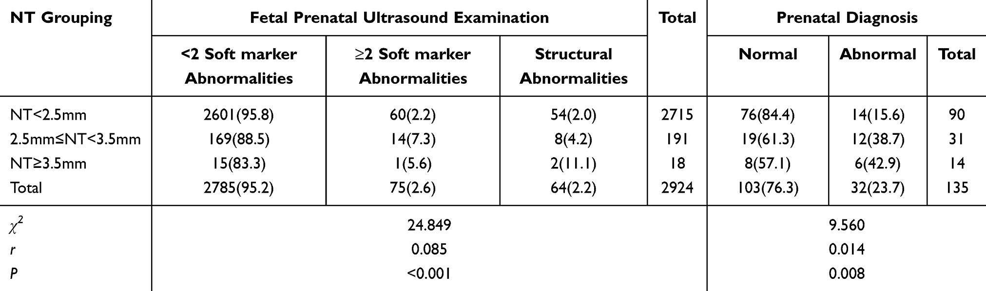 |
Table 3 Correlation Between NT Measurement Categories and Prenatal Ultrasound Findings, Including Soft Marker and Structural Abnormalities [n (%)] |
Comparison of pregnancy outcomes between NT examination alone and NT combined with prenatal ultrasound demonstrated improved diagnostic performance for the detection of surface structural abnormalities when both modalities were used together. Among 97 cases with confirmed surface structural abnormalities, NT examination alone detected 22 cases (22.7%), prenatal ultrasound alone detected 34 cases (35.1%), and the combined assessment identified 48 cases (49.5%). The combined use of NT and prenatal ultrasound significantly improved diagnostic yield compared to either modality alone (p < 0.001). In terms of sensitivity, specificity, and overall accuracy, the combined examination demonstrated superior performance, as shown in Table 4.
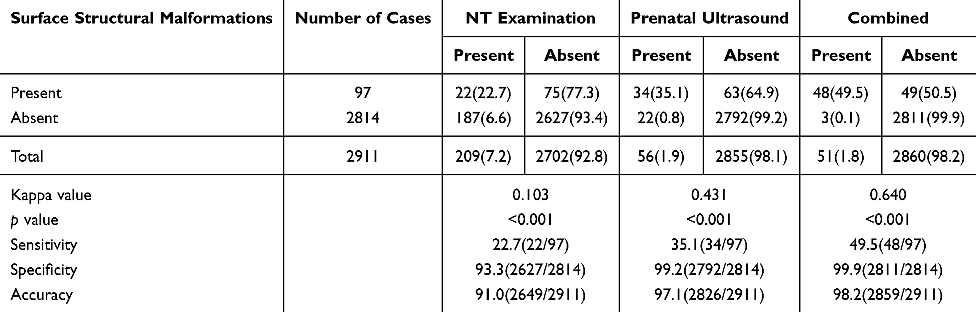 |
Table 4 Diagnostic Performance of Various Prenatal Screening Protocols in Terminated Fetuses and Liveborn Neonates [n (%)] |
These findings support the clinical value of integrating NT measurement with prenatal ultrasound to enhance the detection of fetal surface structural abnormalities.
Correlation Between NT Thickening and Adverse Pregnancy Outcomes
Among the 2,993 pregnant women enrolled in the study: in the NT < 2.5 mm group, 2,234 cases (81.4%) experienced normal delivery, while 512 cases (18.6%) experienced adverse pregnancy outcomes; in the 2.5 mm ≤ NT < 3.5 mm group, 139 cases (68.5%) experienced normal delivery and 64 cases (31.5%) experienced adverse outcomes; in the NT ≥ 3.5 mm group, 11 cases (25.0%) experienced normal delivery and 33 cases (75.0%) had adverse outcomes.
Statistical analysis indicated a significant positive correlation between increased NT thickness and adverse pregnancy outcomes (χ2 = 101.653, r = 0.145, p < 0.001). The incidence of adverse outcomes increased proportionally with NT thickening. When compared to the NT < 2.5 mm group, the OR for adverse outcomes in the 2.5 mm ≤ NT < 3.5 mm group was 2.009 (95% CI: 1.472 – 2.743, χ2 = 19.957, p < 0.001), and the OR in the NT ≥ 3.5 mm group was 13.090 (95% CI: 6.571 – 26.074, χ2 = 87.499, p < 0.001), indicating a significantly increased risk with greater NT values.
Further analysis of specific adverse pregnancy outcomes across NT categories demonstrated statistically significant associations between increased NT and the occurrence of structural abnormalities (χ2 = 116.177, p < 0.001), miscarriage (χ2 = 13.142, p = 0.002), and chromosomal abnormalities (χ2 = 55.549, p < 0.001). However, no statistically significant correlations were found between NT and the following outcomes: stillbirth (χ2 = 2.142, p = 0.352), preterm birth (χ2 = 1.727, p = 0.435), fetal growth restriction (FGR) (χ2 = 0.306, p = 0.844), and macrosomia (χ2 = 5.346, p = 0.059). These results are presented in Table 5.
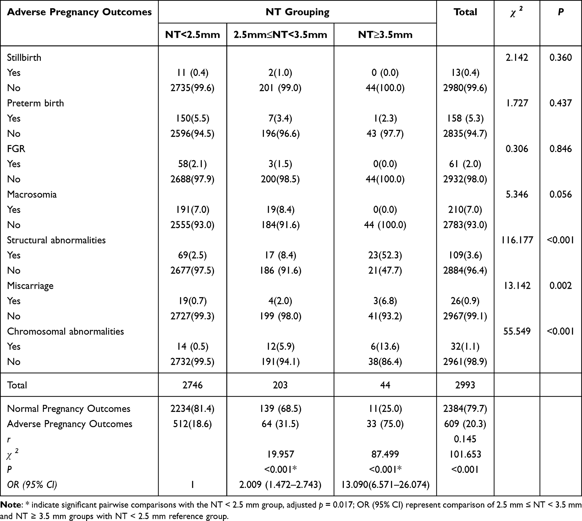 |
Table 5 Association Between NT Measurement Categories and Adverse Pregnancy Outcomes [n (%)] |
High-Risk Factors
Correlation Between Other Influencing Factors and Pregnancy Outcomes
Univariate analysis identified increased NT measurements, HDP, and advanced maternal age as significant risk factors associated with adverse pregnancy outcomes. The findings of this study indicate that age, NT, and HDP are independent risk factors affecting pregnancy outcomes. Among pregnant women diagnosed with HDP, 13 cases (4.8%) resulted in FGR and 37 cases (13.8%) in preterm birth. In comparison, among those without HDP, 48 cases (1.8%) resulted in FGR and 121 cases (4.4%) in preterm birth. HDP was significantly correlated with both FGR and preterm birth.
Compared with normotensive pregnant women, those with HDP demonstrated a substantially increased risk of FGR (χ2FGR = 11.562, rFGR = 0.062, PFGR < 0.001, ORFGR: 2.831, 95% CI: 1.514–5.295), preterm birth (χ2preterm birth = 42.462, rpreterm birth = 0.119, Ppreterm birth < 0.001, ORpreterm birth: 3.431, 95% CI: 2.318–5.077). With respect to maternal age, 86 cases (3.4%) of fetal structural abnormalities were identified among those younger than 35 years, whereas 28 cases (5.7%) were identified among those aged 35 years or older. Advanced maternal age was significantly associated with an increased risk of fetal structural abnormalities (χ2 = 5.693, r = 0.044, p = 0.017, OR: 1.695, 95% CI: 1.094–2.626). These findings are summarized in Table 6. Multivariate stepwise logistic regression analysis identified maternal age (OR: 1.422, 95% CI: 1.131–1.789, P=0.003), NT value (OR: 1.697, 95% CI: 1.497–1.923, P<0.001), and HDP (OR: 1.768, 95% CI: 1.329–2.350, P<0.001) as factors significantly associated with pregnancy outcomes. These results indicated that maternal age, NT value and HDP were independent risk factors for adverse pregnancy outcomes, as shown in Table 7.
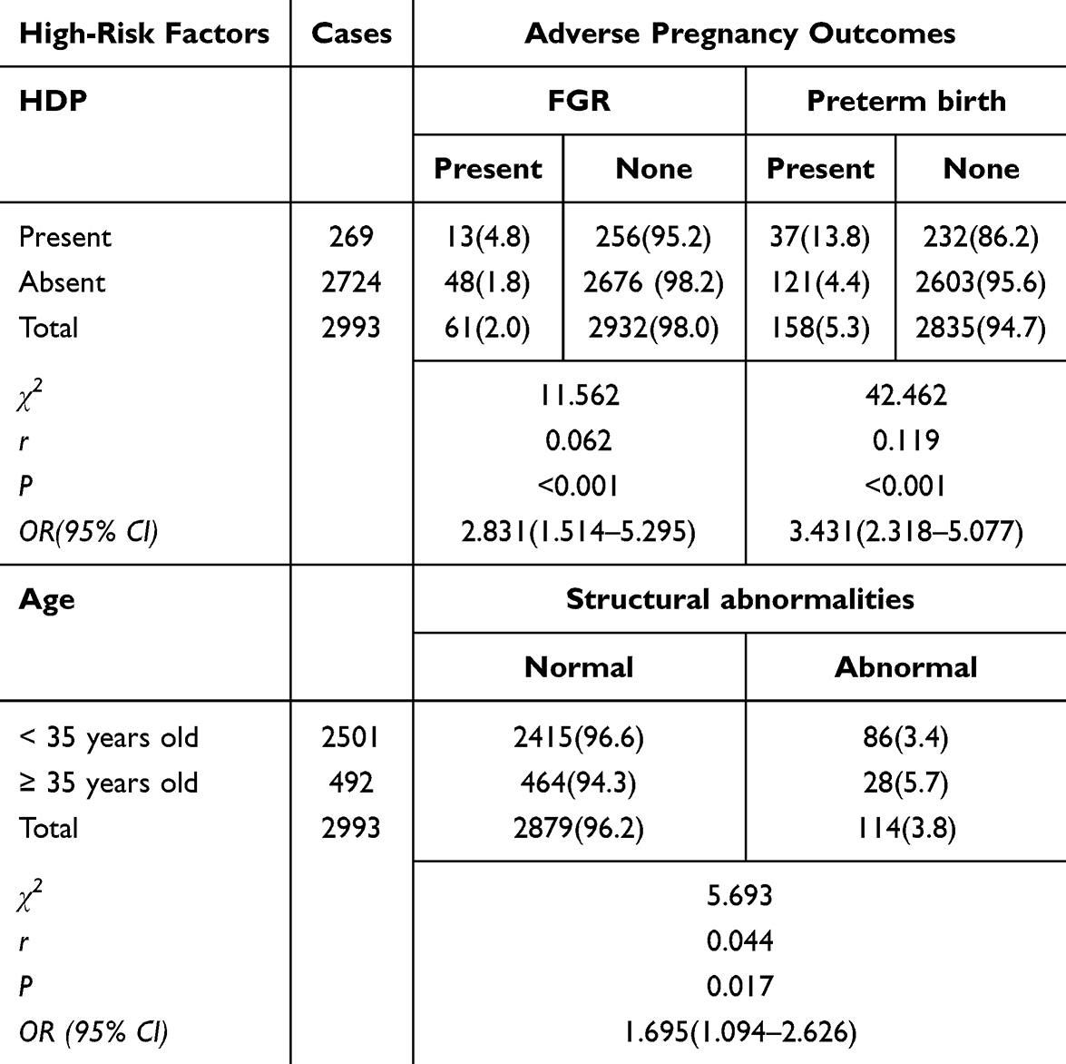 |
Table 6 Correlation of HDP with Fetal Growth Restriction and Preterm Birth, and Correlation of Maternal Age with Fetal Structural Abnormalities [n (%)] |
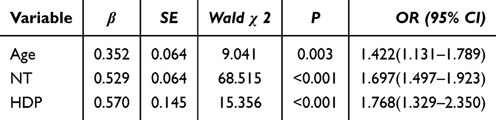 |
Table 7 Stepwise Logistic Regression Analysis of Multiple Factors Influencing Pregnancy Outcomes |
Correlation Between Maternal Age and Pregnancy Complications
Among women younger than 35 years, the incidence of GDM was 490 cases (19.6%), HDP 219 cases (8.8%), and pregnancy complicated by hypothyroidism 412 cases (16.5%). In comparison, among women aged 35 years or older, the incidence rates were: GDM 142 cases (28.9%), HDP 50 cases (10.2%), and pregnancy complicated by hypothyroidism 71 cases (14.4%).
Statistical analysis demonstrated a significant positive correlation between advanced maternal age and the incidence of GDM (χ2 = 21.208, r = 0.113, p < 0.001), indicating that women aged 35 years or older had a higher risk of developing GDM. However, no significant associations were observed between maternal age and HDP (χ2HDP = 0.994, PHDP = 0.343) or pregnancy complicated by hypothyroidism (χ2pregnancy complicated by hypothyroidism = 1.267, Ppregnancy complicated by hypothyroidism = 0.260). These findings are presented in Table 8.
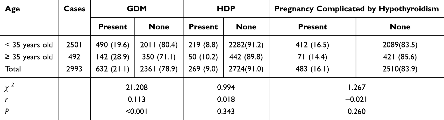 |
Table 8 Association Between Maternal Age and Incidence of GDM, HDP, and Pregnancy Complicated by Hypothyroidism [n (%)] |
Discussion
Birth defects continue to pose a major public health challenge in China, with substantial implications for population health and quality. As a result, the implementation of comprehensive prenatal screening and accurate prenatal diagnosis has become a core strategy in reducing the incidence of congenital anomalies.13
NT measurement plays a crucial role in early screening for fetal abnormalities and serves as an important indicator for assessing the risk of fetal chromosomal abnormalities.14 Studies have shown that increased NT thickness is strongly associated with chromosomal abnormalities, structural malformations, and adverse pregnancy outcomes.15,16 Integration of NT measurements with additional screening and diagnostic modalities can substantially enhance the detection of fetal anomalies, thereby supporting informed clinical decision-making.
Cut-off Value for NT Thickening
Findings from this study indicate that an NT cutoff value of 2.5 mm offers superior predictive performance compared with other thresholds. The determination of an appropriate NT cutoff must take into account regional differences, availability of medical resources, and variations in diagnostic infrastructure, equipment, and practitioner expertise. From a statistical standpoint, the cut-off value of 2.6 mm yields the highest Youden index. However, in the clinical context of prenatal screening, our priority was to maximize the identification of abnormal fetuses and minimize missed diagnoses, even if this comes at the cost of a slightly higher false-positive rate. The selection of the 2.5 mm cut-off was based precisely on this “ensitivity-first” screening strategy. In our internal validation, the 2.5 mm cut-off demonstrated higher sensitivity for detecting severe structural abnormalities and chromosomal anomalies (54%) compared to the 2.6 mm cut-off (48%). This higher sensitivity held more direct clinical significance for initiating subsequent diagnostic evaluations. This choice reflected a deliberate strategy of combining statistical optimality with specific clinical practice needs.
In conclusion, the final adoption of the 2.5 mm cutoff in this study does not represent a simple rejection of the statistically optimal solution. Rather, it is the result of a careful trade-off between statistical performance and specific clinical practice needs—namely, the core mission of a screening test—made after fully understanding its implications. This reflects a research approach that translates epidemiological evidence into decisions tailored to the local clinical reality.
Increasing the NT cutoff value may reduce the number of women referred for invasive testing, but this carries the risk of missed diagnoses in fetuses with chromosomal abnormalities. Such delays can lead to postponed pregnancy termination, potentially resulting in increased physical, psychological, and financial burden for affected women. Conversely, lowering the NT threshold may reduce the likelihood of missed diagnoses but can also increase the number of pregnant women subjected to invasive procedures. This approach may cause unnecessary anxiety among pregnant women and their families, lead to overuse of medical resources, and increase the risk of iatrogenic pregnancy termination. Prior studies have shown that while reducing the NT threshold may modestly improve the detection rate of chromosomal abnormalities, it significantly increases the false positive rate and decreases the positive predictive value.5
NT Thickening and Pregnancy Outcomes
NT thickening has also been identified as a relevant predictor of structural abnormalities. The present study corroborates earlier findings by demonstrating that increased NT values are associated with a higher prevalence of ≥ 2 ultrasound soft marker abnormalities and fetal structural anomalies.17 Furthermore, the combination NT measurement with detailed second-trimester ultrasound significantly improved sensitivity, specificity, and diagnostic accuracy in detecting structural abnormalities when compared to either modality used independently. These findings support the clinical utility of integrating early and mid-pregnancy ultrasound screening strategies to enhance the detection of fetal anomalies and provide an evidence-based foundation for clinical intervention and genetic counseling.
NT thickening is frequently associated with chromosomal aneuploidies, including trisomy 21, trisomy 18, trisomy 13, as well as sex chromosome abnormalities.18,19 Consistent with prior studies, the present findings demonstrate a significant correlation between increased NT measurements and chromosomal abnormalities. As NT thickness increases, so does the detection rate of chromosomal abnormalities. NT thickening identified during the first trimester may serve as an early indicator of fetal chromosomal anomalies, thereby facilitating timely consideration of further diagnostic procedures such as chorionic villus sampling, amniocentesis, or non-invasive prenatal screening. Early identification enables affected women and their families to make informed decisions, which may contribute to reducing the incidence of births affected by chromosomal abnormalities and improving population health outcomes.
NT thickening is also associated with a range of adverse pregnancy outcomes. These outcomes are primarily linked to chromosomal abnormalities, fetal structural anomalies, FGR, and intrauterine fetal demise, with chromosomal abnormalities posing the highest risk, as extensively reported in both Chinese and international literature.20
In this study, NT thickness was found to be significantly positively correlated with adverse pregnancy outcomes (χ2 = 101.653, r = 0.145, p < 0.001). The incidence of adverse outcomes increased progressively with higher NT measurements, consistent with previous research findings.21 It is noteworthy that although previous studies have reported associations between increased NT and outcomes such as fetal growth restriction, stillbirth, or preterm birth, our regional study did not observe a statistically significant correlation between these outcomes and NT thickness after stratification by NT value. This discrepancy may be attributable to factors such as the single-center design and the limited sample size in the NT ≥ 3.5mm subgroup. We found that NT thickening was more strongly correlated with structural abnormalities, chromosomal anomalies, and miscarriage. The three most prevalent adverse pregnancy outcomes observed in this cohort were macrosomia, preterm birth, and structural abnormalities. These findings highlight the importance of prioritizing these outcome categories in clinical screening and management protocols. Focused monitoring and early intervention strategies targeting these specific conditions may contribute to reducing the burden of adverse pregnancy outcomes. However, no statistically significant difference was observed between NT groups and the incidence of macrosomia in our findings (p > 0.05). This may be due to factors such as dietary habits in the Shihezi region of Western China and the generally higher body mass index among pregnant women in this population, leading to an overall higher baseline incidence of macrosomia.
Comparative analysis in this study demonstrated that although NT < 2.5 mm is generally considered indicative of low risk, adverse pregnancy outcomes may still occur within this range. Therefore, it is essential to interpret NT findings in conjunction with subsequent prenatal screening results and other high-risk factors to develop individualized screening or diagnostic protocols, thereby minimizing the risk of missed diagnoses.
In the 2.5 mm ≤ NT < 3.5 mm group, the incidence rates of macrosomia, structural anomalies, chromosomal abnormalities, miscarriage, and stillbirth were significantly higher than those observed in the NT < 2.5 mm group. Although the incidence of adverse outcomes in the 2.5 mm ≤ NT < 3.5 mm group was lower than that in the NT ≥ 3.5 mm group, it remained elevated compared with the reference (normal) group. These findings indicate that NT measurements within this intermediate range still carry a clinically meaningful risk, particularly for macrosomia, structural abnormalities, chromosomal abnormalities, and miscarriage, and should not be overlooked during risk assessment. When NT values reached ≥ 3.5 mm, the risk of fetal structural abnormalities, chromosomal abnormalities, and miscarriage increased markedly, exhibiting a clear upward trend. These results align closely with previous studies, further validating the reliability and clinical significance of the present findings.22,23
The present study demonstrated that increasing NT thickness was associated with higher incidences of structural abnormalities, chromosomal abnormalities, and miscarriage. Based on these findings, an NT measurement ≥ 3.5 mm is recommended as a critical threshold for recommending invasive prenatal diagnostic procedures. This should be combined with systematic fetal ultrasound screening, including fetal echocardiography, to maximize the detection of chromosomal and structural abnormalities and reduce the likelihood of missed diagnosis. For pregnant women with NT measurements ranging from 2.5 mm to less than 3.5 mm, although the risk is lower than in those with NT ≥ 3.5 mm, it remains elevated relative to the general population. Therefore, personalized prenatal screening strategies should be developed for this cohort, incorporating additional high-risk factors to more effectively prevent birth defects and improve overall pregnancy outcomes.
High-Risk Factors
HDP encompasses a group of conditions characterized by elevated maternal blood pressure during gestation. Hypertension is among the most common complications of pregnancy and poses substantial risks to both maternal and neonatal health.24,25 The incidence of HDP varies globally and is influenced by multiple factors. In resource-limited settings, insufficient access to prenatal care often contributes to higher HDP prevalence and increased rates of adverse pregnancy outcomes.26,27 Consistent with prior studies, we found that maternal hypertension was significantly associated with increased risk of FGR and preterm birth.28,29
Over the past decade, the proportion of advanced maternal age (≥ 35 years) has risen significantly worldwide. Maternal age, as a core biological factor affecting pregnancy outcomes, directly influences maternal and infant health.30 Our study found a positive correlation between age and structural abnormalities, with pregnant women of advanced maternal age (≥ 35 years) facing a significantly increased risk of fetal structural abnormalities, which is consistent with previous research findings. The incidence of GDM and HDP also increases significantly with maternal age.31,32 Our study data further revealed a significant positive correlation between age and GDM. Therefore, pregnant women of advanced maternal age should be particularly vigilant in monitoring and managing their blood glucose levels. Specifically, for those with abnormal fasting blood glucose in early pregnancy, an oral glucose tolerance test is recommended as early as possible to facilitate timely detection and intervention. Therefore, in women of advanced maternal age, particular attention should be directed toward early monitoring and management of blood glucose levels, especially among those with elevated fasting glucose values in early pregnancy. Early administration of an oral glucose tolerance test is recommended to facilitate timely diagnosis and intervention.
Limitations
1.This study was a single-center retrospective analysis and thus had inherent limitations. First, the data derived from a single institution may introduce selection bias, and the retrospective nature of the study limited the ability to control for all potential confounding factors. These issues may affect the external validity of the findings. Second, regarding methodology, although all sonographers had completed standardized training, the retrospective design precluded a centralized, blinded re-evaluation of all historical measurement images. The variability that may exist between observers in this process could have influenced the consistency of NT measurements. Based on these considerations, we propose the following future research directions: Prospective, multicenter cohort studies could be conducted to further validate the generalizability of our conclusions. Furthermore, implementing a standardized real-time quality control process during the data collection phase could enhance the homogeneity and reliability of the measurement data, thereby improving the rigor and clinical applicability of future research findings.
2.The postpartum follow-up period was set at 42 days. While this duration was sufficient to capture most adverse pregnancy outcomes, it may be inadequate for detecting certain delayed-onset birth defects, monogenic disorders, and some genetic metabolic diseases. Future studies should extend the follow-up period to obtain more comprehensive data on long-term outcomes.
3.The number of cases with an NT ≥ 3.5 mm was limited (n = 44). Consequently, the risk estimates for this subgroup may be unstable Therefore, conclusions drawn from this subgroup should be interpreted with caution and await further validation in larger-scale studies.
In summary, based on this single-center retrospective study, we propose using an NT value of 2.5 mm as the cut-off for defining NT thickening in the Shihezi region. It must be emphasized that this value requires validation in other studies, and consideration of the specific study populations and environmental contexts will contribute to a more balanced conclusion. NT thickening was significantly associated with adverse pregnancy outcomes, particularly structural abnormalities, chromosomal abnormalities, and miscarriage. However, NT alone cannot serve as an independent risk factor for predicting adverse pregnancy outcomes and must be evaluated in conjunction with prenatal screening ultrasound findings and other maternal high-risk factors. From a clinical standpoint, for women with NT measurements ranging from 2.5 mm to less than 3.5 mm, prenatal screening protocols should be tailored based on a comprehensive evaluation of high-risk factors and serological screening results. For those with NT ≥ 3.5 mm, invasive prenatal diagnostic procedures are recommended to confirm chromosomal or structural anomalies. This stratified approach helps to minimize both missed diagnoses and unnecessary interventions, thereby supporting the delivery of more precise and effective prenatal care. The findings of this study provide localized data to support risk assessment and stratified management following first-trimester NT measurements in the Shihezi region. The data accumulated in this study will contribute to promoting the standardization of local genetic counseling protocols.
Data Sharing Statement
The datasets used and/or analysed during the current study available from the corresponding author on reasonable request.
Ethics Approval and Consent to Participate
This study was conducted with approval from the Ethics Committee of the First Affiliated Hospital of Shihezi University (No.KJ2022-220-01) and registered in the Chinese Clinical Trial Register website (www.chictr.org.cn, ChiCTR2400091345, 2024-10-27). This study was conducted in accordance with the declaration of Helsinki. Written informed consent was obtained from all participants.
Acknowledgments
Ya-juan Gu is the primary corresponding author, Chao Deng is the secondary corresponding author for this study. We would like to acknowledge the hard and dedicated work of all the staff that implemented the intervention and evaluation components of the study.
Funding
This work was supported by “Tianshan Talents” High-Level Medical and Health Talent Program - Young and Middle-aged Backbone Medical Talents (No.TSYC202401B194) and Supported by Science and Technology Program of Xinjiang Production and Construction Corps (No.2022ZD077).
Disclosure
The authors declare that they have no competing interests in this work.
References
1. Sofia-Gonçalves A, Guedes-Martins L. Nuchal translucency and congenital heart defects. Curr Cardiol Rev. 2024;20(2):1–15. doi:10.2174/011573403X264963231128045500
2. Minnella GP, Crupano FM, Syngelaki A, et al. Diagnosis of major heart defects by routine first-trimester ultrasound examination: association with increased nuchal translucency, tricuspid regurgitation and abnormal flow in ductus venosus. Ultrasound Obstetr Gynecol. 2020;55(5):637–644. doi:10.1002/uog.21956
3. Kelley J, McGillivray G, Meagher S, et al. Increased nuchal translucency after low-risk noninvasive prenatal testing: what should we tell prospective parents. Prenatal Diagnosis. 2021;41(10):1305–1315. doi:10.1002/pd.6024
4. Hui L, Pynaker C, Bonacquisto L, et al. Reexamining the optimal nuchal translucency cutoff for diagnostic testing in the cell-free DNA and microarray era: results from the victorian perinatal record linkage study. Ame J Obstetr Gynecol. 2021;225(5):527.e521–527.e512. doi:10.1016/j.ajog.2021.03.050
5. Han MP, Ferreira AT, Elhindi J, et al. How useful is nuchal translucency in detecting chromosomal abnormalities missed by genome-wide NIPT and what measurement threshold should be used. Prenatal Diagnosis. 2025;45(2):147–154. doi:10.1002/pd.6742
6. Huang Y, Zhong J, Lu Z, et al. Study on the relationship between fetal NT thickness, maternal age, and fetal chromosomal abnormalities in foshan area. China Prac Med. 2019;55–58.
7. Kagan KO, Sonek J, Kozlowski P. Antenatal screening for chromosomal abnormalities. Arch Gynecol Obstetr. 2022;305(4):825–835. doi:10.1007/s00404-022-06477-5
8. Bellai-Dussault K, Dougan SD, Fell DB, et al. Ultrasonographic fetal nuchal translucency measurements and cytogenetic outcomes. JAMA Network Open. 2024;7(3):e243689. doi:10.1001/jamanetworkopen.2024.3689
9. Çorbacıoğlu ŞK, Aksel G. Receiver operating characteristic curve analysis in diagnostic accuracy studies: a guide to interpreting the area under the curve value. Turk J Emerg Med. 2023;23(4):195–198. doi:10.4103/tjem.tjem_182_23
10. Li S, Hua X. Standardized ultrasound scanning techniques and diagnostic criteria for fetuses in the early pregnancy period of 11-13+6 weeks. Chinese J Med Ultrasound. 2014;6–12.
11. Bao H, Zhang J. Outcome analysis of karyotyping combined with microarray technology in prenatal diagnosis of fetal soft index abnormalities. Chinese J Clin Obstetr Gynecol. 2024;25(03):245–248.
12. Ding L. Exploring the clinical value of nuchal translucency ultrasound examination in early pregnancy fetal screening. J Imaging Res Med Appl. 2022;6(13):134–136.
13. Lipinski RJ, Krauss RS. Gene-environment interactions in birth defect etiology: challenges and opportunities. Curr Topics Develop Biol. 2023;152:1–30. doi:10.1016/bs.ctdb.2022.10.001
14. Matarrelli B, Khalil A, Bernassola M, et al. Outcome of fetuses with early increased nuchal translucency: systematic review and meta-analysis. Ultrasound Obstetr Gynecol. 2025;66(2):147–154. doi:10.1002/uog.29260
15. Zheng JL, Wang TT, Sun HL, et al. Genetic correlation between fetal nuchal translucency thickening and cystic hygroma and exploration of pregnancy outcome. Sci Rep. 2024;14(1):27191. doi:10.1038/s41598-024-76628-y
16. Girolamo RD, Rizzo G, Khalil A, et al. Whole exome sequencing in fetuses with isolated increased nuchal translucency: a systematic review and meta-analysis. J Maternal-Fetal Neonatal Med. 2023;36(1):2193285. doi:10.1080/14767058.2023.2193285
17. Li XF, Xuan YH, Wang JJ, et al. Crown-rump length discordance, increased nuchal translucency, and detection of fetal structural anomalies in twin pregnancies in the first trimester: 5 years of experience in a tertiary hospital in China. J Ultrasound Med. 2022;41(4):971–982. doi:10.1002/jum.15784
18. Xiao ZQ, Li DZ. Fetal exome sequencing for increased nuchal translucency: how we do it. Ame J Obstetr Gynecol. 2025;233:e218–e219. doi:10.1016/j.ajog.2025.06.028
19. Gadsbøll K, Brix N, Sandager P, et al. Increased nuchal translucency thickness and normal chromosomal microarray: Danish nationwide cohort study. Ultrasound Obstetr Gynecol. 2025;65(4):462–469. doi:10.1002/uog.29198
20. Lugthart MA, Bet BB, Elsman F, et al. Increased nuchal translucency before 11 weeks of gestation: reason for referral. Prenatal Diagnosis. 2021;41(13):1685–1693. doi:10.1002/pd.6054
21. Jiang F, Li D-Z. Benefits of nuchal translucency measurement prior to 11-14 weeks’ gestation. Ultrasound Obstetr Gynecol. 2024;64(2):267–268. doi:10.1002/uog.27715
22. Yin DS, Chen L, Wang L, et al. Does isolated nuchal translucency from 2.5 to 2.9 mm increase the risk of fetal chromosome disease. Molecular Genetics Genomics. 2022;297(6):1643–1648. doi:10.1007/s00438-022-01948-5
23. Zhang HJ, Wang S, Feng CL, et al. Chromosomal abnormalities and structural defects in fetuses with increased nuchal translucency at a Chinese tertiary medical center. Front Med. 2023;10:1158554. doi:10.3389/fmed.2023.1158554
24. Espinoza J, Vidaeff A, Pettker CM, et al. Gestational hypertension and preeclampsia: acog practice bulletin, number 222. Obstetr Gynecol. 2020;135(6):e237–e260. doi:10.1097/AOG.0000000000003891
25. Xue BJ, Gumusoglu SB, Tiarks G, et al. Gestational hypertension increases risk of seizures in children and mice. Clin Invest. 2025;135(12):e183393. doi:10.1172/JCI183393
26. Garovic VD, Dechend R, Easterling T, et al. Hypertension in pregnancy: diagnosis, blood pressure goals, and pharmacotherapy: a scientific statement from the American heart association. Hypertension. 2022;79(2):e21–e41. doi:10.1161/HYP.0000000000000208
27. Wu P, Green M, Myers Jenny E. Hypertensive disorders of pregnancy. BMJ. 2023;381:e071653. doi:10.1136/bmj-2022-071653
28. Rosenberg EA, Seely EW. Update on preeclampsia and hypertensive disorders of pregnancy. Endocrinol Metabol Clin North Ame. 2024;53(3):377–389. doi:10.1016/j.ecl.2024.05.012
29. Stepan H, Galindo A, Hund M, et al. Clinical utility of sflt-1 and plgf in screening, prediction, diagnosis and monitoring of pre-eclampsia and fetal growth restriction. Ultrasound Obstetr Gynecol. 2023;61(2):168–180. doi:10.1002/uog.26032
30. Yu B, Guo F, Yang Y, et al. Steroidomics of pregnant women at advanced age. Front Endocrinol. 2022;13:796909. doi:10.3389/fendo.2022.796909
31. Correa-de-Araujo R, Yoon Sung SS. Clinical outcomes in high-risk pregnancies due to advanced maternal age. J Women’s Health. 2021;30(2):160–167. doi:10.1089/jwh.2020.8860
32. Wang C, Jin L, Tong MK, et al. Prevalence of gestational diabetes mellitus and its determinants among pregnant women in Beijing. J Maternal-Fetal Neonatal Med. 2022;35(7):1337–1343. doi:10.1080/14767058.2020.1754395
 © 2026 The Author(s). This work is published and licensed by Dove Medical Press Limited. The
full terms of this license are available at https://www.dovepress.com/terms
and incorporate the Creative Commons Attribution
- Non Commercial (unported, 4.0) License.
By accessing the work you hereby accept the Terms. Non-commercial uses of the work are permitted
without any further permission from Dove Medical Press Limited, provided the work is properly
attributed. For permission for commercial use of this work, please see paragraphs 4.2 and 5 of our Terms.
© 2026 The Author(s). This work is published and licensed by Dove Medical Press Limited. The
full terms of this license are available at https://www.dovepress.com/terms
and incorporate the Creative Commons Attribution
- Non Commercial (unported, 4.0) License.
By accessing the work you hereby accept the Terms. Non-commercial uses of the work are permitted
without any further permission from Dove Medical Press Limited, provided the work is properly
attributed. For permission for commercial use of this work, please see paragraphs 4.2 and 5 of our Terms.