
Back to Journals » International Journal of Nanomedicine » Volume 20
Antimicrobial Mechanisms of Metal-Based Nanomaterials
Authors Liao Y, Wang X, Chen P, Zhong Q  , Gong J
Received 1 August 2025
Accepted for publication 10 October 2025
Published 24 October 2025 Volume 2025:20 Pages 12879—12887
DOI https://doi.org/10.2147/IJN.S557649
Checked for plagiarism Yes
Review by Single anonymous peer review
Peer reviewer comments 2
Editor who approved publication: Professor Eng San Thian
Yiqun Liao,1 Xiaoling Wang,1 Puwen Chen,2 Qi Zhong,2 Junjie Gong1
1Department of Laboratory Medicine, First Affiliated Hospital of Gannan Medical University, Ganzhou, People’s Republic of China; 2The First School of Clinical Medicine, Gannan Medical University, Ganzhou, People’s Republic of China
Correspondence: Junjie Gong, Department of Laboratory Medicine, First Affiliated Hospital of Gannan Medical University, NO. 23 Qingnian Road, Zhanggong District, Ganzhou, Jiangxi, 341000, People’s Republic of China, Email [email protected]
Abstract: With the progress of nanotechnology and nano-medicine, a wide range of antimicrobial agents based on nanomaterials have been extensively developed and applied in anti-infection therapy. Metal-based nanomaterials, due to their unique physicochemical properties and outstanding biocompatibility, are extensively used in bioanalysis, drug delivery systems, disease diagnosis, and treatment. Compared with traditional antimicrobial agents, metal-based nanomaterials demonstrate advantages including superior stability, broad-spectrum antibacterial activity, potent antimicrobial effects, and lower risk of antibiotic resistance, making them a promising candidate for combating infections and drug-resistant bacteria. This article elucidates the antimicrobial mechanisms of metal-based nanomaterials, such as Au, Ag, Cu, ZnO, primarily focusing on physical damage, chemical activity, and biological function interference. Furthermore, it discusses synergistic antibacterial strategies to provide robust support for advancing research on the antimicrobial applications of metal-based nanomaterials.
Keywords: metal nanoparticles, antimicrobial mechanism, nanomaterials, antimicrobial agents, drug-resistant bacteria
Introduction
Antibiotics and chemical drugs remain the most widely used antimicrobial agents in clinical practice. For over half a century, antibiotic therapy has been the most effective approach to combat pathogenic bacteria infections. However, the excessive use of broad-spectrum antibiotics has accelerated the evolution and spread of drug-resistant bacteria, leading to the emergence of multidrug-resistant (MDR) bacteria, such as methicillin-resistant Staphylococcus aureus (MRSA), carbapenem-resistant Acinetobacter baumannii (CRAB), and carbapenem-resistant Enterobacteriaceae (CRE). CHINET surveillance data show that the resistance rates of Klebsiella pneumoniae (K. pneumoniae) to imipenem and meropenem have increased from 3.0% and 2.9% in 2005 to 25.0% and 26.3% in 2018. The resistance rates of Acinetobacter species to imipenem and meropenem were as high as 67.5% and 68.1%, respectively.1 These MDR bacteria pose significant challenges to clinical anti-infection therapy and severely threaten public health.
The World Health Organization (WHO) estimates that 4.95 million deaths in 2019 were related to antimicrobial resistance (AMR). Of these, 1.27 million deaths were directly attributed to drug-resistant bacteria, primarily caused by Escherichia coli (E. coli), K. pneumoniae, and Staphylococcus aureus (S. aureus).2 In China, over 600,000 deaths were related to AMR, with CRAB, MRSA, and CRE accounting for the top three deaths.3 In 2014, Jim O’Neill, commissioned by the UK government, published a review report on AMR. The report projected that if AMR continues to rise, annual deaths could reach 10 million by 2050.4 AMR has become one of the major threats to global public health. The WHO warns that current antibiotic stockpiles are insufficient to meet future challenges. Therefore, developing new antimicrobial agents and strategies has become an urgent priority.
Advancements in nanomaterials and technologies have revolutionized antimicrobial drug development nowadays. Nanomaterials are materials with structural dimensions between atoms, molecules, and macroscopic substances. At least one dimension of these materials exists within the nanoscale range (1–100 nm) or is composed of nanoscale building blocks. Compared to conventional materials, nanomaterials exhibit unique characteristics. They have smaller size, a larger specific surface area, quantum size effects, and macroscopic quantum tunneling effects. These features enhance intermolecular interactions. Metal-based nanomaterials, as emerging antibacterial agents, demonstrate several advantages. They show excellent antibacterial stability, superior catalytic activity, controllable drug release, and low risk of antibiotic resistance,5 holding significant potential and offering broad application prospects in antibacterial therapy. This article provides a comprehensive review of the antibacterial mechanisms of metal-based nanomaterials, including physical damage, chemical activity, and biological function interference. It further discusses novel nanosystems and their synergistic antibacterial strategies.
Antimicrobial Mechanisms
In recent years, with the advancement of nanotechnology, metal-based nanomaterials have experienced rapid development in antimicrobial therapy. Common metal-based nanomaterials include single-metal nanoparticles (AuNPs, AgNPs, CuNPs), metal oxides (ZnO, CuO) nanomaterials, metal-organic frameworks (MOFs), and metal-loaded nanomaterials. Nanomaterials inherently possess antibacterial properties. In particular, metal-based nanomaterials can exert antibacterial effects by disrupting bacterial cellular components and triggering their release through mechanisms such as physical damage, chemical activity, and biological function interference (Figure 1).
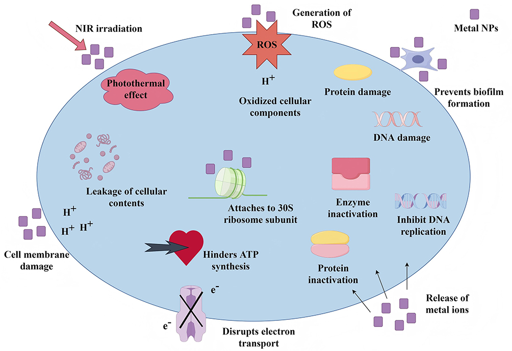 |
Figure 1 Schematic illustration of antibacterial mechanisms of metal nanoparticles. (By FigDraw). Abbreviations: NPs, nanoparticles; NIR, near-infrared; ROS, reactive oxygen species. |
Mechanism of Physical Damage
Cell Membrane Damage
Metal-based nanomaterials, due to their unique dimensions and geometric structures, can directly disrupt bacterial cell membranes through sharp edges, causing cellular content leakage and ultimately leading to cell inactivation.6 Additionally, a mass of negative charge on bacterial surfaces facilitates electrostatic interactions between positively charged metal-based nanomaterials and them, thereby destroying cell membranes to exert their antibacterial effects. Vukomanovic et al synthesized amino acid-functionalized AuNPs, which can accumulate on the surface of Pseudomonas aeruginosa and tightly contact the outer side of the bacterial cell walls, thereby promoting cell membrane disintegration and subsequent bacterial death.7 Haghniaz et al synthesized ZnFe2O4 NPs via a modified co-precipitation method, and investigated their antibacterial activity through membrane permeability and protein leakage assays. The results demonstrated that both E. coli and S. aureus treated with ZnFe2O4 NPs (100 µg/mL) showed significant disruption of the bacterial cell membranes, and the protein leakage levels reached 0.6 µg/mL and 0.7 µg/mL, respectively, representing 3.0-fold and 2.5-fold higher than those in the untreated group. Field-emission scanning electron microscopy (FE-SEM) images showed that most of the ZnFe2O4 NPs were spherical in shape and smooth on the surface, enabling easy contact with bacterial cell walls. This facilitated membrane penetration, leading to protein leakage and subsequent cell death.8
Furthermore, it was found that the concentration of NPs and their exposure duration were correlated with the cellular damage severity. Xiang et al developed silver-gold bimetallic alloy (AgAu@BSA) NPs coated with bovine serum albumin (BSA), achieving precise control over particle size and dispersion through optimized silver-to-gold material ratios. In vitro antimicrobial assays demonstrated that AgAu@BSA NPs exhibited minimum inhibitory concentrations of 128 μg/mL and 256 μg/mL against E. coli and S. aureus, respectively. As NPs concentration and exposure time increased, nucleic acid and protein leakage also increased, while cellular destruction further intensified.9
Photothermal Effect
Photothermal therapy (PTT) is a medical treatment technology that utilizes photothermal agents to convert light energy into thermal energy. Under the action of near-infrared (NIR), photothermal agents generate localized high temperatures that inactivate phospholipids and enzymes within bacterial cells and denature proteins, causing irreversible cell damage or even cell death.10 Metal NPs, particularly gold, silver, and copper, possess strong NIR absorption capabilities due to their localized surface plasmon resonance effect.11 This enables effective conversion of light energy into thermal energy, achieving the purpose of eliminating bacteria.
Wu et al developed a type of silica-coated gold-silver nanocages (Au-Ag@SiO2 NCs) that integrated the photothermal properties of AuNPs with the sustained release capability of Ag+, enabling antibacterial effect under NIR light irradiation.12 The study found that when the mass concentration of Au-Ag@SiO2 NCs was 50 mg/mL, NIR light exposure for 10 minutes, the photothermal temperature rose from 20.7°C to 57.4°C. In vitro and in vivo assays confirmed that Au-Ag@SiO2 NCs effectively inhibited the growth of S. aureus and E. coli under NIR light irradiation. Moreover, applying SiO2 coating on the surface of gold-silver nanocages not only extended the drug action duration, but also enhanced biocompatibility. He et al investigated a vehicle-free antimicrobial polymer polyhexamethylene biguanide (PHMB) hybrid AuNPs platform PHMB@Au NPs.13 This platform exhibited an excellent synergistic effect to enhance the photothermal bactericidal effect on S. aureus under NIR light irradiation. Additionally, PHMB@Au NPs effectively inhibited the formation of biofilms and rapidly eliminated bacteria through PTT, which not only promoted wound healing, even mediated macrophage transition from M1 to M2 type while accelerating tissue angiogenesis. Therefore, PTT is considered an effective strategy for antibacterial therapy.
Mechanism of Chemical Activity
Oxidative Stress
Oxidative stress refers to the pathological biochemical reactions caused by increased reactive oxygen species (ROS) produced by aerobic metabolism or decreased self-cleaning of ROS, which exert toxic effects on cells and lead to damage of cellular DNA, proteins, and lipids. Studies indicate that ROS are generated when oxygen in cells is reduced to superoxide anions, hydrogen peroxide, and hydroxyl radicals.14 Metal NPs can induce oxidative stress by generating ROS, leading to destruction of cell walls, cell membranes, and internal cell structures, as well as affecting gene expression and regulation and protein translation functions.15,16 Normally, ROS generation and cellular antioxidant capacity are maintained in balance. However, NPs can disrupt this balance to accelerate oxidative stress within cells, leading to excessive accumulation of ROS in pathogenic bacteria and ultimate cell death.
The elevated intracellular ROS levels are mainly attributed to the cellular uptake of NPs and the release of intracellular metal ions.17 Wunnoo et al successfully synthesized bio-AgNPs using aqueous Eucalyptus camaldulensis leaf extract. The study revealed that the bio-AgNPs demonstrated a strong inhibitory effect on Candida biofilms and could penetrate to the extracellular matrix to destroy yeast cell morphology, ultimately leading to cell death. Molecular biology studies on biofilms confirmed down-regulation in the expression of genes encoding hyphal growth and biofilm development, as well as those involved in hydrolytic enzymes.18 Zhang et al synthesized a composite silver nanomaterial (Ag2O@UMOPs) by integrating microporous organic polymers (MOPs) with Ag2O NPs.19 This composite material demonstrated potent antibacterial activity against both E. coli and S. aureus, attributed to the synergistic effects of sustained release of Ag+ and enhanced generation of ROS. Xi et al designed two kinds of copper/carbon nanozymes with distinct antimicrobial mechanisms. For the CuO-modified copper/carbon nanozymes, the released Cu2+ caused membrane damage, lipid peroxidation, and DNA degradation of gram-negative bacteria, whereas, for Cu-modified copper/carbon nanozymes, the generation of ROS via peroxidase-like catalytic reactions was the determining factor against both gram-positive and gram-negative bacteria.20 Sharma et al also demonstrated that the leakage of cytoplasmic components, loss of membrane permeability, and ROS generation were the primary causes of CuNPs-induced bacterial cell death.21 This indicates that ROS-induced oxidative stress plays a crucial role in the process of metal NPs destroying cellular components and affecting gene expression.
Metal Ion Release
Dissolution and release represent a critical mechanism through which metal NPs exert antibacterial activity. In physiological environments, metal NPs gradually undergo oxidation and dissolution, releasing bioactive metal ions such as Ag+ and Cu2+. These ions combine with sulfhydryl groups in bacterial proteins, forming unstable complexes that lead to protein denaturation and functional inactivation, thereby disrupting cellular metabolic processes and resulting in bacterial cell death.22 Furthermore, Ag+ can bind to phosphate groups in DNA, interfering with DNA replication and transcription, which hinders bacterial growth and proliferation.23 Additionally, the interaction between Ag+ with membrane proteins alters cell membrane permeability, facilitating intracellular accumulation of Ag+. The elevated intracellular concentration of Ag+ can interact with proteins involved in the respiratory chain, disrupting electron transfer and uncoupling oxidative phosphorylation, thereby inhibiting adenosine triphosphate (ATP) synthesis within bacterial cells.24 Gomma investigated the effects of AgNPs on protein and reducing sugar leakage, as well as respiratory chain dehydrogenase activity in S. aureus and E. coli. The results demonstrated that 50 mg/mL AgNPs could completely inhibit bacterial cell growth, disrupt bacterial membrane permeability, and depress the activity of certain membranous enzymes, ultimately leading to bacterial death.25 This demonstrates that metal ions released from nanomaterials can inactivate cells through interactions with cellular components including proteins, enzymes, and genetic material.
Mechanism of Biological Function Interference
Metabolic Pathway Inhibition
Metal NPs exert antibacterial effects by contacting bacteria and penetrating their cellular structures. These NPs selectively target critical metabolic enzymes and electron transport chains within bacteria, thereby inhibiting the activity of enzymes essential for peptidoglycan and protein synthesis, ultimately hindering their energy production.26,27
Cui et al found that AuNPs can disrupt cell membrane potential after contact bacteria, inhibit the activity of ATP synthase, and thus hinder the synthesis of ATP as the primary energy carriers. Meanwhile, AuNPs can also inhibit ribosome subunits from binding to transfer Ribonucleic Acid (tRNA), further promoting bacterial apoptosis. The synergistic effects of these two pathways enable AuNPs to exert antibacterial effects.28 Zhang et al reported that Ni-ZnO@C nanocomposites modified with photosensitizers generated ROS under the efficient photocatalytic action. This composite damaged bacterial cell membranes, and inhibited ATP synthesis through photoinduced electron transfer, effectively eliminating S. aureus biofilms. Moreover, the Ni-ZnO@C composite demonstrated excellent biocompatibility, further promoting wound healing in the cortex.29 Dai et al demonstrated that the silver nanocomposites (64 μg/mL) coated with high-molecular-weight polymers could eradicate 80% of an established drug-resistant bacterial biofilm and effectively inhibit intracellular enzyme activity, ultimately leading to bacterial death.30 Therefore, the inhibition of ATP synthesis and protein activity represents another crucial antibacterial mechanism associated with metal NPs.
Novel Nanosystems and Antimicrobial Strategies
Stimulus-Responsive Nanomaterials
Stimulus-responsive materials are a type of “intelligent” materials that respond to both exogenous and endogenous stimuli. They possess exceptional sensitivity to environmental or pathological signals, enabling them to convert external stimuli into dynamic structural or morphological changes through multiple pathways, thereby achieving adjustable physical or chemical properties.31,32 Stimulus-responsive metal-based nanomaterials are a frontier branch of stimulus-responsive materials, involving exogenous stimuli, such as light, ultrasound, and magnetic field, while endogenous stimuli include pH, redox, and enzyme activity.33 Researchers have successively developed photo-activated antibacterial strategies, magnetic-activated antibacterial strategies, and ultrasound-activated antibacterial strategies, further optimizing the antibacterial efficiency and controllability of the antimicrobial system. Fang et al designed a photothermal responsive polymer (TRB-ZnO@G) derived from MOFs and constructed a novel NIR-triggered antibacterial platform based on this polymer.34 This platform featured flexible two-dimensional nanostructures, high photothermal activity, sustained Zn2+ release, and switchable phase size conversion capabilities. It achieved a 99.5% eradication rate of biofilms in vivo, which was far superior to using PTT or ZnONPs alone. More importantly, the NIR-triggered synergistic antibacterial strategy demonstrated excellent biosafety and could effectively control inflammatory responses without damaging healthy tissues.
Additionally, antimicrobial strategies that respond to changes in the bacterial microenvironment have also attracted significant attention. In a recent study, Mei et al developed a system combining copper-doped polyoxometalate clusters (Cu-POM) responsive to the biofilm infection microenvironment with mild PTT for the treatment of biofilm-associated infections.35 This system promoted bacterial cuproptosis-like death via metabolic interference, while destroying bacterial biofilms and eliminating planktonic bacteria, achieving biofilm-associated infection clearance at all stages.
Synergistic Antimicrobial Strategies
Nanomaterials and Antibiotics Synergy
The combination of metal NPs with antibiotics can generate synergistic antibacterial effects.36,37 NPs, serving as carriers for natural antimicrobial agents, can not only deliver these agents safely and effectively to infection sites but also actively contribute to bacterial eradication. They enhance the antibacterial efficacy of drugs by minimizing drug degradation, promoting increased drug accumulation at the infected site, and reducing systemic toxicity.38 Intelligent drug delivery systems based on nanomaterials not only achieve controllable drug release through optimized pharmacokinetics, but also ensure the spatiotemporal release of drugs at targeted sites, thereby achieving antibacterial therapy.
Song et al engineered a zinc MOF (ZIF-8) by incorporating a photochemical reagent and used it as a gate for controllable drug release. Using Rifampicin as the drug molecule, they achieved precise release of antibiotics and Zn2+ through ultraviolet light stimulation, demonstrating synergistic antimicrobial effects while enabling controllable degradation of the drug carrier.39 Nadia Ghaffar et al developed a method to synthesize AgNPs, ZnONPs, and CuONPs using castor leaf extract.40 When Streptomycin was combined with these NPs, it significantly enhanced antibacterial activity against S. aureus, indicating synergistic interactions between the NPs and antibiotics. Notably, S. aureus was resistant to Streptomycin; however, S. aureus showed high sensitivity to Streptomycin when it was combined with NPs. This finding opens up new ideas for combating drug-resistant bacterial infections.
Immune Regulation and Synergistic Antibacterial Effects
Metal NPs exhibit anti-inflammatory properties primarily through inhibiting the release of inflammatory response factors to reduce inflammation. Studies have demonstrated that AgNPs, AuNPs, and Ag-Au alloy NPs are capable of reducing the pro-inflammatory cytokine levels in macrophages (IL-1β, IL-6, TNF-α) and NK cells (IFN-γ) to exert anti-inflammatory effects.41 In addition, AgNPs can achieve anti-inflammatory effects by inhibiting inflammatory cell infiltration, down-regulating TNF-α, IL-8, and lactate dehydrogenase (LDH), and suppressing the NF-κB and p38 signal pathways.42 The combination of metal NPs with other functional nanomaterials also demonstrates synergistic antibacterial properties. Zuo et al developed a halofuginone-silver nano thermosensitive hydrogel (HTPM&AgNPs-gel) via a physical swelling method, which effectively promoted skin fibroblast healing and inhibited the proliferation of E. coli and S. aureus.43 Compared with single materials such as AgNPs or hydrogels, this composite material demonstrates a more significant antibacterial effect and the ability to promote wound repair, while inhibiting inflammatory responses during the wound healing process.
Discussion
Metal-based nanomaterials represent promising therapeutic agents in combating drug-resistant bacterial infections in the 21st century. Through synergistic mechanisms including physical damage, oxidative stress, ion release, and metabolic interference, these materials exhibit broad-spectrum and highly efficient antibacterial properties, offering versatile strategies to address AMR. Furthermore, metal NPs can enhance antibacterial efficacy by conjugating with conventional antibiotics and modulating immune responses, thereby enabling synergistic approaches to overcome drug-resistant bacteria.
However, metal-based nanomaterials still present their limitations. Firstly, the synthesis and characterization technologies for these materials are not yet fully developed. They are associated with high production costs, low standardization, and poor stability,44 which significantly hinder their widespread applications. Secondly, the biosafety of metal-based nanomaterials is crucial for biomedical applications. The long-term toxicity of NPs remains unclear. They may trigger immune responses, cause cytotoxicity, or even affect gene expressions,45 thereby posing potential risks to human health. Consequently, further research is required to address the biocompatibility and potential toxicity of metal-based nanomaterials in order to ensure their long-term stability and safety in vivo. Lastly, biosafety evaluation methods for these nanomaterials remain inadequate, with a lack of systematic and standardized assessment criteria and protocols,46 which greatly restrict their translation into clinical applications. The antibacterial mechanisms, along with the advantages and limitations of metal NPs, are summarized in Table 1.
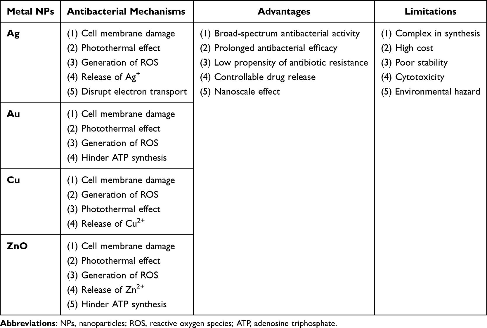 |
Table 1 The Antibacterial Mechanisms, Advantages and Limitations of Metal NPs |
To address these challenges, several key issues remain to be focused on in the design and development of novel nanocomposites: (1) Optimize the preparation methods of metal-based nanomaterials to enhance their synthesis efficiency and stability while reducing production costs; (2) Conduct in-depth research on the biosafety of metal-based nanomaterials, including their biocompatibility, toxicity, and degradability, to promote the development of sustainable and environmentally friendly nanomaterials; (3) Establish a comprehensive safety evaluation system for nanomaterials, and implement standardized assessments and regulatory oversight, to ensure the safety of their practical applications.
Conclusion
The antibacterial application of metal-based nanomaterials is undergoing rapid advancement. These materials exhibit unique antibacterial mechanisms, demonstrating superior antimicrobial properties including broad-spectrum activity, prolonged efficacy, and low propensity for inducing antibiotic resistance. Thus, they offer significant potential and promising prospects in addressing microbial resistance. Nevertheless, the clinical translation of nanobiomaterials necessitates a comprehensive and systematic evaluation of key biological safety parameters, including cytotoxicity, biocompatibility, and biodegradability. Ongoing research efforts are focused on the development of multifunctional nanocomposites, the exploration of synergistic interactions between nanomaterials and conventional antibiotics, and the enhancement of antimicrobial performance. Furthermore, the design of stimuli-responsive intelligent nanomaterials with controllable drug release capabilities is expected to facilitate the clinical translation and application of nanomaterials.
Funding
This work is supported by the Science and Technology Project of the Health Commission of Ganzhou, Jiangxi, China (No. 2022-2-69).
Disclosure
The authors declare that there is no conflicts of interest regarding the publication of this article.
References
1. Guo Y, Hu FP, Zhu DM, et al. CHINET surveillance of bacterial resistance: results of 2023. Chin J Infect Chemother. 2024;24(6):627–637. doi:10.16718/j.1009-7708.2024.06.001
2. Murray CJL, Ikuta KS, Sharara F, Antimicrobial Resistance Collaborators. Global burden of bacterial antimicrobial resistance in 2019: a systematic analysis. Lancet. 2022;399(10325):629–655. doi:10.1016/S0140-6736(21)02724-0
3. Zhang C, Fu X, Liu Y, et al. Burden of infectious diseases and bacterial antimicrobial resistance in China: a systematic analysis for the global burden of disease study 2019. Lancet Reg Health West Pac. 2023;43:100972. doi:10.1016/j.lanwpc.2023.100972
4. O’Neill J. Antimicrobial Resistance: tackling a crisis for the health and wealth of nations. London: review on Antimicrobial Resistance. 2014. Available from: https://amr-review.org/sites/default/files/AMR%20Review%20Paper%20-%20Tackling%20a%20crisis%20for%20the%20health%20and%20wealth%20of%20nations_1.pdf.
5. Guo Z, Chen Y, Wang Y, et al. Advances and challenges in metallic nanomaterial synthesis and antibacterial applications. J Mater Chem B. 2020;8(22):4764–4777. doi:10.1039/d0tb00099j
6. Zhang W, Wu Y, Liu L, et al. The membrane-targeting mechanism of host defense peptides inspiring the design of polypeptide-conjugated gold nanoparticles exhibiting effective antibacterial activity against methicillin-resistant Staphylococcus aureus. J Mater Chem B. 2021;9(25):5092–5101. doi:10.1039/d1tb00533b
7. Vukomanovic M, Cendra MDM, Baelo A, et al. Nano-engineering stable contact-based antimicrobials: chemistry at the interface between nano-gold and bacteria. Colloids Surf B Biointerfaces. 2021;208:112083. doi:10.1016/j.colsurfb.2021.112083
8. Haghniaz R, Rabbani A, Vajhadin F, et al. Anti-bacterial and wound healing-promoting effects of zinc ferrite nanoparticles. J Nanobiotechnology. 2021;19(1):38. doi:10.1186/s12951-021-00776-w
9. Xiang LY, Wang YL, Ren GC, et al. Antibacterial activity and mechanism of AgAu@BSA particles to E. coli and S. aureus. J Nanchang Univ. 2025;47(3):217–226. doi:10.13764/j.cnki.ncdg.20250703.001
10. Zhao L, Yang Q, Guo W, et al. Co 2.67 S 4 -based photothermal membrane with high mechanical properties for efficient solar water evaporation and photothermal antibacterial applications. ACS Appl Mater Interfaces. 2019;11(23):20820–20827. doi:10.1021/acsami.9b04452
11. Wang Y, Meng HM, Li Z. Near-infrared inorganic nanomaterial-based nanosystems for photothermal therapy. Nanoscale. 2021;13(19):8751–8772. doi:10.1039/d1nr00323b
12. Wu S, Li A, Zhao X, et al. Silica-coated gold-silver nanocages as photothermal antibacterial agents for combined anti-infective therapy. ACS Appl Mater Interfaces. 2019;11(19):17177–17183. doi:10.1021/acsami.9b01149
13. He X, Dai L, Ye L, et al. A vehicle-free antimicrobial polymer hybrid gold nanoparticle as synergistically therapeutic platforms for Staphylococcus aureus infected wound healing. Adv Sci. 2022;9(14):e2105223. doi:10.1002/advs.202105223
14. Slavin YN, Asnis J, Häfeli UO, et al. Metal nanoparticles: understanding the mechanisms behind antibacterial activity. J Nanobiotechnology. 2017;15(1):65. doi:10.1186/s12951-017-0308-z
15. Rezvani E, Rafferty A, McGuinness C, et al. Adverse effects of nanosilver on human health and the environment. Acta Biomater. 2019;94:145–159. doi:10.1016/j.actbio.2019.05.042
16. Yuan YG, Wang YH, Xing HH, et al. Quercetin-mediated synthesis of graphene oxide-silver nanoparticle nanocomposites: a suitable alternative nanotherapy for neuroblastoma. Int J Nanomed. 2017;12:5819–5839. doi:10.2147/IJN.S140605
17. Horie M, Tabei Y. Role of oxidative stress in nanoparticles toxicity. Free Radic Res. 2021;55(4):331–342. doi:10.1080/10715762.2020.1859108
18. Wunnoo S, Paosen S, Lethongkam S, et al. Biologically rapid synthesized silver nanoparticles from aqueous Eucalyptus camaldulensis leaf extract: effects on hyphal growth, hydrolytic enzymes, and biofilm formation in Candida albicans. Biotechnol Bioeng. 2021;118(4):1597–1611. doi:10.1002/bit.27675
19. Zhang Y, Tang Y, Liao Q, et al. Silver oxide decorated urchin-like microporous organic polymer composites as versatile antibacterial organic coating materials. J Mater Chem B. 2024;12(8):2054–2069. doi:10.1039/d3tb02619a
20. Xi J, Wei G, An L, et al. Copper/carbon hybrid nanozyme: tuning catalytic activity by the copper state for antibacterial therapy. Nano Lett. 2019;19(11):7645–7654. doi:10.1021/acs.nanolett.9b02242
21. Sharma P, Goyal D, Chudasama B. Antibacterial activity of colloidal copper nanoparticles against Gram-negative (Escherichia coli and Proteus vulgaris) bacteria. Lett Appl Microbiol. 2022;74(5):695–706. doi:10.1111/lam.13655
22. Armendariz Ontiveros M, Quintero Y, Llanquilef A, et al. Anti-biofouling and desalination properties of thin film composite reverse osmosis membranes modified with copper and iron nanoparticles. Materials. 2019;12(13):2081. doi:10.3390/ma12132081
23. Lin Y, Betts H, Keller S, et al. Recent developments of metal-based compounds against fungal pathogens. Chem Soc Rev. 2021;50(18):10346–10402. doi:10.1039/d0cs00945h
24. Bruna T, Maldonado-Bravo F, Jara P, et al. Silver nanoparticles and their antibacterial applications. Int J Mol Sci. 2021;22(13):7202. doi:10.3390/ijms22137202
25. Gomaa EZ. Silver nanoparticles as an antimicrobial agent: a case study on Staphylococcus aureus and Escherichia coli as models for gram-positive and gram-negative bacteria. J Gen Appl Microbiol. 2017;63(1):36–43. doi:10.2323/jgam.2016.07.004
26. Godoy-Gallardo M, Eckhard U, Delgado LM, et al. Antibacterial approaches in tissue engineering using metal ions and nanoparticles: from mechanisms to applications. Bioact Mater. 2021;6(12):4470–4490. doi:10.1016/j.bioactmat.2021.04.033
27. Franco D, Calabrese G, Guglielmino SPP, et al. Metal-based nanoparticles: antibacterial mechanisms and biomedical application. Microorganisms. 2022;10(9):1778. doi:10.3390/microorganisms10091778
28. Cui Y, Zhao Y, Tian Y, et al. The molecular mechanism of action of bactericidal gold nanoparticles on Escherichia coli. Biomaterials. 2012;33(7):2327–2333. doi:10.1016/j.biomaterials.2011.11.057
29. Zhang R, Chen Z, Li Y, et al. Enhanced photodynamic therapy efficacy of Ni-doped/oxygen vacancy double-defect Ni-ZnO@C photosensitizer in bacteria-infected wounds based on ROS damaged and ATP synthesis inhibition. J Math Sci Technol. 2024;192:173–189. doi:10.1016/J.JMST.2024.01.018
30. Dai X, Chen X, Zhao J, et al. Structure-activity relationship of membrane-targeting cationic ligands on a silver nanoparticle surface in an antibiotic-resistant antibacterial and antibiofilm activity assay. ACS Appl Mater Interfaces. 2017;9(16):13837–13848. doi:10.1021/acsami.6b15821
31. Wang X, Shan M, Zhang S, et al. Stimuli-responsive antibacterial materials: molecular structures, design principles, and biomedical applications. Adv Sci. 2022;9(13):e2104843. doi:10.1002/advs.202104843
32. Said SS, Campbell S, Hoare T. Externally addressable smart drug delivery vehicles: current technologies and future directions. Chem. Mater. 2019;31(14):4971–4989. doi:10.1021/acs.chemmater.9b01798
33. Astaneh ME, Hashemzadeh A, Fereydouni N. Recent advances in sodium alginate-based dressings for targeted drug delivery in the context of diabetic wound healing. J Mater Chem B. 2024;12(40):10163–10197. doi:10.1039/d4tb01049c
34. Fang J, Wan Y, Sun Y, et al. Near-infrared-activated nanohybrid coating with black phosphorus/zinc oxide for efficient biofilm eradication against implant-associated infections. Chem Eng J. 2022;435(P1):134935. doi:10.1016/J.CEJ.2022.134935
35. Mei J, Xu D, Wang L, et al. Biofilm microenvironment-responsive self-assembly nanoreactors for all-stage biofilm associated infection through bacterial cuproptosis-like death and macrophage re-rousing. Adv Mater. 2023;35(36):e2303432. doi:10.1002/adma.202303432
36. Namdar N, Nayeri Fasaei B, Shariati P, et al. Mesoporous silica nanoparticles co-loaded with lysozyme and vancomycin for synergistic antimicrobial action. Sci Rep. 2024;14(1):29242. doi:10.1038/s41598-024-78922-1
37. Zade BM, Abdollahi S, Raoufi Z, et al. Synergistic antibacterial and wound healing effects of chitosan nanofibers with ZnO nanoparticles and dual antibiotics. Int J Pharm. 2024;666:124767. doi:10.1016/J.IJPHARM.2024.124767
38. Hetta HF, Ramadan YN, Al-Harbi AI, et al. Nanotechnology as a promising approach to combat multidrug resistant bacteria: a comprehensive review and future perspectives. Biomedicines. 2023;11(2):413. doi:10.3390/biomedicines11020413
39. Song Z, Wu Y, Cao Q, et al. pH-responsive, light-triggered on-demand antibiotic release from functional metal-organic framework for bacterial infection combination therapy. Adv Funct Mater. 2018;28(23):1800011–1800017. doi:10.1002/adfm.201800011
40. Ghaffar N, Javad S, Farrukh MA, et al. Metal nanoparticles assisted revival of Streptomycin against MDRS Staphylococcus aureus. PLoS One. 2022;17(3):e0264588. doi:10.1371/journal.pone.0264588
41. Amina M, Al Musayeib NM, Alarfaj NA, et al. Antibacterial and immunomodulatory potentials of biosynthesized ag, au, ag-au bimetallic alloy nanoparticles using the asparagus racemosus root extract. Nanomaterials. 2020;10(12):2453. doi:10.3390/nano10122453
42. Li H, Yang T, Zhou H, et al. Emodin combined with nanosilver inhibited sepsis by anti-inflammatory protection. Front Pharmacol. 2017;7:536. doi:10.3389/fphar.2016.00536
43. Zuo RN, Gong JH, Gao XG, et al. Using halofuginone-silver thermosensitive nanohydrogels with antibacterial and anti-inflammatory properties for healing wounds infected with Staphylococcus aureus. Life Sci. 2024;339:122414. doi:10.1016/j.lfs.2024.122414
44. Yang F, Tong Y, Gu N. Research progress and development trend of nanobiomaterials. China Basic Science. 2022;24(01):46–56. doi:10.3969/j.issn.1009-2412.2022.01.006
45. Kothari D, Kumar A. Metallic, carbon-based, and polymeric nanomaterials: transforming dairy farming practices for sustainability. Food Chem X. 2025;29:102640. doi:10.1016/j.fochx.2025.102640
46. Allan J, Belz S, Hoeveler A, et al. Regulatory landscape of nanotechnology and nanoplastics from a global perspective. Regul Toxicol Pharmacol. 2021;122:104885. doi:10.1016/j.yrtph.2021.104885
 © 2025 The Author(s). This work is published and licensed by Dove Medical Press Limited. The
full terms of this license are available at https://www.dovepress.com/terms
and incorporate the Creative Commons Attribution
- Non Commercial (unported, 4.0) License.
By accessing the work you hereby accept the Terms. Non-commercial uses of the work are permitted
without any further permission from Dove Medical Press Limited, provided the work is properly
attributed. For permission for commercial use of this work, please see paragraphs 4.2 and 5 of our Terms.
© 2025 The Author(s). This work is published and licensed by Dove Medical Press Limited. The
full terms of this license are available at https://www.dovepress.com/terms
and incorporate the Creative Commons Attribution
- Non Commercial (unported, 4.0) License.
By accessing the work you hereby accept the Terms. Non-commercial uses of the work are permitted
without any further permission from Dove Medical Press Limited, provided the work is properly
attributed. For permission for commercial use of this work, please see paragraphs 4.2 and 5 of our Terms.