Back to Journals » Drug Design, Development and Therapy » Volume 19
Anticancer, Antioxidant and Antimicrobial Activity of Elaeagnus angustifolia L. Leaf Extract
Authors Doğan S ![]() , Meşe A, Baran MF, Baran A, Aktepe N
, Meşe A, Baran MF, Baran A, Aktepe N ![]() , Ahmadian E
, Ahmadian E ![]() , Mohamed AJ, Eftekhari A, Aktaş H
, Mohamed AJ, Eftekhari A, Aktaş H
Received 29 December 2024
Accepted for publication 19 August 2025
Published 3 September 2025 Volume 2025:19 Pages 7719—7734
DOI https://doi.org/10.2147/DDDT.S509705
Checked for plagiarism Yes
Review by Single anonymous peer review
Peer reviewer comments 2
Editor who approved publication: Professor Manfred Ogris
Serap Doğan,1 Ahmet Meşe,2 Mehmet Fırat Baran,3 Ayse Baran,1 Necmettin Aktepe,4 Elham Ahmadian,5 Ali Jimale Mohamed,6 Aziz Eftekhari,7 Hüsnü Aktaş1
1Mardin Artuklu University, Kızıltepe Faculty of Agricultural Sciences and Technologies, Department of Field Crops, Mardin, Artuklu, 47200, Türkiye; 2Department of Chemistry, Graduate Education Institute, Mardin Artuklu University, Mardin, 47200, Turkey; 3Department of Food Technology, Vocational School of Technical Sciences, Batman University, Batman, 72000, Turkey; 4Mardin Artuklu University, Faculty of Health Sciences, Department of Nursing, Mardin, 47200, Türkiye; 5Kidney Research Center, Tabriz University of Medical Sciences, Tabriz, 51666-15731, Iran; 6Department of Pharmacology, Faculty of Medicine, Somali National University, Mogadishu, 801, Somalia; 7Department of Biochemistry, Faculty of Science, Ege University, Izmir, 35040, Turkey
Correspondence: Ali Jimale Mohamed, Email [email protected] Mehmet Fırat Baran, Email [email protected]
Objective: This study was conducted to determine and compare the antioxidant, cytotoxic, and antimicrobial effects of spindle leaves of Elaeagnus angustifolia L. (E. angustifolia) (oleaster) leaves.
Methods: Total phenolic content was measured using the Folin-Ciocalteu method, phenolic compound analysis by liquid chromatography-electrospray ionization-tandem mass spectrometry (LC-ESI-MS/MS) and antimicrobial effect by the minimum inhibition concentration (MIC) method. The free radical scavenging activity was determined by the (2,2-Diphenyl-1-picrylhydrazyl) DPPH method, the free radical scavenging activity was determined by the ABTS method, and cytotoxicity assays were performed by the MTT method in human retinal epithelium cells (RPE-1), human osteosarcoma cells (U2OS), and prostate cancer cells (DU-145) cell lines.
Results: High amounts of gallic acid, protocatechuic acid, and o-coumaric acid were identified as phenolic compounds. E. angustifolia was found to have a good antioxidant capacity and high free radical scavenging capacity. In this study, for the first time, E. angustifolia leaf extract was used to investigate cytotoxic effects on human retinal epithelium (RPE-1), human osteosarcoma cells (U2OS), and prostate cancer (DU-145) cells and antimicrobial effects on Listeria monocytogenes American Type Culture Collection (ATTC) 7644, Staphylococcus aureus ATCC 29213, Escherichia coli ATCC 25922, Klebsiella pneumonia ATCC 11774, and Candida albicans ATCC 10231 microorganisms. The highest cytotoxic effect was observed in the DU-145 cell line, and the highest antimicrobial effect was observed in Listeria monocytogenes ATTC 7644 and Candida albicans ATCC 10231. The leaf extract of the plant contains some important phenolic compounds and has high free radical scavenging capacity, a good anticancer effect, and effective antimicrobial activity on yeast species such as C. albicans.
Conclusion: Our study will contribute greatly to the search for anticancer and antimicrobial agents, especially from a pharmacological perspective, by examining biological activity using three different methods.
Keywords: E. angustifolia antioxidant, cytotoxicity, phenolics, antimicrobial activity
Introduction
Reactive oxygen species (ROS) are by-products of metabolites produced during metabolism in the cytosol, oxidative phosphorylation, and electron leakage from mitochondrial electron carriers and enzymes. In addition, some defence cells in the human immune system contribute to ROS production. Sunlight, radiation, ultraviolet rays, and environmental pollution are other sources of ROS.1 Reactive oxygen species (ROS) and lipid peroxidation are etiologically associated with many diseases. Lipid peroxidation can alter the vital membrane protein structure and function, and if left unchecked, can lead to cellular dysfunction and widespread tissue damage.2 As a result, biomolecules, such as body proteins, lipids, enzymes, RNA, and DNA, are damaged, resulting in oxidative stress. Following oxidative damage, they play a role in the development and progression of cancer, atherosclerosis, cardiovascular diseases, neurodegenerative diseases, and many other diseases.3
Bioactive compounds have strong antioxidant and scavenging activities, most of which are produced by plants under conditions of oxidative stress against physical effects and chemicals that can damage the plant, such as microorganisms, pests, and reactive oxygen species. Plants produce a large number of secondary metabolites, that is, bioactive compounds containing a phenol group in their structure, to protect their lives from various adverse conditions. Some of these compounds are chemically termed as phenolic compounds. Some of these compounds are soluble in organic solvents, and some are soluble in water, whereas large polymers, such as lignin are insoluble.4 Phenolic compounds are important plant compounds with radical-scavenging abilities, owing to their hydroxyl groups. Therefore, they exhibit antioxidative, antimutagenic, and anticarcinogenic properties.5 When humans ingest these compounds as food through plants, they have been proven to have significant health benefits for the prevention of many diseases such as infectious diseases, cancer, and cardiovascular diseases.6
Antioxidants prevent uncontrolled formation of ROS in cells or stop their reactions with biological structures. The antioxidant defence system consists of bioactive compounds that are either enzymatic or non-enzymatic. Active chemicals synthesized in the cytosol, phenolic compounds, and flavonoids, which have different biological activities that naturally form the defense system in plants, can inhibit free radicals. In addition to their antioxidant and free radical scavenging properties, phenolic compounds also exhibit antimicrobial, anti-radioactivity, immunomodulatory, anti-cancer, and anti-inflammatory activities.7
The hallmark of osteosarcoma (OS), a high-grade primary skeletal cancer, is the deposition of immature osteoid matrix by spindle cells of mesenchymal origin. Osteosarcoma, along with osteochondroma and Ewing family tumors, are the three most common bone tumors observed in patients under 20 years of age. The most common are osteosarcomas. The genetic profile of this tumor is quite diverse, and there is no single consistent factor that contributes to the pathophysiology of osteosarcoma. Current disease management strategies include surgical resection of all clinically visible tumors or high-dose chemotherapy.8 OS has remained unchanged for decades despite the development of an increasing number of targeted medicines and improvements in the survival rate of other malignancies. More attention should be paid to complementary and alternative treatments in light of these disappointing outcomes. Owing to their strong anticancer activity and low toxicity in healthy tissues, dietary supplements and phytotherapeutic substances have emerged as promising treatment options that merit further research in this area.9
Prostate cancer (PCa) is a malignant change in the prostate, an important component of the male reproductive system, and is one of the most common cancers in older men. Prostate cancer is the most commonly diagnosed neoplasm in men, and the second leading cause of cancer-related mortality in the United States. An estimated 2.6 million new cancer cases are diagnosed annually in Europe. Prostate cancer accounts for 11% of cancers in men and is the cause of 9% of all cancer-related deaths in men.10 Chemotherapy and radiotherapy are the most common and effective methods for prostate cancer treatment.11 However, owing to the high cost of these treatments and their negative effects on patients and the country’s economy, there is a need to investigate new nationally sourced compounds with high cytotoxic effects.
E. angustifolia is a fruit tree from the Elaeagnaceae family that can grow in harsh and diverse environmental conditions in regions extending from Europe and Asia to the Himalayas, is better adapted than other tree species, has brownish elliptical fruits, and is not currently in danger of extinction.3 The leaves and fruits of the plants were covered with dense silverscales. E. angustifolia has a wide range of indications for use in various countries and folk medicine. Some studies have reported that it contains various phytochemical compounds with various pharmacological and biological activities.12 According to previous studies, needle leaves have antimicrobial, anti-inflammatory, antioxidant, and antioxidant activities. It has anti-mutagenic and anti-cancer properties that reduce gastrointestinal spasms, increase blood flow in the coronary arteries, and promote wound closure and healing.13
E. angustifolia is used worldwide in folk medicine in many countries. Its pharmacological effects have been reported in several studies. This study aimed to determine the total phenolic compounds in oleaster (E. angustifolia) leaves, which are known to have medicinal properties, and to determine their phytochemical components, as well as their antioxidant, cytotoxic, and antimicrobial properties. We believe that less studied cytotoxic and antimicrobial research on plants may shed light on future new drug studies. Thus, the aim is to create resources for the pharmaceutical industry using bioactive compounds that can be obtained from plants for the treatment of various diseases.
Materials and Method
Obtaining the Plant and Preparing the Extract
The plant was obtained from the leaves of an E. angustifolia L. plant growing in its natural environment in the rural area of Yayla village in the center of Mardin Province in June and was biologically identified as a species. The plant still grows in a wide geographical area and is a tree species with economic value. E. angustifolia L. leaves were described botanically and morphologically by Mardin Artuklu University Plants Department plant taxonomist Dr. Fatma Mungan Kılıç. (Figure 1). The plant MARIUM 42 was registered at the Mardin Artuklu University Herbarium. They were then dried away from the sun in the laboratory. After drying, leaves were ground and weighed to 50 g. The weighed sample was placed in a Soxhlet cartridge and 300 mL of Merck’s 99.7% pure methanol was added to one of the flasks to obtain the extract. The extract was filtered through Whatman filter paper. The solvent was evaporated using a rotary evaporator (Figure 2). The obtained extract was stored in a deep freezer at −22°C for future study.14
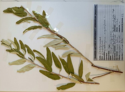 |
Figure 1 E. angustifolia L. plant and leaves (Mardin Artuklu University Herbarium MARIUM 42). |
 |
Figure 2 E. angustifolia L. Laboratory procedures. |
Determination of Bioactive Components via LC-MS/MS
LC-MS/MS was used to determine the composition of the E. angustifolia.
Chemicals Used in Analysis
All chemicals and standards used for LC-MS/MS were of analytical grade and were obtained from Sigma-Aldrich (Germany). Ultrapure water was used for all measurements. All solutions used in the measurements were filtered using a polypropylene-protected nylon membrane syringe filter and analyzed. Commercial polyphenol compounds were used in the development of this method. The compounds found in solid form were also dissolved in methanol and used to prepare the main stock solution.
Optimizing Device Conditions
The components were evaluated using a high-performance liquid chromatography (HPLC) system equipped with a tandem mass spectrometer. A 1260 degasser, a column oven, and binary pumps were integrated into the HPLC device. The chromatographic conditions were optimized to facilitate the separation of the compounds. In addition, optimization was performed to prevent suppression effects.15
Total Phenolic Content Measurement
Folin-Ciocalteu reagent (Folin Phenol Reagent or Folin-Denis reagent) is a mixture of phosphomolybdate and phosphotungstate and was used in the colorimetric determination of phenolic and polyphenolic antioxidants.16 Phenolic compounds formed colored complexes with Folin-Ciocalteu reagent in alkaline medium, and the maximum absorbance of the purple-violet complex formed after 2 hours of incubation at 700 nm was measured. The values obtained were calculated with the gallic acid equivalence (mg GAE/g extract) equation. In order to obtain the calibration graph required to calculate gallic acid equivalence, gallic acid was prepared at 5 different concentrations, and the calibration graph was obtained.
Determination of Free Radical and Antioxidant Activity
DPPH Free Radical Scavenging Activity Determination Experiment
Antioxidant activity determinations of all prepared samples were made according to Wu et al.17 This method is based on the principle of scavenging 2,2-diphenyl-1-picrylhydrazyl (DPPH), a stable free radical, in the presence of antioxidant chemicals that donate electrons and hydrogen atoms, and spectrophotometrically determining the opening of the characteristic purple color. First of all, 4 mL of 0.0004% (w/v) methanolic DPPH solution and 1 mL of extract solutions prepared at different concentrations (1.0, 2.0, 4.0, 8.0 mg/mL) were mixed, and the absorbance values were measured at 517 nm after 30 minutes of incubation at room temperature in the dark. Using the absorbance values of the samples;

The % inhibition value was calculated according to the formula above. The obtained inhibition values were plotted against the extract concentrations determined as mg/mL, and the concentrations of each extract providing 50% color lightening were calculated as the 50% inhibition (IC50) value. Butylated hydroxyanisole (BHA) was used as a positive control.
The ABTS Free Radical Scavenging Activity Determination Experiment
A 2,2-azino-bis(3-ethylbenzothiazoline-6-sulfonic acid) (ABTS) free radical scavenging activity experiment was carried out according to the method of Re et al.18 First, ABTS and potassium peroxodisulphate solutions were mixed and kept at room temperature for 12–18 hours in a light-proof environment. Before starting the experiment, it was diluted until the absorbance value was read as 0.700 using ethanol at a 734 nm wavelength in the spectrophotometer. Then, samples prepared at different concentrations (0.05, 0.10, 0.15, 0.20, 0.25 mg/mL) and the prepared ABTS solution were mixed and incubated for 30 min at room temperature. After this period, the absorbance values of the samples were measured at 734 nm. BHA solutions prepared at 5 different concentrations (0.01, 0.02, 0.03, 0.04, 0.05 mg/mL) were used as a positive control. After finding the percentage values of antioxidant activities, the corresponding IC50 values were calculated.
CUPRAC (Copper(II) Ion Reduction Capacity) Test
The CUPRAC experiment was performed according to the method performed by Apak et al.19 Prepared CUPRAC solution was placed in microplate wells, and 30 µL of extract solution dissolved in its own solvent at 1 mg/mL was added to them. After incubation at room temperature for 30 minutes, it was read at 450 nm. The absorbance values obtained were calculated in the Trolox equivalence (mg TE/g extract) equation. In order to obtain the calibration graph required to calculate Trolox equivalence, Trolox was prepared at 4 different concentrations, and the calibration graph was obtained.
Metal Chelation Test
The determination of metal chelating capacity was carried out according to Dinis et al.20 Plant extracts were dissolved in their own solvent, and the positive control ethylenediaminetetraacetic acid (EDTA) was dissolved in methanol at a concentration of 1 mg/mL. About 3.2 mL of distilled water was added to the extract solutions, and the first 100 µL of FeCl2 and then 200 µL of ferrozine were added. It was mixed well and left to incubate for 10 min at room temperature. Then, measurements were made at 562 nm in a spectrophotometer. The percentage antioxidant activity values of the obtained data were calculated by substituting them into the equation.
Cell Culture and Cytotoxic Activities via MTT Assay
MTT [3-(4,5- dimethyl thiazol-2-yl)-2,5-diphentyl tetrazolium bromide] assay is a widely used enzymatic test for evaluating the cytotoxicity of plant samples.21 The MTT assay was used to determine the toxic effects of E. angustifolia extracts at different time intervals.
Cytotoxicity activity applications on cells was assessed in osteosarcoma (U2OS) and human prostate carcinoma (DU-145) cancer cell lines obtained from the American Type Culture Collection (ATCC) and healthy human retinal pigment epithelial-1 (RPE-1) cell lines at the Dicle University Faculty of Veterinary Medicine Cell Culture Laboratory.
The cell lines used in this study were incubated in T75 culture flasks in DMEM (Gibco 41965039, UK) medium with 10% FBS, 100 U/mL penicillin, and 100 U/mL streptomycin in 5% CO2 at 37°C.
When the cells reached 80–90% of the adequate number, they were removed from the flasks and their numbers were determined using the hemocytometric method. The counted cells were seeded in 96-well plates in 90 µL medium in each well with RPE-1, U2OS (5 × 10³) and DU-145 (8 × 10³) cells in each well and two plates (to perform two separate time applications, 24 h and 48 h) in three replicates. The next day, E. angustifolia leaf extract was prepared at various concentrations (1, 31.25, 62.50, 125, 250, 250, 500, 1000, and 2000 µg/mL) and applied to the cultured plates, and ultrapure water was applied to the cells in the control group. At 24 and 48 h after treatment, an MTT assay was performed to determine changes in cell viability.
To each well, 10 µL of the prepared MTT (5 mg mL) solution was added and cells were incubated for 3 h at 37°C in a humidified atmosphere containing 5% CO2. After 3 h, the medium in the wells was removed and replaced with 100 µL DMSO. The assays were repeated three times, and the results were averaged.
Determination of Antimicrobial Activity
The antimicrobial effects of E. angustifolia extract on gram-negative and gram-positive bacterial strains and Candida albicans fungi were investigated using the microdilution method.15 This method was applied to gram-positive Listeria monocytogenes ATTC 7644 (Lm), Staphylococcus aureus ATCC 29213 (Sa), gram-negative Escherichia coli ATCC 25922 (Ec), Klebsiella pneumonia ATCC 11774 (Kp), and Candida albicans ATCC 10231 (Ca). Vancomycin was used as the positive control for L. monocytogenes and S. aureus strains, colistin was used for K. pneumonia and E. coli strains, and fluconazole was used as the C. albicans yeast control. The microorganisms used in this study were obtained from Mardin Artuklu University Turkey.
A total of 96 microplates were used in the experiments. Appropriate media for each strain were distributed into the wells. Extracts prepared at varying concentrations were distributed into wells in a series of dilutions, starting from the first well. Some wells were used for the growth and sterilization steps. After these procedures, microorganisms prepared according to McFarland 0.5 turbidity criterion were distributed into the wells. The same procedure was performed for each strain using an appropriate antibiotic (to compare the effects of the extract). Vancomycin was used for gram-positive strains, colistin for gram-negative strains, and fluconazole for yeast. Plates prepared for examination of antibiotic effects by microdilution were incubated in an oven at 25–37°C for 24 h. The growth was then checked, and the minimum inhibitory concentration (MIC) was determined.
Findings and Discussion
Total Phenolic Content Measurement Experiment Results
A calibration graph was obtained using 5 different concentrations of gallic acid (Figure 3), and the gallic acid equivalence (mg GAE/g) of the plant extracts was calculated using this graph. The total phenolic substance amount of the methanol extract of the oleaster plant at a concentration of 1 mg/mL was found to be 2.59 ± 0.014 mg GAE/g (Table 1).
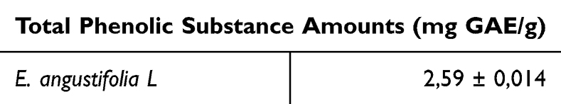 |
Table 1 Total Phenolic Substance Amount Results |
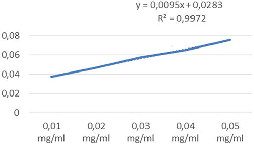 |
Figure 3 Gallic acid calibration graph. |
Berktaş and Çam conducted a study to determine the phenolic contents of the fruits and leaves of the oleaster (E. angustifolia L.) plant and found phenolic content of 18.32 mg GAE/g in oleaster fruits and 3.62 mg GAE/g in oleaster leaves. Leaf content is close to the phenolic content value in our study.22
In a study investigating the phenolic contents of some non-traditional edible plants, it was reported that it was 0.64 ± 0.07 mg GAE/g in narrow-leaf banana (Plantago lanceolata); 1.81 ± 0.16 mg GAE/g in white clover (Tifolium repens); 0.68 ± 0.07 mg GAE/g in perennial ryegrass (Lolium perenne) and 0.89 ± 0.04 mg GAE/g in tall fescue (Festuca arundinacea).23 (Iqbal et al, 2022). (23). It is seen that the phenolic contents of these plants are lower than the phenolic content of the oleaster plant subject to our study. In the study conducted with some plants native to Northern Thailand, the phenolic content of the ethanolic extract of Glochidion hirsutum (Roxb). Voigt (3.02 ± 0.25) was found to be the highest. The lowest phenolic content was observed in Hapaline benthamiana Schott (0.40 ± 0.00) plant.24
The phenolic bioactive substance content in the leaves of E. angustifolia was determined using the Folin-Ciocalteu spectrophotometric method. Component analyses of the two extracts were performed by LC-ESI-MS/MS at aqueous concentrations. Total phenolic contents (TPC) are listed in Table 2. The concentrations of total phenolic constituents, according to the Folin-Ciocaltaeu method, were calculated from the calibration graph of gallic acid on a mg/mL gallic acid equivalent (GAE) basis. Accordingly, the GAE value for E. angustifolia was calculated at 10,20 µg/mL.
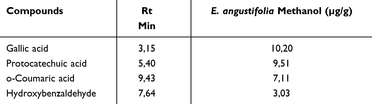 |
Table 2 Total Phenolic Content of E. angustifolia L. Plant Leaf |
In our study, gallic acid, protocatechuic acid (PCA) (3,4-dihydroxybenzoic acid), o-coumaric acid, and hydroxybenzaldehyde were detected in high amounts, as shown in Table 2 and Figure 4. Each of these four phenolic acids is a bioactive component derived from hydroxycinnamic acid.25 Shikimic acid derivatives, which are precursors of the flavonoid biosynthesis pathway through photosynthesis in plant leaves, are more abundant in leaves than in other plant organs. This is another explanation for the higher flavonoid content in the leaves than in the flowers and other organs (Saboonchian ve ark. Saltan ve ark. determined high amounts of gallic acid, PCA, o-coumaric acid, and hydroxybenzaldehyde phenolics in the HPLC analysis of E. angustifolia leaf extract.5,26 Gallic acid was found at its highest concentration (10,20 µg/g). Gallic acid is a benzoic acid, a secondary metabolite of phenolic compounds found in many plants, with hydroxyls at positions 3, 4, and 5 of its carbon units.27 It has been shown to have antioxidant, antitumor, anti-inflammatory, antiallergic, antimicrobial, neuroprotective, cardioprotective, and gastroprotective effects, especially in the health and protecting of biological cells.28 The scientific community has recently shown significant interest in the potential applications of gallic acid, which protects against various oxidative damage disorders, owing to its antioxidant and free radical-scavenging properties. Gallic acid has been shown to treat many cancer cells in many ways, such as by acting as a prooxidant29 causes caspase enzyme activation,30 induces apoptosis31 and exhibits selective cytotoxic effects.32 It has been reported that gallic acid can prevent the release of ROS in heart cells resulting from advanced glycation end products through its antioxidant effect.33 Gallic acid has also been shown to play a protective role in neurodegenerative diseases such as Alzheimer’s34 and Parkinson’s35 PCA was the main metabolite of anthocyanin. PCA is synthesized from phenylalanine via the shikimic acid pathway36 PCA is found in many foods, especially vegetables and fruits, especially in our daily foods, and is a very common phenolic derivative in nature. It is structurally similar to gallic acid, vanillic acid, and caffeic acid and has strong antioxidant properties37 PCA is recognized as an active component of most traditional Chinese herbal medicines, and has properties that are highly beneficial for the progression and prevention of many diseases. In vitro and in vivo studies of PCA and its derivatives for the treatment or prophylaxis of a wide range of diseases caused by oxidative stress damage have shown good results.38 Studies have shown that PCA exerts antioxidant, anti-inflammatory, antihyperglycemic, anticancer, and antimicrobial activities by inducing apoptosis.39 In addition, antiulcer, antiviral, analgesic, antiatherosclerotic, hepatoprotective, and neuroprotective properties have also been shown in the analysis of phenolic compounds in the methanol extract of E. angustifolia leaves, which obtained PCA values close to those in our study.3,24,40 Ayaz and Bertoft found PCA at a high levels of PCA in the phenolic analysis of spindled fruits.41 The second-most bioactive compound detected in the plant extracts was o-coumaric acid (7.11 µg/g). Coumaric acid is a hydroxylated derivative of cinnamic acid that is widely distributed in nature and plays an important role. It is abundant in vegetables, such as tomatoes, carrots, and cereals, and in the seed coats of cereal grains.42 Coumaric acid can be absorbed in all parts of the gastrointestinal system as a nutrient and is known to be a bioactive compound with very high bioavailability. Studies have shown that coumaric acid has antioxidant, anticancer, antimutagenesis, antidiabetic, anti-hyperpigmentation, and antiulcerative properties.43 In addition, it can be used in the treatment of arthritis and some neurological disorders owing to its anti-melanogenic, anti-inflammatory, and strong free-radical-scavenging properties.44 In their phenolic compound research on E. angustifolia leaves. Karkar and Şahin, Saltan et al, Carradori et al, and Lee et al found high amounts of coumaric acid in the methanol extract of E. angustifolia leaf extract, as in the findings of our study.3,26,45,46 It is the third most abundant hydroxybenzaldehyde in plant extracts. Hydroxybenzaldehyde belongs to the phenolic aldehyde family and is widely synthesized in many plants. Vanillin (4-hydroxy-3-methoxybenzaldehyde), an important flavoring agent widely used commercially, is a p-hydroxybenzaldehyde derived from ferulic acid. Shyamala et al showed that hydroxybenzaldehyde exhibits high antioxidant properties.47 Yuan et al showed that hydroxybenzaldehyde alleviated motor impairment in time-dependent cerebral ischemia-reperfusion in rats in experimental research.48 Zhu et al showed that it inhibited superoxide anion formation and elastase release by human neutrophils.49 In their experimental research on rats, Kang et al proved that hydroxybenzaldehyde promotes both wound closure and healing.50
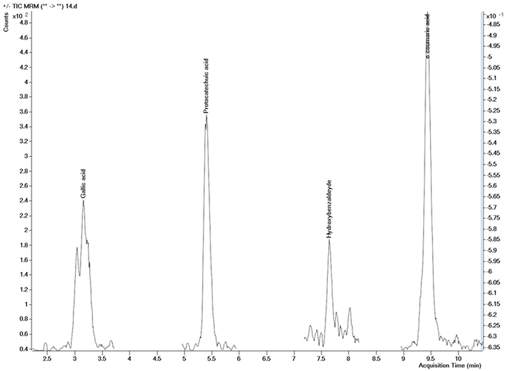 |
Figure 4 LC-ESI-MS/MS chromatogram of E. angustifolia plant leaf. |
Determination of DPPH Free Radical Scavenging Activity Experiment Results
Since the above-ground extract of the oleaster plant showed low activity, it was calculated at concentrations of 1, 2, 4, and 8 mg/mL. Four intermediate concentrations were created from the main stocks at different rates, and the IC50 value of the absorbances obtained in the spectrophotometer at the end of the incubation period was calculated. According to the calculations, the IC50 value of the oleaster plant is given in Table 3 together with BHA used as a positive control. The decrease in the IC50 value is an indication that the antioxidant activity has increased.51
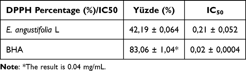 |
Table 3 DPPH Free Radical Scavenging Activity Results (1 mg/mL % ± Standard Error, (mg/mL, IC50)) |
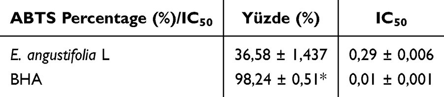 |
Table 4 ABTS Free Radical Scavenging Activity Results (0.25 mg/mL % ± Standard Error, (mg/mL, IC50)) |
An increase in DPPH free radical scavenging activities was observed as the concentrations of the plant extracts used in the study increased. Although the results were lower compared to BHA at the determined concentrations (Table 3), a regular increase in biological activity was observed depending on the concentration.
Akgül et al reported the percentage of DPPH free radical scavenging activity of the ethanol extract of the plant at a concentration of 2 mg/mL as 68.721 ± 1.694 in their study with Euphorbia eriphora.52 It is lower than the standard at the same concentration (96.388 ± 0.6708). The percentage scavenging value of the oleaster plant used in our study at the same concentration (69.78 ± 1.564) was determined to be higher than the Euphorbia eriphora extract. In the study investigating the antioxidant capacity of the Sisymbrium officinale plant extract, the percentage of DPPH free radical scavenging activity was reported as 193.7 ± 3.4 μmol Vitamin C/g. The results show that the plant extract has high antioxidant activity.53
Results of the ABTS Free Radical Scavenging Activity Determination Experiment
Five intermediate concentrations were created from the main stock at different rates, and the IC50 value of the absorbances obtained in the spectrophotometer at the end of the incubation period was calculated. According to the calculations, the IC50 value of the oleaster plant is given in Table 4 together with BHA used as the positive control. It can be said that the closer the IC50 value is to the positive control BHA values, the higher the free radical scavenging activity of the plant. An increase in the values of the plant extracts was observed simultaneously with the increase in BHA values due to the increase in concentration. The free radical scavenging activity of ABTS at a concentration of 0.25 mg/mL was determined as 36.58 ± 1.437%. It was observed that this value was lower than the positive control BHA value.
In the ABTS free radical scavenging activity test carried out to determine the antioxidant activity of the parts of Myrtus communis L. (Mersin), it was determined that the flower buds showed the highest activity (924.12 ± 4.20), and these results are significantly higher than the oleaster plant.54 The ABTS free radical scavenging activities of the materials obtained as a result of different extractions of the leafy stem of Flamingia faginea Guill. and Perr. (Barker), a medicinal plant used in the endogenous treatment of hypertension, were compared, and the IC50 value of the extracts obtained in the form of an aqueous decoction of the plant was reported as 252.36 ± 1.26; the IC50 value of the extracts obtained as a decoction residue fraction was reported as 258.70 ± 0.26.55 These results show lower activity than the oleaster plant.
CUPRAC (Copper(II) Ion Reduction Capacity) Experiment Results
A calibration graph was obtained using 4 different concentrations of trolox (Figure 5), and the trolox equivalence of plant extracts (mg TE/g) was calculated using this graph. Copper(II) ion reduction capacity of oleaster plant methanol extract at 1 mg/mL concentration was found to be 2.77 ± 0.004 mg TE/g (Table 5).
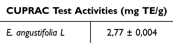 |
Table 5 CUPRAC Experiment Results |
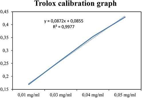 |
Figure 5 Trolox calibration graph. |
They determined the copper(II) ion reducing capacity of the methanol extract obtained from the fruits of Hippophae rhamnoides L. (Wild Oleaster) as 23.41 mg TE/g.56 It is seen that this value is higher than the CUPRAC value of the oleaster plant, which is the subject of our study. In their study, Kesim and Yıldıztekin examined the copper(II) ion reducing capacities of Inula graveolens and Inula viscosa species, and decreases occurred in the 48-hour measurements compared to the 24-hour measurements.57 The highest CUPRAC activity of the Inula graveolens species was determined in the hexane extract (331.3 ± 0.31 mg TE/g), and the highest CUPRAC activity of the Inula viscosa species was determined in the hexane extract (291.8 ± 1.21 mg TE/g). It is seen that the methanol extracts of the mentioned plants are lower at 17.61 ± 0.18 and 12.14 ± 0.27 mg TE/g, respectively. Compared to the value of the methanol extract of the oleaster plant, the copper(II) ion reduction capacities of the Inula graveolens and Inula viscosa species are quite high.
The metal chelating antioxidant activity result of the methanol extract of the oleaster plant at a concentration of 0.1 mg/mL was found to be 38.32 ± 0.73. The metal chelating antioxidant activity value of the oleaster plant extract is lower than the value of the synthetic antioxidant EDTA (90.39 ± 0.36 mg/L) (Table 6).
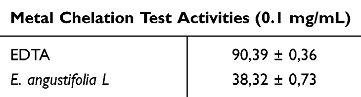 |
Table 6 Metal Chelation Test Results (%) |
It was stated that while the ethyl acetate fractions of onion from Crocus mathewii, one of the forgotten angiosperms of Turkey, did not show any bioactivity in the metal chelation experiment, the aerial parts fraction showed lower antioxidant activity (IC50 591.71 ± 142.10 mg/L) compared to EDTA (IC50 3.47 ± 0.35 mg/L).58 The metal chelation results with leaf and fruit extracts of the Elaeagnus angustifolia plant were reported as the highest: 2.97 ± 0.16 and 18.25 ± 0.44 mg EDTAE/g (Carradori et al, 2020).(44). When the antioxidant activity studies were evaluated together, an increase in DPPH and ABTS free radical scavenging activities was observed depending on the increase in concentration; phenolic content was determined; CUPRAC copper ion reducing power and metal chelating antioxidant activity were demonstrated. When the results were compared with the literature, they showed low or high activity depending on the plant species.
Cytotoxic Activity
The rapid increase in the number of cancer patients in recent years has led researchers to develop new treatment methods. With these methods, interest in products that can be obtained naturally has increased to reduce the harmful chemical effects of currently used drugs. In previous studies, plants with high antimicrobial and antioxidant content, especially in terms of strengthening the immune system, have attracted attention. However, a detailed examination is required to determine the effective doses of these plants and to correctly use them for treatment purposes.59 (Zimmermann-Klemd ve ark., 2022).(58) The main reasons why herbal products and their derivatives are preferred in treatment are that they are compatible with the body’s immune system and, when found to be effective, they help increase the body’s resistance without harming healthy cells.60 Anticancer drugs used in classical treatments are classified as mitosis inhibitors, alkylating compounds, antimetabolites, cytostatic antibiotics, hormones and hormone antagonists, other cytostatics (asparaginase, cisplatin, and carboplatin), radioactive isotopes, interferons, and tyrosine kinase inhibitors.61 Prostate cancer is a multifactorial disease, with aging and a family history being important risk factors. In prostate cancer, in addition to the stage and spread of the disease, the patient’s condition and genetic factors should be considered when planning the treatment method. Drugs such as Paclitaxel, Docetaxel, and Prednisone, or their combinations, are used in prostate cancer-specific treatment. Unfortunately, resistance to these drugs is also increasing; therefore, research on alternative drugs is must.62
The bioactive secondary metabolites of plants used in folk medicine are an important, effective, and rich source for the treatment of many diseases, such as cancer, infectious diseases, and chronic diseases. E. angustifolia plant is used as an antipyretic, antitussive, expectorant, antioxidant, anti-rheumatoid, asthma, anti-inflammatory analgesic, antipyretic, and herbal medicine.5 In this study, the cytotoxic activity of E. angustifolia, the methanol extract of E. angustifolia was studied using the MTT assay in three different human cancer cell lines, DU-145, RPE1, and U2OS. The cytotoxic activity of E. angustifolia plant leaf extract As shown in Figure 6, DU-145 cells were shown to be the most sensitive cell line to E. angustifolia plant leaf methanol extract compared to other cells. The highest cytotoxicity was observed in DU-145 cells at 2000 µg/mL after 24 and 48 h. It was observed that E. angustifolia leaf extract did not decrease the viability rate and did not have a cytotoxic effect on RPE1 and U2OS cells at the concentrations tested in cell culture.
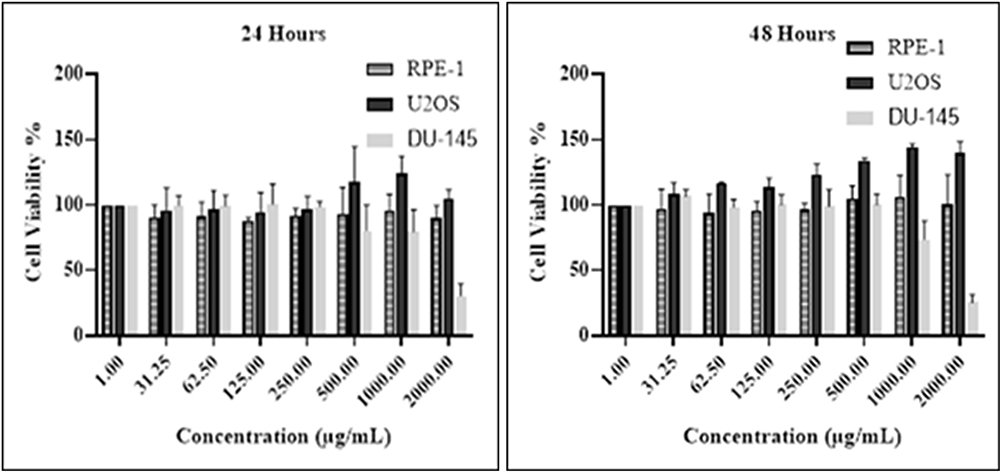 |
Figure 6 Cytotoxic analysis of E. angustifolia plant leaf extract applied to RPE-1, U2OS, and DU-145 cell lines. |
Similarly, Ghanghareh and Zare showed that E. angustifolia leaf extract had a highly cytotoxic effect on liver carcinoma cells (HepG2 cells).63 This effect was directly related to the plant extract and induced apoptosis. In another study, Liao et al showed strong cytotoxic activity in non-small cell lung cancer A549 cells using phenolic compounds obtained from E. angustifolia plant leaf extract.64 Kaur et al investigated several cancer cell lines, including DU 145, and found that gallic acid decreased the viability of both neoplastic and xenograft cells, but not of non-neoplastic cell lines.65 Gallic acid has also been found to have pro-apoptotic and pro-proliferative effects on prostate cancer cell lines and xenografts, as well as inducing apoptotic death and restricting xenograft growth. Similar results have been observed in other studies using comparable cell lines and xenografts.
Antimicrobial Activity
There is a direct proportional relationship between the total phenolic matter composition of plant extracts and their antioxidant and antimicrobial properties. Medicinal plants, fruits, and vegetables are important for activity studies because they contain many antioxidant compounds. Recently, owing to the increase in the antibiotic resistance of pathogenic bacteria and fungi, the search for new antibiotics has accelerated. Therefore, biological activity studies were carried out to determine the antimicrobial activity of components that can reduce the presence of pathogenic microbes. For this purpose, we determined the MIC values of E. angustifolia extract and antibiotics effective in suppressing the growth of microorganisms using the microdilution method. The extract suppressed the growth of C. albicans and L. monocytogenes at the lowest concentrations compared with the reference antibiotics. These concentrations were lower than those of the antibiotics used in the application. Accordingly, it showed a better effect than antibiotics. The extract did not exhibit antimicrobial effects against S. aureus, E. coli, or K. pneumonia. (Table 7).
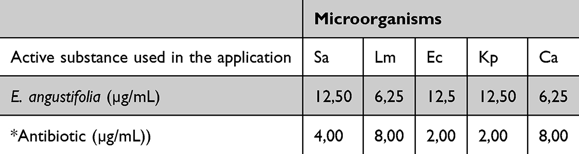 |
Table 7 MIC Values Were Obtained as a Result of the Antimicrobial Effects of E. angustifolia Extract and Antibiotics on Microorganisms |
*Listeria monocytogenes ATTC 7644 (Lm), Staphylococcus aureus ATCC 29213 (Sa), Escherichia coli ATCC 25922 (Ec), Klebsiella pneumonia ATCC 11774 (Kp), and Candida albicans (Ca) were obtained from the American Type Culture Collection (ATCC). Vancomycin was used for the Lm and Sa strains, colistin for the Kp and Ec strains, and fluconazole for the Ca strain.
Different structural groups of phenolic compounds exert antimicrobial effects on various pathogenic bacteria, mainly by inhibiting cell wall synthesis and exerting bactericidal and bacteriostatic effects. Phenolic compounds with antimicrobial effects include phenolic acids, flavonoids, stilbenoids, tannins, quinones, and coumarins.66 PCA and coumaric acid obtained from E. angustifolia are phenolic compounds with proven antimicrobial effects.67,68
As shown in Figure 7, E. angustifolia leaf extract showed a more effective antimicrobial effect than antibiotic application on C. albicans and gram-positive (G+) L. monocytogenes bacteria. In their antibacterial activity test with E. multiflora extract, Cho et al showed that it was effective against L. monocytogenes. Cho et al (2018) and Różalska et al showed that the antimycotic effect of E. rhamnoides extract was weak in antimicrobial tests on various bacteria, including C. albicans.69,70 In our study, its effect on C. albicans was obvious.
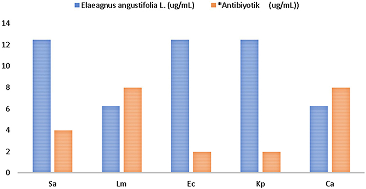 |
Figure 7 MIC values of E. angustifolia extract and antibiotics that suppress the growth of microorganisms. *(Ca: (C) albicans, Lm: (L) monocytogenes, Sa: (S) aureus, Ec:E. coli, Kp: (K) pneumonia). |
Conclusion
E. angustifolia is a tree species that grows in most parts of the world, is resistant to harsh environmental conditions, and the fruits and leaves of the plant, and in some cases, the bark, are frequently used in folk medicine. The leaves of the plant contain important bioactive compounds, as they are the center of metabolism, and may have beneficial health effects by preventing or reducing the onset and development of many diseases. In this study, for the first time, E. angustifolia leaf extract was used to investigate its cytotoxic effects on human retinal epithelium RPE-1, U2OS, and DU-145 cells and its antimicrobial effects against L. monocytogenes, S. aureus, E. coli, K. pneumonia, and C. albicans. Our study will contribute greatly to the search for anticancer and antimicrobial agents, especially in terms of pharmacological aspects, by examining biological activity using three different methods. In our study, the free radical scavenging capacity and antimicrobial activity of phenolic compounds were determined using a methanolic extract of the E. angustifolia plant. The leaves of the plant contain some important phenolic compounds, have a high free radical scavenging capacity, and have effective antimicrobial activity, especially in yeast species such as C. albicans. Phenolic compounds are secondary metabolites synthesized at different concentrations in all plants according to their species. As it is known, the side effects of synthetic anticancer and antimicrobial drugs are quite high. However, plant secondary metabolites have fewer side effects, and plants have difficulty forming resistance against these compounds, which has led the scientific world to use these compounds in the production of new drugs. An in vitro study showed that E. angustifolia has medicinal properties. It is believed that this effect provided by the bioactive components in the extract structure will greatly contribute to the pharmaceutical industry, especially with a more comprehensive examination of this effect in future in vivo studies.
Ethical Committee Approval
This study did not include any animal or human studies that would have required an ethics committee.
Acknowledgment
This study was supported by the Master’s thesis of the first author. We would like to thank Fatma Mungan Kılıç for plant identification.
Funding
No support was received for this study.
Disclosure
The authors declare no conflicts of interest related to this article.
References
1. Brenneisen P, Steinbrenner H, Sies H. Selenium, oxidative stress, and health aspects. Mol Aspect Med. 2005;26(5):256–267. doi:10.1016/j.mam.2005.07.004
2. Ramana KV, Srivastava S, Singhal SS. Lipid peroxidation products in human health and disease. Oxid Med Cell Longev. 2013;1(2013):1–3. doi:10.1155/2017/2163285
3. Karkar B, Şahin S. Determination of phenolic compounds profiles and antioxidant properties of oleaster (Elaeagnus angustifolia L.) grown in Turkey. Eur Food Res Technol. 2022;248(1):219–241. doi:10.1007/s00217-021-03875-y
4. Naczk M, Shahidi F. Phenolics in cereals, fruits and vegetables: occurrence, extraction and analysis. J Pharm Biomed Anal. 2006;41(1):1523–1542. doi:10.1016/j.jpba.2006.04.002
5. Saboonchian F, Jamei R, Sarghein SH. Phenolic and flavonoid content of Elaeagnus angustifolia L. (leaf and flower). Avicenna J Phytomed. 2014;4(4):231–238. doi:10.22038/AJP.2014.1975
6. Metsämuuronen S, Sirén H. Bioactive phenolic compounds, metabolism and properties: a review on valuable chemical compounds in Scots pine and Norway spruce. Phytochem Rev. 2019;18(1):623–664. doi:10.1007/s11101-019-09630-2
7. Bhuyan DJ, Basu A. Phenolic compounds potential health benefits and toxicity. In: Utilisation of Bioactive Compounds From Agricultural and Food Production Waste (Pp. 27-59). CRC Press. A.B.D; 2017. doi10.1201/9781315151540
8. Durfee RA, Mohammed M, Luu HH. Review of osteosarcoma and current management. Rheumatol ther. 2016;3:221–243. doi:10.1007/s40744-016-0046-y
9. Cimmino A, Fasciglione GF, Gioia M, Marini S, Ciaccio C. Multi-anticancer activities of phytoestrogens in human osteosarcoma. Int J Mol Sci. 2023;24(17):13344. doi:10.3390/ijms241713344
10. Rawla P. Epidemiology of prostate cancer. World J Oncol. 2019;10(2):63. doi:10.14740/wjon1191
11. Zhou CK, Check DP, Lortet‐Tieulent J, et al. Prostate cancer incidence in 43 populations worldwide: an analysis of time trends overall and by age group. Int J Cancer. 2016;138(6):1388–1400. doi:10.1002/ijc.29894
12. Farzaei MH, Bahramsoltani R, Abbasabadi Z, Rahimi R. A comprehensive review on phytochemical and pharmacological aspects of Elaeagnus angustifolia L. J Pharm Pharmacol. 2015;67(11):1467–1480. doi:10.1111/jphp.12442
13. Hamidpour R, Hamidpour S, Hamidpour M, et al. Russian olive (Elaeagnus angustifolia L.): from a variety of traditional medicinal applications to its novel roles as active antioxidant, anti-inflammatory, anti-mutagenic and analgesic agent. J Trad Complementary Med. 2017;7(1):24–29. doi:10.1016/j.jtcme.2015.09.004
14. Aktepe N, Baran A, Atalar MN, et al. Analysis of bioactive compounds using LC–ESI–MS/MS, cytotoxic, antimicrobial effects, and enzyme activities from Cyclotrichium origanifolium. Chem Biol Drug Des. 2023;101(3):740–748. doi:10.1111/cbdd.14177
15. Baran A, Keskin C. Determination of constituents of extract of Celtis tournefortii Lam. by LC-MS/MS, investigation of enzyme inhibition, antimicrobial and anticancer effects. Int J Pure Appl Sci. 2023;9(1):56–65. doi:10.29132/ijpas.1168200
16. Singleton VL, Rossi JA. Colorimetry of total phenolics with phosphomolybdic-phosphotungstic acid reagents. Am J Enol Vitic. 1965;16(3):144–158. doi:10.5344/ajev.1965.16.3.144
17. Wu C, Chen F, Wang X, et al. Antioxidant constituents in feverfew (Tanacetum parthenium) extract and their chromatographic quantification. Food Chem. 2006;96(2):220–227. doi:10.1016/j.foodchem.2005.02.024
18. Re R, Pellegrini N, Proteggente A, Pannala A, Yang M, Rice-Evans C. Antioxidant activity applying an improved ABTS radical cation decolorization assay. Free Radic Biol Med. 1999;26(9–10):1231–1237. doi:10.1016/S0891-5849(98)00315-3
19. Apak R, Güçlü K, Özyürek M, Esin Karademir S, Erça E. The cupric ion reducing antioxidant capacity and polyphenolic content of some herbal teas. Int J Food Sci Nutr. 2006;57:292–304. doi:10.1080/09637480600798132
20. Dinis TC, Madeira VM, Almeida LM. Action of phenolic derivatives (Acetaminophen, salicylate, and 5-aminosalicylate) as inhibitors of membrane lipid peroxidation and as peroxyl radical scavengers. Arch Biochem Biophys. 1994;315(1):161–169. doi:10.1006/abbi.1994.1485
21. Tolosa L, Donato MT, Gómez-Lechón MJ. General cytotoxicity assessment by means of the MTT assay. Protocols in Vitro Hepatocyte Res. 2015;333–348. doi:10.1007/978-1-4939-2074-7_26
22. Berktaş S, Mustafa ÇAM. İğde (Elaeagnus angustifolia L.) meyve ve yapraklarının antioksidan ve antidiyabetik özellikleri. Akademik Gida. 2020;18(3):270–278. doi:10.24323/akademik-gida.818125
23. Iqbal Y, Ponnampalam EN, Cottrell JJ, Suleria HA, Dunshea FR. Extraction and characterization of polyphenols from non-conventional edible plants and their antioxidant activities. Food Res Int. 2022;157:111205. doi:10.1016/j.foodres.2022.111205
24. Dedvisitsakul P, Watla-Iad K. Antioxidant activity and antidiabetic activities of Northern Thai indigenous edible plant extracts and their phytochemical constituents. Heliyon. 2022;8(9). doi:10.1016/j.heliyon.2022.e10740
25. Marchiosi R, Dos Santos WD, Constantin RP, et al. Biosynthesis and metabolic actions of simple phenolic acids in plants. Phytochem Rev. 2020;19(1):865–906. doi:10.1007/s11101-020-09689-2
26. Saltan FZ, Okutucu B, Canbay HS, Özel D. In vitro alpha-Glucosidase and alpha-amylase enzyme inhibitory effects in Elaeagnus angustifolia Leaves Extracts. Eurasian J Anal Chem. 2017;12(2):117–126. doi:10.12973/ejac.2017.00158a
27. Fernandes FHA, Salgado HRN. Gallic acid: review of the methods of determination and quantification. Crit Rev Anal Chem. 2016;46(3):257–265. doi:10.1080/10408347.2015.1095064
28. Kahkeshani N, Farzaei F, Fotouhi M, et al. Pharmacological effects of gallic acid in health and diseases: a mechanistic review. Iran J Basic Med Sci. 2019;22(3):225.
29. Maruszewska A, Tarasiuk J. Antitumour effects of selected plant polyphenols, gallic acid and ellagic acid, on sensitive and multidrug‐resistant leukaemia HL60 cells. Phytother Res. 2019;33(4):1208–1221. doi:10.1002/ptr.6317
30. Lin ML, Chen SS. Activation of casein Kinase II by gallic acid induces BIK–BAX/BAK-mediated ER Ca++-ROS-dependent apoptosis of human oral cancer cells. Front Physiol. 2017;8:761. doi:10.3389/fphys.2017.00761
31. Russell LH, Mazzio E, Badisa RB, et al. Autoxidation of gallic acid induces ROS-dependent death in human prostate cancer LNCaP cells. Anticancer Res. 2012;32(5):1595–1602. doi:10.21873/anticanres.17241
32. Wang R, Ma L, Weng D, Yao J, Liu X, Jin F. Gallic acid induces apoptosis and enhances the anticancer effects of cisplatin in human small cell lung cancer H446 cell line via the ROS-dependent mitochondrial apoptotic pathway. Oncol Rep. 2016;35(5):3075–3083. doi:10.3892/or.2016.4690
33. Prince PSM, Priscilla H, Devika PT. Gallic acid prevents lysosomal damage in isoproterenol induced cardiotoxicity in Wistar rats. Eur J Pharmacol. 2009;615(1–3):139–143. doi:10.1016/j.ejphar.2009.05.003
34. Naghizadeh B, Mansouri M. Protective effects of gallic acid against streptozotocin-induced oxidative damage in rat striatum. Drug Res. 2014;65(10):515–520. doi:10.1055/s-0034-1377012
35. Mansouri MT, Naghizadeh B, Ghorbanzadeh B, et al. Gallic acid prevents memory deficits and oxidative stress induced by intracerebroventricular injection of streptozotocin in rats. Pharmacol Biochem Behav. 2013;111:90–96. doi:10.1016/j.pbb.2013.09.002
36. Khan AK, Rashid R, Fatima N, et al. Pharmacological activities of protocatechuic acid. Acta Pol Pharm. 2015;72(4):643–650.
37. Kakkar S, Bais S. A review on protocatechuic acid and its pharmacological potential. 2014. Scholarly Res Notices. 2014;2014(1):1–10. doi:10.1155/2014/952943
38. Gutierrez-Zetina SM, Gonzalez-Manzano S, Perez-Alonso JJ, Gonzalez-Paramas AM, Santos-Buelga C. Preparation and characterization of protocatechuic acid sulfates. Molecules. 2019;24(2):307–325. doi:10.3390/molecules24020307
39. Semaming Y, Pannengpetch P, Chattipakorn SC, Chattipakorn N. Pharmacological properties of protocatechuic acid and its potential roles as complementary medicine. Evid Based Complement Alternat Med. 2015;2015(1):1–11. doi:10.1155/2015/593902
40. Cho KM, Joo OS. Quality and antioxidant charactistics of Elaeagnus multiflora wine through the thermal processing of juice. Food Sci Preservation. 2014;21(2):206–214. doi:10.11002/KJFP.2014.21.2.206
41. Ayaz FA, Bertoft E. Sugar and phenolic acid composition of stored commercial oleaster fruits. J Food Comp Anal. 2001;14(5):505–510. doi:10.1006/jfca.2001.1004
42. Boz H. p‐Coumaric acid in cereals: presence, antioxidant and antimicrobial effects. Int J Food Sci Technol. 2015;50(11):2323–2328. doi:10.1111/ijfs.12898
43. Pei K, Huang J, Ou J, Ou S. p-Coumaric acid and its conjugates: dietary sources, pharmacokinetic properties and biological activities. J Sci Food Agric. 2016;96(9):2952–2962. doi:10.1002/jsfa.7578
44. Roychoudhury S, Sinha B, Choudhury BP, et al. Scavenging properties of plant-derived natural biomolecule para-coumaric acid in the prevention of oxidative stress-induced diseases. Antioxidants. 2021;10(8):1205–1223. doi:10.3390/antiox10081205
45. Carradori S, Cairone F, Garzoli S, et al. Phytocomplex characterization and biological evaluation of powdered fruits and leaves from Elaeagnus angustifolia. Molecules. 2020;25(9):1–33. doi:10.3390/molecules25092021
46. Lee JH, Seo WT, Cho KM. Determination of phytochemical contents and biological activities from the fruits of Elaeagnus multiflora. Preventive Nutr Food Sci. 2011;16(1):29–36. doi:10.3746/jfn.2011.16.1.029
47. Shyamala BN, Naidu MM, Sulochanamma G, Srinivas P. Studies on the antioxidant activities of natural vanilla extract and its constituent compounds through in vitro models. J Agri Food Chem. 2007;55(19):7738–7743.
48. Yuan Y, Liu L, Du Y, Fan R, Zhang R, Zhou N. p-hydroxy benzaldehyde revitalizes the microenvironment of peri-infarct cortex in rats after cerebral ischemia-reperfusion. Phytomedicine. 2022;105(1):154379. doi:10.1016/j.phymed.2022.154379
49. Zhu YP, Li X, Du Y, Zhang L, Ran L, Zhou NN. Protective effect and mechanism of p-hydroxybenzaldehyde on blood-brain barrier. Zhongguo Zhong Yao Za Zhi. 2018;43(5):1021–1027. doi:10.19540/j.cnki.cjcmm.20171113.014
50. Kang CW, Han YE, Kim J, Oh JH, Cho YH, Lee EJ. 4-Hydroxybenzaldehyde accelerates acute wound healing through activation of focal adhesion signalling in keratinocytes. Sci Rep. 2017;7(1):14192.
51. Gupta S, Finelli R, Agarwal A, Henkel R. Total antioxidant capacity—Relevance, methods and clinical implications. Andrologia. 2021;53(2):e13624. doi:10.1111/and.13624
52. Akgül H, Mohammed FS, Kına E, Uysal İ, Sevindik M, Doğan M. Total antioxidant and oxidant status and DPPH free radical activity of euphorbia eriophora. Turk J AgricFood Sci Technol. 2022;10(2):272–275. doi:10.24925/turjaf.v10i2.272-275.4685
53. Khalid M, Amayreh M, Sanduka S, et al. Assessment of antioxidant, antimicrobial, and anticancer activities of Sisymbrium officinale plant extract. Heliyon. 2022;8(9):e10477. doi:10.1016/j.heliyon.2022.e10477
54. Guzelmeric E, Ugurlu P, Celik C, et al. Myrtus communis L. (Myrtle) plant parts: comparative assessment of their chemical compositions and antioxidant, anticancer, and antimutagenic activities. S Afr J Bot. 2022;150(1):711–720. doi:10.1016/j.sajb.2022.07.043
55. Ouedraogo WRC, Belemnaba L, Nitiéma M, et al. Phytochemical study, antioxidant and vasodilatation activities of leafy stem extracts of Flemingia faginea Guill. & Perr. (Barker), a medicinal plant used for the traditional treatment of arterial hypertension. Pharmacol Res Mod Chin Med. 2023;7(1):100231. doi:10.1016/j.prmcm.2023.100231
56. Hayta B, Gülaboğlu M, Kutlu Z. Hippophae Rhamnoides L.(Yabani İğde) Bitkisinin Meyve Ekstraklarının İn Vitro Antioksidan Özelliklerinin Araştırılması. J Inst Sci Technol. 2021;11(4):2992–3002. doi:10.21597/jist.938349
57. Kesim H, Yıldıztekin M. The biological activity features and mineral element analyses of some Inula L. species exhibit natural spread in Mugla (Turkiye). Int J Agri Environ Food Sci. 2023;7(2):316–325. doi:10.31015/jaefs.2023.2.9
58. Yildiztekin F, Nadeem S, Erol E, Yildiztekin M, Tuna AL, Ozturk M. Antioxidant, anticholinesterase and tyrosinase inhibition activities, and fatty acids of Crocus mathewii–A forgotten endemic angiosperm of Turkey. Pharm Biol. 2016;54(9):1557–1563. doi:10.3109/13880209.2015.1107746
59. Zimmermann-Klemd AM, Reinhardt JK, Winker M, Gründemann C. Phytotherapy in integrative oncology—an update of promising treatment options. Molecules. 2022;27(10):3209. doi:10.3390/molecules27103209
60. Lopes CM, Dourado A, Oliveira R. Phytotherapy and nutritional supplements on breast cancer. Biomed Res Int. 2017;2017(1):7207983. doi:10.1155/2017/7207983
61. Pucci C, Martinelli C, Ciofani G. Innovative approaches for cancer treatment: current perspectives and new challenges. ecancermedicalscience. 2019;13. doi:10.3332/ecancer.2019.961
62. Omabe K, Paris C, Lannes F, Taïeb D, Rocchi P. Nanovectorization of prostate cancer treatment strategies: a new approach to improved outcomes. Pharmaceutics. 2021;13(5):591. doi:10.3390/pharmaceutics13050591
63. Ghanghareh M, Zare M. Cytotoxic Effects of Hydro-Alcoholic Extract of the Leaf of Elaeagnus angustifolia in Hepatocellular Carcinoma Cell Line (HepG2). Jentashapir J Cell Mol Biol. 2020;11(3):e108505. doi:10.5812/jjcmb.108505
64. Liao CR, Kuo YH, Ho YL, et al. Studies on cytotoxic constituents from the leaves of Elaeagnus oldhamii Maxim. in non-small cell lung cancer A549 cells. Molecules. 2014;19(7):9515–9534. doi:10.3390/molecules19079515
65. Kaur M, Agarwal C, Agarwal R. Anticancer and cancer chemopreventive potential of grape seed extract and other grape-based products. J Nutr. 2009;139(9):1806S–1812S. doi:10.3945/jn.109.106864
66. Oulahal N, Degraeve P. Phenolic-rich plant extracts with antimicrobial activity: an alternative to food preservatives and biocides. Front Microbiol. 2022;12(2022):753518. doi:10.3389/fmicb.2021.753518
67. Jalali O, Best M, Wong A, Schaeffer B, Bauer B, Johnson L. Protocatechuic acid as a topical antimicrobial for surgical skin antisepsis: preclinical investigations. JBJS Open Access. 2020;5(3):e19. doi:10.2106/jbjs.oa.19.00079
68. Ojha D, Patil KN. p-Coumaric acid inhibits the Listeria monocytogenes RecA protein functions and SOS response: an antimicrobial target. Biochem Biophys Res Commun. 2019;517(4):655–661. doi:10.1016/j.bbrc.2019.07.093
69. Cho KM, Hwang CE, Kim SC, Joo OS. Physicochemical properties, phytochemicals, and biological activities of heat-treated Elaeagnus multiflora juice and vinegar. Food Sci Preservation. 2018;25(1):52–61. doi:10.11002/kjfp.2018.25.1.52
70. Różalska B, Sadowska B, Żuchowski J, et al. Phenolic and nonpolar fractions of Elaeagnus rhamnoides (L.) A. Nelson extracts as virulence modulators in vitro study on bacteria, fungi, and epithelial cells. Molecules. 2018;23(7):1498. doi:10.3390/molecules23071498
 © 2025 The Author(s). This work is published and licensed by Dove Medical Press Limited. The
full terms of this license are available at https://www.dovepress.com/terms
and incorporate the Creative Commons Attribution
- Non Commercial (unported, 4.0) License.
By accessing the work you hereby accept the Terms. Non-commercial uses of the work are permitted
without any further permission from Dove Medical Press Limited, provided the work is properly
attributed. For permission for commercial use of this work, please see paragraphs 4.2 and 5 of our Terms.
© 2025 The Author(s). This work is published and licensed by Dove Medical Press Limited. The
full terms of this license are available at https://www.dovepress.com/terms
and incorporate the Creative Commons Attribution
- Non Commercial (unported, 4.0) License.
By accessing the work you hereby accept the Terms. Non-commercial uses of the work are permitted
without any further permission from Dove Medical Press Limited, provided the work is properly
attributed. For permission for commercial use of this work, please see paragraphs 4.2 and 5 of our Terms.