Back to Journals » Journal of Experimental Pharmacology » Volume 12
Antibacterial Activity and Phytochemical Components of Leaf Extracts of Agave americana
Received 5 May 2020
Accepted for publication 16 October 2020
Published 3 November 2020 Volume 2020:12 Pages 447—454
DOI https://doi.org/10.2147/JEP.S258605
Checked for plagiarism Yes
Review by Single anonymous peer review
Peer reviewer comments 3
Editor who approved publication: Professor Bal Lokeshwar
Tewodros Shegute,1 Yared Wasihun2
1Kotebe Metropolitan University, Menelik II Health and Medical Science College, Department of Pharmacy, Addis Ababa, Ethiopia; 2Yekatit 12 Hospital Medical College, Addis Ababa, Ethiopia
Correspondence: Tewodros Shegute
Kotebe Metropolitan University, Menelik II Health and Medical Science College, Department of Pharmacy, Addis Ababa, Ethiopia
Tel +251 913579630
Email [email protected]
Background: Ethiopian flora is a source of innumerable cures for several infections. The medicinal potential of A. americana has been evaluated in some studies. The current study aimed to investigate the antimicrobial effect of A. americana leaf extracts on selected bacterial strains and to determine the phytochemical components.
Purpose: To determine the phytochemical constituents and in vitro antibacterial activity of leaf extracts of Agave americana against Staphylococcus aureus, Pseudomonas aeruginosa, Klebsiella pneumoniae, Salmonella species, and Eshercia coli.
Methods: The macerated and Soxhlet crude extracts of Agave americana were further fractionated to petroleum ether, chloroform, acetone, and methanol fractions. The agar well diffusion method and disc diffusion methods were used to test the antibacterial effect and determine the minimum inhibitory concentration (MIC) of the plant extract. Standard methods of determination were used to determine the phytochemical components of Agave americana.
Results: The percentage yield of crude extracts of A. americana was 15.11%. Alkaloids, saponins, tannins, polyphenols, and flavonoids were identified as phytochemical constituents of A. americana. The crude and solvent fractions of A. americana have an antibacterial activity comparable to gentamycin, with zones of inhibition ranging from 17 to 40mm and minimum inhibitory concentration (MIC) of 2.5 mg mL for S. aureus, P. aeruginosa, and S. typhi strains and 10 mg mL for E. coli strains. S. aureus and E. coli were the most and least susceptible bacteria among the four bacterial test strains tested.
Conclusion: The crude and solvent fractions of A. americana have antibacterial activity against S. aureus, P. aeruginosa, and S. thyphi, comparable to gentamycin.
Keywords: antibacterial activity, phytochemical screening, Agave americana, minimum inhibitory concentration, zones of inhibition
Introduction
Diseases of infectious origin are among the most common causes of human mortality in the globe. Even though a higher burden is observed in countries around the tropics, developed countries are also affected significantly. The problem is further complicated by the development of antimicrobial resistance.1 One way to tackle this problem is to search for new potential antimicrobials from plants.2 Plants have been used as human medicines since antiquity.3 A report by the World Health Organization (WHO) indicated the primary use of drugs from herbal sources by more than 80% of people in the world, especially those living in rural areas.4 Several studies have identified herbs as sources of development for the majority of currently used conventional drugs and potential for the future discovery of novel agents against a range of disorders.5,6
Approximately 12% of endemic plants are found in 6500–7000 species of Ethiopian flora. They have been sources of innumerable cures, making up 95% of folk remedies used throughout the country.7
The majority of drugs that are currently in use are derivatives of secondary plant metabolites identified from crude extracts. Secondary metabolites have multiple non-essential, but regulatory roles for the plants. The presence of these secondary metabolites is responsible for the therapeutic effectiveness of most medicinal plants.8 Secondary metabolites are classified into different phytochemical groups based on their physical and chemical characteristics.9 Among the range of these phytochemical groups’ alkaloids, flavonoids, saponins (steroidal sapogenins), polyphenolics, and glycosides (spirostanol and furostanol) with a range of pharmacological activities, including antimicrobial, antioxidant, anti-inflammatory and cytotoxic activities, were identified and characterized in the agave genus.10–12
Agave americana is a plant in the Asparagaceae family with a global occurrence, except the Frigid Zone growing in areas with a range of climates and altitudes.13 Twenty species and 6 genera of this plant inhabit Ethiopia among 200–300 species found worldwide.14–16 Even though it is considered an offensive plant in southern parts of Africa, Agave americana is widely cultivated for ornamental purposes because of its pale yellow margin leaves and ease of breeding and cultivation. Seasonal and age-wise variation of some ingredients of the leaves was identified earlier.13,15
Several previous studies have reported the potential Agave americana to be used in the management of a range of disorders. A study by Thaker VS and Maharshi AR showed that 80% methanolic extracts of five species of Agave plant, including Agave americana, had an inhibitory effect on the hyphal growth of common pathogenic fungi.16 The presence of saponin-based components with antifungal activity against conidial germination of A. brassicae in the leaf extracts of Agave americana was reported by Guleria S and Kumar A.17 In another study, Santos et al identified significant antifungal activity of hydroethanolic extracts of A. sisalana leaves against C. albicans.18 Bouaziz et al formulated a method to extract insoluble fibers from Agave americana, which can be used to treat and prevent a range of disorders, including colon cancer, constipation, diabetes, and coronary heart disease.19 The ability of Agave fourcroydes powder to increase the growth performance and serum concentration of IgG in rabbits when used as a dietary supplement was also reported.20 The potential antioxidant and cytotoxic activity of A. sisalana and antioxidant and the antibacterial activities of six other Agave species have been identified earlier.21,22 Another study by Alcázar et al reported that steroidal saponins from Agave durangensis had a strong growth inhibitory effect on two yeast strains, S. cerevisiae and K. marxianus.23 The beneficial physiometabolic effect was observed in Wistar rats upon their exposure to fructans extracted from Agave salmiana.24 In this study, we evaluated the in vitro antibacterial activity of leaf extracts of Agave americana against Staphylococcus aureus, Pseudomonas aeruginosa, Klebsiella pneumoniae, Salmonella species, and E. coli.
Materials and Methods
Plant Material
The leaves of A. americana were collected in February 2018 in Addis Ababa, and plant identification and authentication was done with the help of local floras. The specimen was preserved in the Addis Ababa University herbarium with a voucher number of 064988. Fresh leaves of A. americana were dried under a shed and powdered with a mechanical grinder. The powder was then passed through a sieve with sieve No. 40 and stored in an airtight container until extraction.
Preparation of Extracts
An 80% methanolic extract of 100 g leaf powder made by maceration for 48 h, which was performed five times with continuous stirring, filtration using filter paper, and a combination of the five portions. The final extract was then concentrated under reduced pressure in a rota vapor at 40 °C, and further dried for 48 h in an oven at 40 °C to produce dry powder, packed in a vial, and kept in a desiccator containing silica until the in vitro experiments were performed. Similarly, 100 g of A. americana leaf powder was used to produce petroleum ether, chloroform, acetone, and methanol fractions by sequential injection of the solvents into the extraction section of the Soxhlet apparatus. Concentration, oven drying (24 h), packaging, and storage were performed for the four extracts, similar to the crude extract described above.
Testing for Antibacterial Effects
Standard strains of one gram-positive (S. aureus) and three gram-negative (E. coli, S. typhi, and P. aeruginosa) were used for antibacterial effect determination. The antibacterial activities of the crude and solvent fraction extracts of Agave americana were determined using the agar well diffusion method. The cultures of all the standard strains were grown in 5% sheep red blood agar plates at 37 °C for 18 to 24 h. Four to five colonies of all bacteria were then transferred to TSY liquid broth with a sterilized inoculating loop. The liquid medium containing the colonies of the standard bacteria was then incubated for 6 h at 37 °C until it achieved a turbidity equivalent to 0.5 McFarland turbidity standard.25
A serialized swab was then used to streak a sample of each inoculum of the standard bacteria taken from the liquid broth into nonselective agar plates to assure formation of uniform growth throughout incubation. A sterile cork borer was then used to form bores of 10mm size on the nutrient agar plates and 100 μL of the test extracts or 0.1 mg mL gentamycin solution (positive control) was applied to fill bores using a micropipette. All the plates containing the test extract and the control drug were then kept at room temperature for 1 to 2 h. Finally, the measurement of the diameter of zones of inhibition was performed for each plate after 24 h incubation at 37°C.25–28
Determination of Minimum Inhibitory Concentrations
The minimum inhibitory concentrations of the crude extract and fractions were determined using the tube diffusion method. Concentrations 25, 50, 75, and 100 mg mL of Agave americana extract fractions were formed by dissolving the dried extracts in chloroform (for chloroform and petroleum ether fractions) and methanol (for acetone and methanol fractions). Negative controls of the experiment were formed using 80% chloroform and methanol solvents alone. Antibacterial activity of all samples was determined by measuring the diameter of the zones of inhibition.29,30
Phytochemical Screening
Standard extraction and screening procedures were used to determine the phytochemical components of A. americana crude extract.
Test for Alkaloids
Stirring 500mg of the crude extract and 5mL of 1% HCl in steam and addition of a few drops of Mayer’s and Dragendorff’s reagents to 1mL of the filtrate was performed to confirm the presence of alkaloids. Turbidity and precipitation indicated the presence of alkaloids.31,32
Test for Saponins
Shaking 500 mg of the crude extract with water in a test tube was performed to confirm the presence of saponins. The formation of froth that persists on warming was confirmatory for saponin content.31 Another evidence for the saponin content of the crude extract was also obtained using normal phase thin layer chromatography (TLC). Silica gel was used as the stationary phase and the mobile phase constituted of a mixture of chloroform-glacial acetic acid-methanol-water (64:32:12:8). The formation of blue, blue-violet, red, or yellow-brown zones by spraying vanillin-sulfuric acid indicator on the TLC plates containing the chromatogram of the crude extract was used to identify the saponin content.33
Test for Tannins
Stirring 500 mg of the crude extract of Agave americana in 10 mL of distilled water and filtration was performed to confirm the presence of tannins. The formation of blue, blue-black, green, or blue-green color upon the addition of FeCl3 to the filtrate was used as a tannin confirmatory test.34
Test for Flavonoids
In total, 0.5 grams of the crude extract was dissolved in 2 mL of methanol in a test tube and a few drops of 2% lead acetate were added to the mixture. The formation of orange or yellow color was indicative of flavonoid content.35
Tests for Phenolic Compounds
A few drops of a mixture of 1 mL 1% ferric chloride FeCl3 and 1 mL potassium ferrocyanide were added to 2 mL of the crude extract dissolved in distilled water. The formation of green-blue color was a positive result for phenolic compounds.36
Test for Anthraquinones
The test for anthraquinones was performed by stirring 5 g of the crude extract in 10 mL of benzene. The mixture was filtered and 5 mL of 10% ammonium hydroxide was added to the filtrate. The formation of red, violet, or pink color at the bottom of the test tube upon shaking was a positive test for anthraquinones.37
Results
The percentage yields of A. americana crude extracts indicated in Table 1 (15. 11%), the leaves of the plant lose a significant amount of moisture upon drying. However, the yields obtained adequate for further analysis. The results of antibacterial activity tests on the crude extracts of A. americana on the selected bacterial strains are shown in Table 2. The percentage yields of the different fractions of A. americana extracts are presented in Table 3. The antibacterial effects of petroleum ether, chloroform, acetone, and methanol fractions of A. americana extracts on the selected bacterial strains are shown in Table 4. The minimum inhibitory concentration (MIC) values of A. americana crude extracts determined to demonstrate the antibacterial potency are shown in Table 5. Phytochemical screening of A. americana extracts indicated the presence of alkaloids, saponins, phenols, flavonoids, and tannins (Table 6).
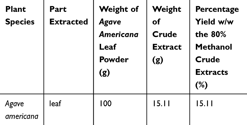 |
Table 1 Percentage Yields of the 80% Methanol Extracts of Dried and Powdered Agave americana, Addis Ababa, June 2018 |
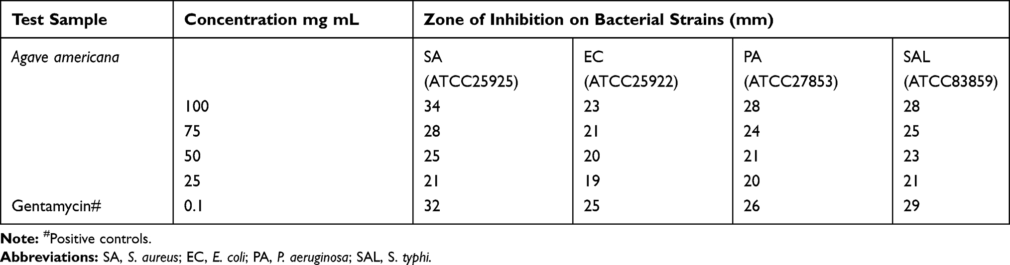 |
Table 2 Antibacterial Effects of Agave americana Crude Extracts on Selected Strains of Bacteria, Addis Ababa, June 2018 |
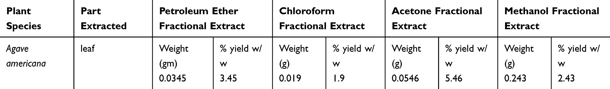 |
Table 3 Percentage Yields of Solvent Fractions of Agave americana, Addis Ababa, June 2018 |
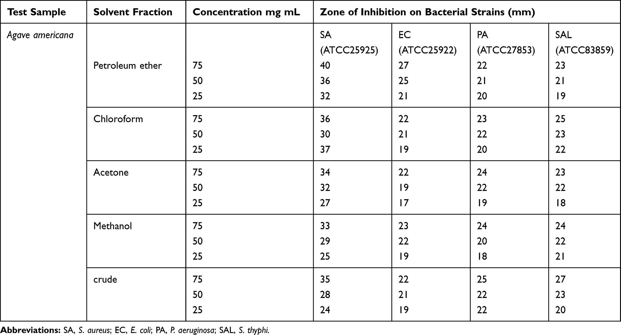 |
Table 4 Antibacterial Effect of Solvent Fractions of Agave americana Against Selected Strains of Bacteria, Addis Ababa, June 2018 |
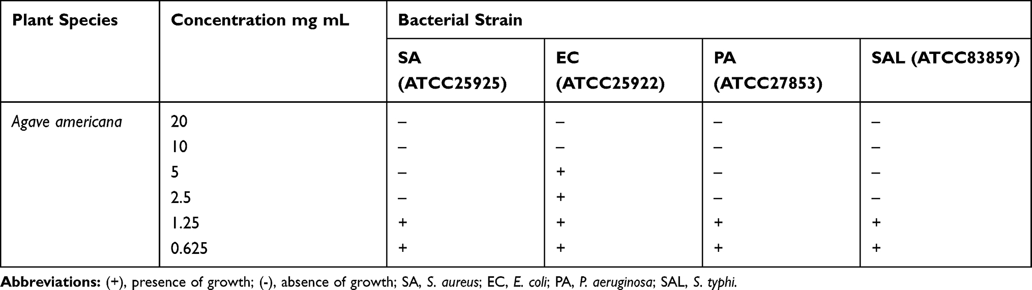 |
Table 5 Minimum Inhibitory Concentration (MIC) Values of Crude (80% Methanol) Extracts of Agave americana on the Tested Strains, Addis Ababa, June 2018 |
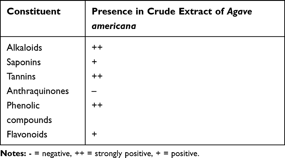 |
Table 6 Phytochemical Constituents of Crude Extract of Agave americana Leaves Using Chemical Test Methods, Addis Ababa, June 2018 |
Discussion
The results of the current study indicate a higher percentage yield of A. americana obtained when extracted with acetone and petroleum ether (Table 3). The positive test results for alkaloids, saponins, and flavonoids phytochemical screening in the current study (Table 6) are supported by the results of another study by Pandey et al However confirmatory test results for polyphenols and tannins in this study contradict the same study. The variation may be due to the difference in climate, soil, and other environmental conditions for plant growth in the current study (Ethiopia) and Nepal.38
Plant extracts having chemicals with antimicrobial activity usually belong to alkaloids, flavonoids, phenolics, saponins, and their derivatives.39,40 The positive test result for alkaloids confirmed in the current study is due to the ability of the metallic atom in the reagent (bismuth and mercury) to form an ion pair with the nitrogen in the alkaloid, leading to the formation of a precipitate.32 The formation of froth that persists on warming due to the saponin content of Agave americana in the saponin confirmatory test is due to the surfactant nature of saponins due to the presence of polar carbohydrate groups and nonpolar sapogenin groups in their structure.41 The color change observed in TLC plates upon addition of vanillin-sulfuric acid indicator used in the phytochemical screening is due to the interaction between triterpene saponins oxidized by sulfuric acid and vanillin.42 The ability of tannins to form tannic acid – ferric complexes upon addition of ferric chloride is the mechanism behind tannin identification test use in the current study.43 The bacterial strains used for the determination of antimicrobial activity in the current study were the common causes of illness in the community. Similar antibacterial effects of the crude extract and the solvent fractions of Agave americana were obtained in the current study against different strains of bacteria. This indicates the intermediate polarity of the component of the plant material responsible for antibacterial activity. The results of the current study indicated that Agave americana produces comparable antibacterial on the test strains of bacteria at a concentration of 75 mg mL and greater activity at 100 mg mL as compared to the positive control (gentamycin) for some strains (S. aureus). No antibacterial activity was observed on the test strains for the negative controls (80% methanol and chloroform). The results of antimicrobial activity screening also identified S. aureus and E. coli as the most and least susceptible bacteria among the four bacterial test strains under study. On the other hand, E. coli was found to be the most insensitive strain among all bacteria. This also indicates the potential effectiveness of the extract of Agave americana on gram-positive bacteria over their gram-negative counterparts. Structural differences between gram-positive and gram-negative bacteria causing variation in antimicrobial penetration leading to the concentration of the potentially antibacterial compound found in Agave americana in gram-positive bacteria could explain such variations in susceptibility.44 The inhibitory effect of the extract on bacterial cell wall synthesis (peptidoglycan crosslinking), which are less concentrated in gram-negative bacteria, may also be responsible for their reduced susceptibility to the extract and solvent fractions compared to gram-positive bacteria. This is also supported by the results from Alcázar et al who reported the growth inhibitory effect of the steroidal saponin components of Agave durangensis and Agave salmiana ssp. crassispina and their ability to change the cell wall structure on S. cerevisiae and K. marxianus.23 The saponin component identified from the extract of A. americana in the current study may be responsible for the antimicrobial effect observed. However, other phytochemical components identified in the crude extract of A. americana such as alkaloids, flavonoids, tannins, and phenolic compounds have also a potential antimicrobial activities. Therefore, further experiments should be performed on the isolated phytochemical components to determine the exact component responsible for the antibacterial activity and perform structural elucidation to obtain a potential antimicrobial agent of the future.
Conclusions
The results of the current study showed that the crude and solvent fractions of A. americana have antibacterial activity against S. aureus, P. aeruginosa, and S. thyphi comparable to gentamycin, indicating the potential of compounds from A. americana leaf extracts to be used as leads for future antimicrobial agents.
Abbreviations
NDA, new drug application; PGE, polarity gradient extraction; MIC, minimum inhibitory concentration.
Data Sharing Statement
The data sets used and/or analyzed during the current study are available from the corresponding author upon reasonable request.
Ethics Approval and Consent to Participate
Not Applicable.
Consent for Publication
Not Applicable.
Acknowledgment
The authors are thankful to the laboratory staff members for their kind support throughout the study.
Author Contributions
Both authors contributed to data analysis, drafting or revising the article, gave final approval of the version to be published, and agree to be accountable for all aspects of the work.
Funding
This research did not receive any specific grant from funding agencies in the public, commercial, or not-for-profit sectors.
Disclosure
The authors declare that they have no competing interests.
References
1. Metrics GH. Global, regional, and national incidence, prevalence, and years lived with disability for 354 diseases and injuries for 195 countries and territories, 1990 – 2017: a systematic analysis for the Global Burden of Disease Study 2017. Lancet. 2018;392:1789–1858. doi:10.1016/S0140-6736(18)32279-7
2. Chowdhury MMH, Kubra K, Ahmed SR. Antimicrobial, phytochemical and toxicological evaluation of lawsonia inermis extracts against clinical isolates of pathogenic bacteria. Ann Clin Microbiol Antimicrob. 2013;12(36):1–8. doi:10.3923/rjmp.2014.187.195
3. Petrovska BB. Historical review of medicinal plants ’ usage. Pharmacog Rev. 2012;6(11):1–6. doi:10.4103/0973-7847.95849
4. Ekor M. The growing use of herbal medicines: issues relating to adverse reactions and challenges in monitoring safety. Front Pharmacol. 2014;4(January):1–10. doi:10.3389/fphar.2013.00177
5. Boopathy NS, Kathiresan K. Anticancer drugs from marine flora: an overview. J Oncol. 2010;214186:1–18. doi:10.1155/2010/214186
6. Newman DJ. Natural products as sources of new drugs from 1981 to 2014. J Nat Prod. 2016;79:629–661. doi:10.1021/acs.jnatprod.5b01055
7. Gabriel T, Guji T. Ethnopharmacological survey of medicinal plants in Agaro District, Jimma Zone, South West Ethiopia. IJPSR. 2014;5(8):3551–3559. doi:10.13040/IJPSR.0975-8232
8. Chatterjee SS. From covalent bonds to eco-physiological pharmacology of secondary plant metabolites. BCP. 2015;98(2):269–277. doi:10.1016/j.bcp.2015.07.037
9. Jamwal K, Bhattacharya S, Puri S. Plant growth regulator mediated consequences of secondary metabolites in medicinal plants. J Appl Res Med Aromat Plants. 2018;9:26–38. doi:10.1016/j.jarmap.2017.12.003
10. Che Hassan NKN, Taher M, Susanti D. Phytochemical constituents and pharmacological properties of Garcinia xanthochymus- a review. Biomed Pharmacother. 2018;106:1378–1389. doi:10.1016/j.biopha.2018.07.087
11. Sidana J, Singh B, Sharma OP. Saponins of Agave: chemistry and bioactivity. Phytochemistry. 2016;130:22–46. doi:10.1016/j.phytochem.2016.06.010
12. Puente-Garza CA, García-Lara S, Gutiérrez-Uribe JA. Enhancement of saponins and flavonols by micropropagation of Agave salmiana. Ind Crops Prod. 2017;105:225–230. doi:10.1016/j.indcrop.2017.05.014
13. De La Torrre L, Cummins IL-HE. andina: key species to andean cultures in ecuador. Bot Sci. 2018;96(2):246–266. doi:10.17129/botsci.1813
14. Rodríguez B, Siverio F, Siverio M, Barone R, Rodríguez A. Nectar and pollen of the invasive century plant Agave americana as a food resource for endemic birds. Bird Study. 2015;62:232–242. doi:10.1080/00063657.2015.1015484
15. Niechayev NA, Jones AM, Rosenthal DM, Davis SC. A model of environmental limitations on production of Agave americana L. grown as a biofuel crop in semi-arid regions. J Exp Bot. 2019;70(22):6549–6559. doi:10.1093/jxb/ery383
16. Maharshi AR, Thaker VS. Antifungal Activity of. Microb Divers Biotechnol Food Secur. 2014;423–430. doi:10.1007/978-81-322-1801-2
17. Guleria SKA. Antifungal activity of Agave americana leaf extract against Alternaria brassicae, causal agent of Alternaria blight of Indian mustard (Brassica juncea) Access details: access Details: [subscription number 909519597]. Arch Phytopathol Plant Prot. 2014;42(Jul4):370–375. doi:10.1080/03235400601121380
18. Santos DGS, Branco A, Silva AF, Pinhiero CSR, et al. Antimicrobial activity of Agave sisalana. African J Biotechnol. 2009;8(22):6181–6184. doi:10.5897/ajb09.862
19. Bouaziz A, Masmoudi M, Kamoun A, Besbes S. Optimization of insoluble and soluble fibres extraction from agave americana L. Using response surface methodology. J Chem. 2014;627103:1–13. doi:10.1155/2014/627103
20. Iser M, Martínez Y, Ni H, et al. The effects of agave fourcroydes powder as a dietary supplement on growth performance, gut morphology, concentration of igg, and hematology parameters in broiler rabbits. Biomed Res Int. 2016;3414319:1–7. doi:10.1155/2016/3414319
21. Araldi RP, Dos Santos MO, Barbon FF, et al. Analysis of antioxidant, cytotoxic and mutagenic potential of Agave sisalana Perrine extracts using Vero cells, human lymphocytes and mice polychromatic erythrocytes. Biomed Pharmacother. 2018;98:873–885. doi:10.1016/j.biopha.2018.01.022
22. Ahumada-Santos YP, Montes-Avila J, de J U-BM, et al. Chemical characterization, antioxidant and antibacterial activities of six Agave species from Sinaloa, Mexico. Ind Crops Prod. 2013;49:143–149. doi:10.1016/j.indcrop.2013.04.050
23. Alcázar M, Kind T, Gschaedler A, et al. Effect of steroidal saponins from Agave on the polysaccharide cell wall composition of Saccharomyces cerevisiae and Kluyveromyces marxianus. LWT - Food Sci Technol. 2016;77:1–37. doi:10.1016/j.lwt.2016.11.048
24. Castillo Andrade AI, Rivera Bautista C, Godínez Hernández C, et al. Physiometabolic effects of Agave salmiana fructans evaluated in Wistar rats. Int J Biol Macromol. 2018;108:1300–1309. doi:10.1016/j.ijbiomac.2017.11.043
25. CLSI. Broth Microdilution Method In: Methods for Dilution Antimicrobial Susceptibility Tests for Bacteria That Grow Aerobically.
26. Tkachenko H, Buyun L, Terech-majewska E, Honcharenko V, Prokopiv A, Osadowski Z. Preliminary in vitro screening of the antibacterial activity of leaf extracts from various Ficus species (Moraceae) against Yersinia ruckeri. Archives of Polish Fisheries. 2019:15–26. DOI:10.2478/aopf-2019-0002
27. Roger T, Pierre-marie M, Igor VK, Patrick VD. Phytochemical screening and antibacterial activity of medicinal plants used to treat typhoid fever in Bamboutos division, West Cameroon. Journal of Applied Pharmaceutical Science.2015;5(06):34–49. doi:10.7324/JAPS.2015.50606
28. Teh CH, Nazni WA, Nurulhusna AH, Norazah A, Lee HL. Determination of antibacterial activity and minimum inhibitory concentration of larval extract of fly via resazurin-based turbidometric assay. BMC Microbiology. 2017;17(36):1–8. DOI:10.1186/s12866-017-0936-3
29. Andrews JM. Determination of minimum inhibitory concentrations. J Antimicrob Chemother. 2001;48:5–16. doi:10.1093/jac/48.suppl_1.5
30. Valgas C, De SSM, Smânia EFA
31. Visweswari G, Christopher R, Rajendra W. Phytochemical screening of active secondary metabolites present in withania somnifera root: role in traditional medicine | international journal of pharmaceutical sciences and research. Int J Pharm Sci Res. 2013;4(43):2770–2776. doi:10.13040/IJPSR.0975-8232.4(7).2770-76
32. Salamah N, Ningsih DS. Total alkaloid content in various fractions of Tabernaemonata sphaerocarpa Bl. (Jembirit) leaves. Mater Sci Eng. 2017;259:1–7. doi:10.1088/1757-899X/259/1/012017
33. Oleszek WA. Chromatographic determination of plant saponins. J Chromatogr. 2002;967:147–162. doi:10.1016/S0021-9673(01)01556-4
34. Gangwar M, Gautam MK, Sharma AK, Tripathi YB, Goel RK, Nath G. Antioxidant capacity and radical scavenging effect of polyphenol rich Mallotus philippenensis fruit extract on human erythrocytes: an in vitro study. Sci World J. 2014;279451:1–12. doi:10.1155/2014/279451
35. Sharief Mohammad N, Geneto M, Abateneh DD, Salahuddin M, Manzar MD, UMR V. Extraction of secondary metabolites from roots of acanthus ilicifolius l and screening for antioxidant and antibacterial activity. Int J Pharm Sci Invent. 2017;6(2):31–36.
36. Siddique AB, Rahman SMM, Hossain MA, Rashid MA. Phytochemical screening and comparative antimicrobial potential of different extracts of Stevia rebaudiana Bertoni leaves. Asian Pacific J Trop Dis. 2014;4(4):275–280. doi:10.1016/S2222-1808(14)60572-7
37. Doughari JH, Ndakidemi PA, Human IS, Benade S. Antioxidant, antimicrobial and antiverotoxic potentials of extracts of Curtisia dentata. J Ethnopharmacol. 2012;141(3):1041–1050. doi:10.1016/j.jep.2012.03.051
38. Pandey BR, Shrestha A, Sharma N, Shrestha BG. Evaluation of phytochemical, antimicrobial, antioxidant activity and cytotoxic potentials of agave americana. Nepal Journal of Biotechnology. 2019; 7(1):30–38.
39. Kurhekar JV. TANNINS – antimicrobial CHEMICAL. Int J Technol Sci. 2017;9(3):5–9.
40. Guil-Guerrero JL, Ramos L, Moreno C, Zúñiga-Paredes JC, Carlosama-Yepez M, Ruales P. Antimicrobial activity of plant-food by-products: A review focusing on the tropics. Livest Sci. 2016;189:32–49. doi:10.1016/j.livsci.2016.04.021
41. Savage GP. Saponins. Encycl Food Sci Nutr. 1993;5095–5098.
42. Cheok CY, Salman HAK, Sulaiman R. Extraction and quantification of saponins: A review. Food Res Int. 2014;59:16–40. doi:10.1016/j.foodres.2014.01.057
43. Phiwchai I, Yuensook W, Sawaengsiriphon N, Krungchanuchat S, Pilapong C. Tannic acid (TA): A molecular tool for chelating and imaging labile iron. Eur J Pharm Sci. 2018;114:64–73. doi:10.1016/j.ejps.2017.12.004
44. Tadeg H, Mohammed E, Asres K, Gebre-mariam T. Antimicrobial activities of some selected traditional Ethiopian medicinal plants used in the treatment of skin disorders. Journal of Ethnopharmacology. 2005;100:168–175. doi:10.1016/j.jep.2005.02.031
 © 2020 The Author(s). This work is published and licensed by Dove Medical Press Limited. The
full terms of this license are available at https://www.dovepress.com/terms
and incorporate the Creative Commons Attribution
- Non Commercial (unported, 3.0) License.
By accessing the work you hereby accept the Terms. Non-commercial uses of the work are permitted
without any further permission from Dove Medical Press Limited, provided the work is properly
attributed. For permission for commercial use of this work, please see paragraphs 4.2 and 5 of our Terms.
© 2020 The Author(s). This work is published and licensed by Dove Medical Press Limited. The
full terms of this license are available at https://www.dovepress.com/terms
and incorporate the Creative Commons Attribution
- Non Commercial (unported, 3.0) License.
By accessing the work you hereby accept the Terms. Non-commercial uses of the work are permitted
without any further permission from Dove Medical Press Limited, provided the work is properly
attributed. For permission for commercial use of this work, please see paragraphs 4.2 and 5 of our Terms.