
Back to Journals » International Journal of Women's Health » Volume 17
Analysis of the Application Value of Low Molecular Weight Heparin Combined with Heparin in Patients with Chorionic Bump in Early Pregnancy
Authors Peng XT, Cai ZH, Shen J, Zhu HY
Received 27 November 2024
Accepted for publication 5 March 2025
Published 7 April 2025 Volume 2025:17 Pages 973—982
DOI https://doi.org/10.2147/IJWH.S507845
Checked for plagiarism Yes
Review by Single anonymous peer review
Peer reviewer comments 2
Editor who approved publication: Dr Matteo Frigerio
Xu-Ting Peng,1 Zhu-Hua Cai,2 Jie Shen,3 Hai-Yan Zhu2
1Department of Gynecology, The Third Affiliated Hospital of Wenzhou Medical University, Wenzhou, People’s Republic of China; 2Department of Gynecology, the First Affiliated Hospital of Wenzhou Medical University, Wenzhou, People’s Republic of China; 3Department of Ultrasound, The Third Affiliated Hospital of Wenzhou Medical University, Wenzhou, People’s Republic of China
Correspondence: Hai-Yan Zhu, Email [email protected]
Objective: To explore the application value of low molecular weight heparin combined with heparin in patients with chorionic bump in early pregnancy.
Methods: This retrospective study collected 86 cases of patients with early pregnancy complicated by chorionic bump treated in our hospital from January 2020 to February 2022. According to the treatment methods, the patients were divided into the combined treatment group (n=41) and the control group (n=45). Patients in the combined treatment group were treated with oral prednisone combined with low molecular weight heparin injection, while patients in the control group received no additional treatment. The changes and differences in chorionic size, serum hormone levels, and uterine artery blood flow parameters before treatment and at 1 month and 2 months after treatment were compared between the two groups. The pregnancy outcomes and adverse reaction rates were also analyzed.
Results: At 1 month and 2 months after treatment, the average size of the chorionic bump in the combined treatment group was significantly smaller than before treatment and the control group (P< 0.05). Two months after treatment, the average levels of E2, P, and LH in the combined treatment group were significantly higher, and the average FSH level was significantly lower than those before treatment and in the control group (P< 0.05). At 1 month and 2 months after treatment, the average RI in the combined treatment group was significantly lower than before treatment and the control group, and although the average S/D ratio was higher than before treatment, it was still lower than that of the control group (P< 0.05). The incidence of adverse pregnancy outcomes in the combined treatment group was significantly lower than that in the control group (χ²/P=5.469/0.019). There was no significant difference in the incidence of adverse reactions between the two groups (χ²/P=0.613/0.434).
Conclusion: Prednisone combined with low molecular weight heparin can effectively reduce the size of the chorionic bump, improve uterine artery blood flow parameters, and ultimately lower the risk of adverse pregnancy outcomes in patients with early pregnancy complicated by chorionic bump. This treatment approach has reliable clinical safety and holds potential for broader application in such patients.
Keywords: early pregnancy, chorionic bump, prednisone, low molecular weight heparin, pregnancy outcomes
Introduction
During embryonic development, trophoblast cells differentiate to form the chorion, which is initially uniformly covered with villous structures. As the embryo grows, the villi adjacent to the basal decidua gradually develop into the main components of the placenta, while the villous structures in other areas of the chorion degenerate due to insufficient blood supply, eventually becoming part of the fetal membrane. This structure can be observed as the chorion during ultrasound examinations.1 In early pregnancy, chorionic bump is a rare abnormal phenomenon of the gestational sac, which can be observed through transvaginal ultrasound as a circular or oval protrusion from the sac wall into the sac cavity.2 Emerging evidence suggests that chorionic bump may not be merely an incidental ultrasound finding; it has been associated with adverse maternal psychological outcomes (eg, pregnancy-related anxiety) and fetal risks, including a 2–3 fold increase in spontaneous miscarriage rates compared to unaffected pregnancies.3 The mechanism of chorionic bump formation remains controversial. Some studies suggest it may represent a form of subchorionic hematoma, but other viewpoints highlight differences in their locations.4 Notably, recent cohort studies have demonstrated that even isolated chorionic bumps may correlate with placental dysfunction, potentially leading to late-onset complications such as preterm birth (12–18% vs 5–10% in controls) and fetal growth restriction (8–12% vs 3–5%).5 With advancements in ultrasound technology, the detection rate of chorionic-related diseases in early pregnancy has been increasing yearly. Despite growing clinical attention on chorionic bump, its specific impact on pregnancy outcomes remains unclear. In particular, conclusions on whether it increases the risk of early miscarriage are still inconclusive.6
This clinical ambiguity poses significant challenges: while some clinicians adopt a “watchful waiting” approach due to perceived benign nature, others advocate proactive interventions to mitigate potential risks. Such divergent practices underscore the urgent need for evidence-based management protocols. Previous studies have shown that patients with isolated chorionic bump in early pregnancy generally have favorable prognoses, whereas those complicated with subchorionic hematoma may face risks of miscarriage, preterm delivery, placental abruption, and intrauterine growth restriction.7 Moreover, the psychological burden on expectant mothers facing this diagnosis—characterized by uncertainty about pregnancy viability and long-term fetal health—remains an underaddressed aspect of maternal care.8
Currently, no consensus has been established in clinical practice regarding systematic and specific treatment protocols for early pregnancy complicated by chorionic bump. However, clinical studies have suggested that prednisone acetate tablets combined with low-molecular-weight heparin may improve pregnancy rates and reduce miscarriage risks by alleviating inflammatory responses, inhibiting immune cell function, and exerting anticoagulant effects.9,10 Given the rarity of chorionic bump (estimated prevalence 0.15–0.7% in first-trimester ultrasounds) and the paucity of randomized trials, retrospective analyses of therapeutic strategies become crucial for optimizing care in this understudied population.11,12
Based on this background, and motivated by the dual imperative to reduce both physical pregnancy risks and maternal psychological distress, this study aims to conduct a retrospective investigation to preliminarily explore the application value of low-molecular-weight heparin combined with heparin in patients with chorionic bump during early pregnancy. Our approach prioritizes bridging the knowledge gap between ultrasound diagnostics and actionable therapeutic interventions, with particular emphasis on preventing the progression from sonographic abnormality to clinical complications.
Materials and Methods
This study The protocol was approved by the ethics committee of the First Affiliated Hospital of Wenzhou Medical University. All patients signed informed consent forms. All the methods were carried out in accordance with the Declaration of Helsinki.
Study Population
This study retrospectively included 86 patients with chorionic bump during early pregnancy treated in our hospital from January 2020 to February 2022. Inclusion criteria were as follows: ① Intrauterine and singleton pregnancy confirmed by ultrasound; ② Gestational age <12 weeks at enrollment; ③ First pregnancy with no history of pregnancy or miscarriage; ④ No anatomical abnormalities or genital tract malformations; ⑤ Presence of irregular protrusions from the chorion-deciduous surface into the gestational sac confirmed by ultrasound. Exclusion criteria were as follows: ① Concurrent gynecological diseases such as uterine fibroids or ovarian cysts; ② Voluntary termination of pregnancy; ③ Failure to undergo routine prenatal examinations or incomplete clinical data; ④ Unclear pregnancy outcomes; ⑤ Failure to follow the treatment protocol specified in the study. Patients were allocated to the combined treatment group (n=41) or control group (n=45) based on chronological order of hospital visits and treatment preferences. No randomization was performed due to the retrospective nature of the study.
Sample size calculation: Based on prior studies showing 40% adverse pregnancy outcomes in untreated chorionic bump cases, we estimated that a sample of 38 patients per group would provide 80% power (α=0.05, β=0.2) to detect a 50% reduction in adverse outcomes. In retrospective studies, sample attrition is typically not a concern since the data has already been collected.
Treatment Methods
The control group adhered to standard dietary therapy without any additional interventions. The combined treatment group received prednisone acetate tablets and low-molecular-weight heparin. Specific operations were as follows: Prednisone acetate tablets (produced by Zhejiang Xianju Pharmaceutical Co., Ltd., National Medicine Approval Number H33021207, 5mg/tablet) were administered orally at a dose of 5mg twice daily, taken with warm water during meals. Dalteparin sodium injection (produced by Hebei Changshan Biochemical Pharmaceutical Co., Ltd., National Medicine Approval Number H20143110, 0.25mL: 5000IU) was administered by subcutaneous injection after skin disinfection within a 5cm radius around the umbilicus, with injection points spaced at least 2cm apart. All patients received continuous treatment for at least two months and underwent monthly prenatal examinations at our hospital. Follow-up continued until either normal pregnancy or adverse pregnancy outcomes occurred. Safety monitoring protocol: Platelet counts were monitored weekly for heparin-induced thrombocytopenia. Treatment was suspended if platelet count dropped below 100×109/L or if severe bleeding (≥500mL/24hr) occurred. Alternative anticoagulation strategies were prepared but not required in this cohort.
Observational Indicators
General Clinical Data
General clinical data collected included age, gestational weeks, gestational sac size, early pregnancy body mass index (BMI), history of hypertension or diabetes, pre-treatment human chorionic gonadotropin (HCG), and the number of chorionic bumps (1 or ≥2). Gestational sac size was calculated as the average of the long, wide, and high diameters measured by ultrasound.
Chorion Size
Chorion size was recorded at enrollment and during prenatal examinations after 1 and 2 months of treatment using ultrasound diagnostic equipment. The average of the long, wide, and high diameters was calculated.
Serum Sex Hormones
Peripheral venous blood (3mL) was collected at enrollment and during prenatal examinations after 1 and 2 months of treatment. Serum was obtained by centrifugation at 1200×g for 10 minutes at room temperature. Enzyme-linked immunosorbent assay and a SuPerMax 3000AL multifunctional microplate reader (produced by Shanghai Shanpu Biotechnology Co., Ltd.) were used to measure the expression levels of estrogen (E2), progesterone (P), luteinizing hormone (LH), and follicle-stimulating hormone (FSH).
Uterine Artery Blood Flow Parameters
Color Doppler ultrasound diagnostic equipment was used to examine uterine artery blood flow at enrollment and after 1 and 2 months of treatment. Patients were instructed to empty their bladders and rest in a calm state for more than 5 minutes. Vaginal ultrasound was used to locate the uterine artery at the junction of the cervix and uterine body, ensuring clear blood flow signals. A 2mm sampling volume was used to continuously record five blood flow spectra for high stability. Resistance index (RI), pulsation index (PI), and peak systolic blood flow velocity/end-diastolic flow velocity (S/D) were recorded.
Pregnancy Outcomes
The incidence of adverse pregnancy outcomes, including missed abortion, inevitable abortion, and stillbirth, was compared between the two groups.
Treatment-Related Adverse Reactions
Treatment-related adverse reactions, including thrombocytopenia, injection site bleeding, vaginal bleeding, and dermatitis, were compared between the two groups from enrollment to the end of follow-up.
Statistical Analysis
All data were statistically analyzed using SPSS 26.0. Measurement data were subjected to normality tests. Data conforming to normal distribution and homogeneity of variance were expressed as mean ± standard deviation ( ). Intergroup comparisons were performed using independent sample t-tests, and repeated measures ANOVA was used to analyze intergroup and temporal effects at different time points. Count data were expressed as frequencies (%) and compared using chi-square tests. A P-value <0.05 was considered statistically significant.
Results
Comparison of General Clinical Data Between the Two Groups
As a retrospective cohort study, we performed propensity score matching (1:1 ratio) to balance baseline characteristics between the combination treatment group (n=41) and control group (n=45). There were no statistically significant differences in general clinical data between the two groups after matching, including mean age, gestational weeks, gestational sac size, early pregnancy BMI, proportion of patients with a history of hypertension, proportion of patients with a history of diabetes, pre-treatment HCG levels, and number of chorionic protrusions (all _P_>0.05). Standardized mean differences (SMD) for all variables were <0.1, confirming adequate balance. See Table 1.
 |
Table 1 Comparison of General Clinical Data Between the Two Groups ( |

Comparison of Chorionic Size Between the Two Groups Before and After Treatment
There was no significant difference in the mean chorionic size between the two groups before treatment (P>0.05). However, at 1 month and 2 months after treatment, the mean chorionic size in the combination treatment group was significantly smaller than before treatment and the control group (P<0.05). There were significant time effects, group effects, and time × group interaction effects in chorionic size changes between the two groups (P<0.05). See Table 2.
 |
Table 2 Comparison of Chorionic Size Between the Two Groups Before and After Treatment ( |

Comparison of Serum Sex Hormone Levels Between the Two Groups Before and After Treatment
Before treatment, there were no significant differences in mean E2, P, LH, or FSH levels between the two groups (P>0.05). After 2 months of treatment, the mean E2, P, and LH levels in the combination treatment group were significantly higher than before treatment and the control group, while the mean FSH level was significantly lower than before treatment and the control group (P<0.05). Significant time effects, group effects, and time × group interaction effects were observed in serum sex hormone level changes between the two groups (P<0.05). See Table 3.
 |
Table 3 Comparison of Serum Sex Hormone Levels Between the Two Groups Before and After Treatment ( |

Comparison of Uterine Artery Blood Flow Parameters Between the Two Groups Before and After Treatment
Before treatment, the mean RI, PI, and S/D of the uterine artery showed no significant differences between the two groups (P>0.05). At 1 month and 2 months after treatment, the mean RI of the combination group was significantly lower than both before treatment and the control group (P<0.05). Although the mean S/D was higher than before treatment, it remained consistently lower than the control group (P<0.05). Significant time effects, group effects, and time × group interaction effects were observed in RI and S/D parameter changes between the two groups (P<0.05). See Table 4.
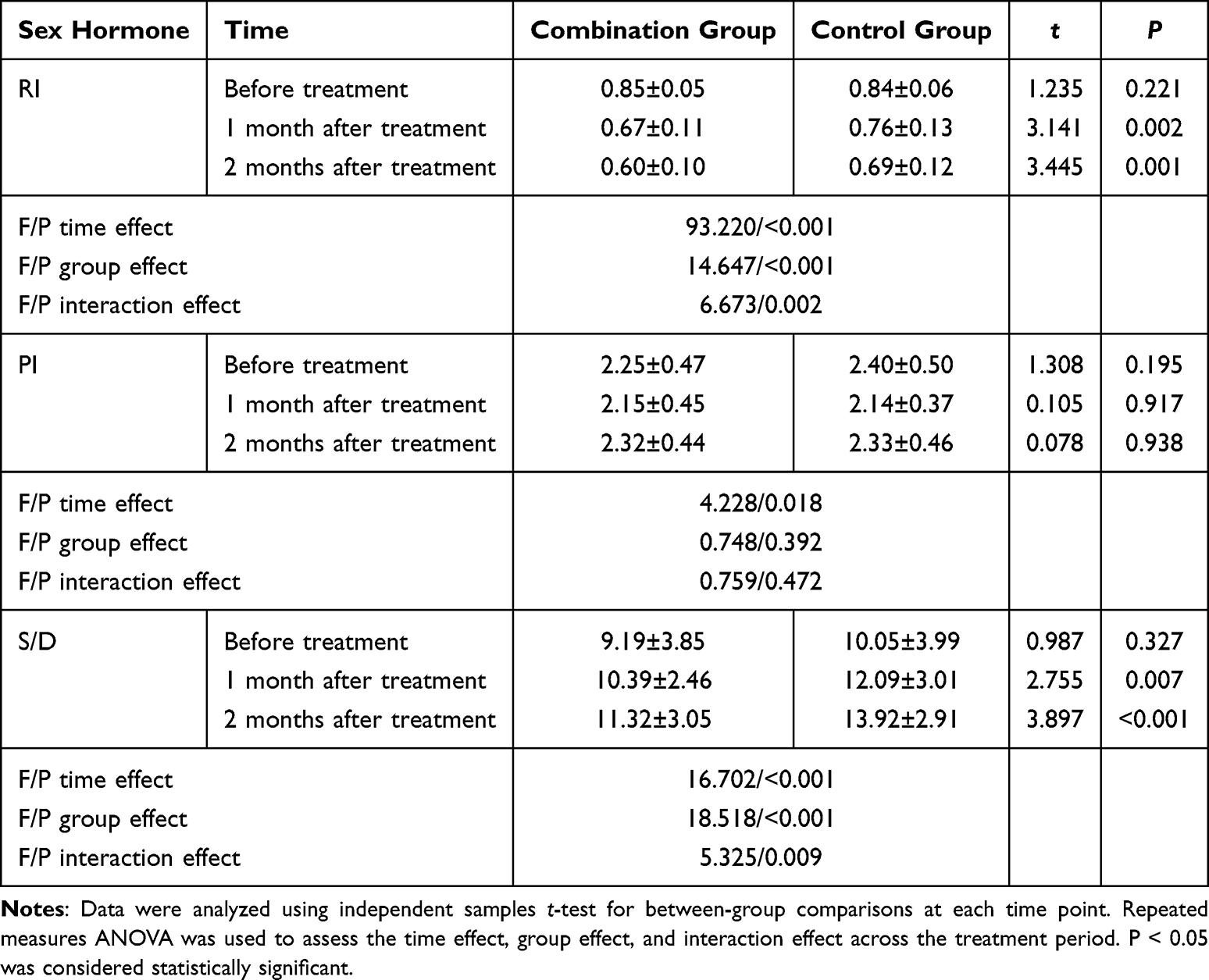 |
Table 4 Comparison of Uterine Artery Blood Flow Parameters Between the Two Groups Before and After Treatment ( |

Comparison of Pregnancy Outcomes Between the Two Groups
Among the enrolled patients, 53 (61.6%) experienced adverse pregnancy outcomes, including 20 (48.8%) in the combination group and 33 (73.3%) in the control group. After adjusting for potential confounders (age, baseline HCG, and chorionic protrusion number) through multivariate logistic regression, the combination group showed significantly lower odds of adverse pregnancy outcomes (adjusted OR=0.39, 95% CI: 0.17–0.89, _P_=0.025).
Comparison of Treatment-Related Adverse Reactions Between the Two Groups
In the combination group, there was 1 case (1.6%) of thrombocytopenia and 1 case (1.6%) of dermatitis, with a total adverse reaction rate of 3.2%. All reactions resolved spontaneously without special intervention. No adverse reactions were observed in the control group. The comparison of adverse reaction rates between the two groups showed no significant difference (χ²=0.613, P=0.434).
Discussion
The early pregnancy period is a critical stage for embryonic development and a key phase for embryogenesis and organ differentiation. During this period, embryos are extremely sensitive to various external factors, with approximately 80% of miscarriages occurring in early pregnancy stages.13 The American Institute of Ultrasound in Medicine first proposed in 2015 that the presence of chorionic bump and subchorionic hematoma during early pregnancy might indicate potential embryonic abnormalities, thereby increasing the risk of miscarriage.14 Although the incidence of chorionic bump is relatively low during early pregnancy, it is closely associated with an increased risk of miscarriage.15 Since pregnant women with chorionic bump in early pregnancy often exhibit no specific clinical symptoms, this condition is usually identified and diagnosed through ultrasound examination.16 Some studies suggest that chorionic bump may result from venous bleeding in the trophoblast layer, where the blood accumulates within the gestational sac as it cannot penetrate the trophoblast layer, leading to the formation of a bump17 This theory supports the etiological differences between chorionic bump and subchorionic hematoma.
Previous studies have also indicated that natural miscarriage rates can reach up to 40% in early pregnancy cases accompanied by chorionic bump, which is four times the risk in healthy pregnancies during early pregnancy. When three or more bumps are present, the miscarriage rate exceeds 70%, highlighting a strong correlation between pregnancy failure and the number of bumps rather than the size of the chorionic bump.5,18 However, a large-scale clinical study revealed that in cases where sex hormones and uterine hemodynamics were normal, isolated chorionic bump did not affect the live birth rate of embryos.19 Additionally, chorionic bump detected in early pregnancy may only be associated with a high risk of early miscarriage but does not appear to increase the risk of complications in mid-to-late pregnancy.20 Nevertheless, the exact etiology of chorionic bump remains unclear, and targeted treatment strategies are still lacking in clinical practice.
In previous studies, for women with unexplained infertility or recurrent miscarriage with high miscarriage rates, a combination treatment of prednisone acetate and low-molecular-weight heparin has been suggested.21 Prednisone acetate tablets suppress the activity of the hypothalamic-pituitary axis, reduce the aggregation of phagocytes and leukocytes at inflammatory sites, inhibit their phagocytic function, and lower the number of monocytes.22 Low-molecular-weight heparin, with its anticoagulant and antithrombotic properties, also suppresses the phagocytic activity of neutrophils, thereby exerting an immunomodulatory effect, improving maternal blood circulation, and alleviating spasms of the uterus and umbilical veins.23,24 The combination of these two drugs is considered clinically safe and shows positive therapeutic effects for unexplained embryonic arrest and natural miscarriage. Thus, this combined treatment may also benefit the pregnancy outcomes of patients with chorionic bump during early pregnancy. In this study, we observed that patients treated with prednisone acetate tablets and low-molecular-weight heparin showed significant reductions in average chorionic size and resistance index (RI), while the mean systolic/diastolic ratio (S/D) increased after treatment. Obstruction of uterine spiral artery dilation is a major cause of abnormal pregnancy, emphasizing the importance of closely monitoring hemodynamic parameters such as RI, pulsatility index (PI), and S/D of the uterine spiral artery to assess pregnancy status.25 The mechanisms of prednisone acetate include reducing the binding affinity of cell surface receptors to immunoglobulins, decreasing lymphoblast transformation, and lowering immunoglobulin concentration, thereby preventing immune-mediated vascular wall damage.26 Low-molecular-weight heparin enhances vascular permeability, improves blood circulation between the fetus, placenta, and mother, and increases fetal blood supply.27 After combined treatment, patients showed significant improvements in uterine arterial hemodynamics, including reduced luminal blood flow pressure and localized hypoxia, ensuring adequate uterine blood perfusion. Additionally, the severity of venous bleeding within the trophoblast layer was alleviated, leading to the shrinkage of the chorionic bump, all of which favor normal embryonic development.
Moreover, in this study, it was observed that after two months of treatment with prednisone acetate tablets and low-molecular-weight heparin, patients in the combined treatment group exhibited significantly higher average levels of estradiol (E2), progesterone (P), and luteinizing hormone (LH) than before treatment and compared to the control group, while average follicle-stimulating hormone (FSH) levels were significantly lower. Previous studies have revealed the role of sex hormones in regulating gonadotropin secretion. For example, E2 and P can downregulate the receptors for FSH on oocytes, thereby improving ovarian function and protecting residual follicles.28 The findings of this study suggest that prednisone acetate tablets and low-molecular-weight heparin treatment may help maintain normal sex hormone levels in early pregnant women, preventing hormone imbalances that could lead to abnormal embryonic development. However, the specific mechanisms by which these two drugs affect sex hormone levels in early pregnancy and their potential direct effects on chorionic bump remain to be further explored.
This study has several limitations. First, as a retrospective cohort study, it may introduce certain biases that affect the reliability of the findings. The small sample size also limits the generalizability of the results, and the single-center design restricts the diversity of the population, potentially failing to represent a broader demographic of patients with chorionic bumps. Future studies should consider increasing the sample size or conducting multi-center clinical studies in collaboration with other hospitals to improve the reliability and external validity of the findings. Additionally, specific strategies for addressing potential biases, such as randomization or propensity score matching, should be explored in future research. The study also did not thoroughly examine potential confounding factors, such as patients’ baseline health conditions or treatment adherence, which may influence the results. A more detailed consideration and reporting of such confounding factors would enhance the rigor and transparency of the study.
Conclusion
This study concludes that the combination of prednisone acetate and low-molecular-weight heparin can significantly reduce the size of chorionic bumps, improve uterine arterial blood flow, and ultimately reduce the risk of adverse pregnancy outcomes in early pregnancy patients with chorionic bump. This treatment approach has demonstrated reliable clinical safety and has the potential for wider application in this patient population.
Disclosure
The authors report no conflicts of interest in this work.
References
1. DeVilbiss EA, Mumford SL, Sjaarda LA. et al. Prediction of pregnancy loss by early first trimester ultrasound characteristics. Am J Obstet Gynecol. 2020;223(2):
2. Galleguillos C, Sepulveda W. Chorionic bump: progression to a subamniotic hematoma. Ultrasound. 2022;30(1):90–93. doi:10.1177/1742271X211008565
3. Giordano C, Miner L. Spontaneous expulsion of an intact gestational sac following an arrested pregnancy. J Obstet Gynaecol Can. 2021;43(1):1. doi:10.1016/j.jogc.2019.06.004
4. Goodday SM, Karlin E, Brooks A, et al. Better Understanding of the Metamorphosis of Pregnancy (BUMP): protocol for a digital feasibility study in women from preconception to postpartum. NPJ Digit Med. 2022;5(1):40. doi:10.1038/s41746-022-00579-9
5. Hamed A, Alhouri R, Allahham N, et al. New insight on etiology: a chorionic bump with partial mole pregnancy case report from Syria. J Clin Ultrasound. 2025;53(1):194–197. doi:10.1002/jcu.23812
6. Hiersch L, Barrett J, Aviram A, et al. Patterns of discordant growth and adverse neonatal outcomes in twins. Am J Obstet Gynecol. 2021;225(2):
7. Ji W, Hou B, Li W, et al. Associations between first-trimester intrauterine hematoma and twin pregnancy outcomes: a retrospective cohort study. BMC Pregnancy Childbirth. 2021;21(1):46. doi:10.1186/s12884-020-03528-0
8. Lisova KM, Kalinovska I, Tokar P. Ultrasound characteristic of embryo, fetal egg and chorionic structures in pregnant women with miscarriage. Wiad Lek. 2022;75(1):75–78. doi:10.36740/WLek202201114
9. Liu E, Noor A, Pereira N. Chorionic bump: an early marker for miscarriage and embryonic aneuploidy? J Obstet Gynaecol Can. 2024;46(7):102576. doi:10.1016/j.jogc.2024.102576
10. D’Ippolito S, Gavi F, Granieri C, et al. Efficacy of corticosteroids in patients with recurrent pregnancy loss: a systematic review and meta-analysis. Am J Reprod Immunol. 2025;93(1):e70037. doi:10.1111/aji.70037
11. Lu Y, Wu Y, Huang F, et al. A single-center retrospective study of the clinical significance of chorionic bump at early stage of gestation. Am J Reprod Immunol. 2021;85(3):e13346. doi:10.1111/aji.13346
12. Huang Q, Wu H, Li M, et al. Prednisone improves pregnancy outcome in repeated implantation failure by enhance regulatory T cells bias. J Reprod Immunol. 2021;143:103245. doi:10.1016/j.jri.2020.103245
13. Mazer Zumaeta A, Wright A, Syngelaki A, et al. Screening for trisomy at 11–13 weeks’ gestation: use of pregnancy-associated plasma protein-A, placental growth factor or both. Ultrasound Obstet Gynecol. 2020;56(3):408–415. doi:10.1002/uog.22140
14. McCarter K, Willson S, Shah N, et al. Chorionic bump in early pregnancy associated with first-trimester miscarriage. BMJ Case Rep. 2020;13(7):e236624. doi:10.1136/bcr-2020-236624
15. Mirghani HM, Wadhwa N, Jahangir F, et al. Case report: chorionic bump detected in the third trimester. J Clin Ultrasound. 2022;50(1):82–85. doi:10.1002/jcu.23027
16. Vena F, Bartolone M, D’Ambrosio V, et al. Pregnancy and perinatal outcomes in pregnancy with diagnosis of chorionic bump on first-trimester sonography: a systematic review and meta-analysis. J Matern Fetal Neonatal Med. 2022;35(25):8897–8904. doi:10.1080/14767058.2021.2006627
17. Yousaf A, Tayyab A, Anil MSU, et al. Chorionic bump: radiologic features and pregnancy outcomes. Cureus. 2020;12(11):e11480. doi:10.7759/cureus.11480
18. Vasilyev SA, Tolmacheva EN, Vasilyeva OY, et al. LINE-1 retrotransposon methylation in chorionic villi of first trimester miscarriages with aneuploidy. J Assist Reprod Genet. 2021;38(1):139–149. doi:10.1007/s10815-020-02003-1
19. Benn P, Grati FR. Aneuploidy in first trimester chorionic villi and spontaneous abortions: windows into the origin and fate of aneuploidy through embryonic and fetal development. Prenat Diagn. 2021;41(5):519–524. doi:10.1002/pd.5795
20. Gil MM, Molina FS, Rodríguez‐Fernández M, et al. New approach for estimating risk of miscarriage after chorionic villus sampling. Ultrasound Obstet Gynecol. 2020;56(5):656–663. doi:10.1002/uog.22041
21. Li J, Gao Y-H, Su J, et al. Diagnostic ideas and management strategies for thrombocytopenia of unknown causes in pregnancy. Front Surg. 2022;9:799826. doi:10.3389/fsurg.2022.799826
22. Palmsten K, Bandoli G, Vazquez-Benitez G, et al. Oral corticosteroid use during pregnancy and risk of preterm birth. Rheumatology. 2020;59(6):1262–1271.
23. Xue H, Guo Q, Yu A, et al. Genetic analysis of chorionic villus tissues in early missed abortions. Sci Rep. 2023;13(1):21719. doi:10.1038/s41598-023-48358-0
24. Sun Y, Cui L, Lu Y, et al. Prednisone vs placebo and live birth in patients with recurrent implantation failure undergoing in vitro fertilization: a randomized clinical trial. JAMA. 2023;329(17):1460–1468. doi:10.1001/jama.2023.5302
25. Tao S, Du J, Chi X, et al. Associations between antenatal corticosteroid exposure and neurodevelopment in infants. Am J Obstet Gynecol. 2022;227(5):
26. Wang Q, Bai W, Li C, et al. Effect of low dose prednisone on glucose metabolism levels in patients with spontaneous abortion: a single-center, prospective cohort study. J Reprod Immunol. 2024;166:104395. doi:10.1016/j.jri.2024.104395
27. Zha X, Fang M, Zhong W, et al. Dose-, stage- and sex- difference of prenatal prednisone exposure on placental morphological and functional development. Toxicol Lett. 2024;402:68–80. doi:10.1016/j.toxlet.2024.11.005
28. Zou Y, Ming L, Ding J, et al. Low dosage of prednisone acetate combined with doxycycline in the treatment of chronic endometritis in patients with repeated implantation failure. Am J Reprod Immunol. 2023;89(6):e13713. doi:10.1111/aji.13713
 © 2025 The Author(s). This work is published and licensed by Dove Medical Press Limited. The
full terms of this license are available at https://www.dovepress.com/terms
and incorporate the Creative Commons Attribution
- Non Commercial (unported, 4.0) License.
By accessing the work you hereby accept the Terms. Non-commercial uses of the work are permitted
without any further permission from Dove Medical Press Limited, provided the work is properly
attributed. For permission for commercial use of this work, please see paragraphs 4.2 and 5 of our Terms.
© 2025 The Author(s). This work is published and licensed by Dove Medical Press Limited. The
full terms of this license are available at https://www.dovepress.com/terms
and incorporate the Creative Commons Attribution
- Non Commercial (unported, 4.0) License.
By accessing the work you hereby accept the Terms. Non-commercial uses of the work are permitted
without any further permission from Dove Medical Press Limited, provided the work is properly
attributed. For permission for commercial use of this work, please see paragraphs 4.2 and 5 of our Terms.