
Back to Journals » International Medical Case Reports Journal » Volume 19
An Uncommon Case of Recurring Pulmonary, Lymphatic, and Skeletal Tuberculosis Accompanied by Myositis: Is It Associated with Particular Resistance?
Authors Sugiri YJR, Fahmi K, Tantular R, Hastuti NDP, Winaris N  , Fitri LE , Maksum IP, Yudhanto HS, Karliasari L
Received 11 December 2025
Accepted for publication 9 April 2026
Published 22 May 2026 Volume 2026:19 584317
DOI https://doi.org/10.2147/IMCRJ.S584317
Checked for plagiarism Yes
Review by Single anonymous peer review
Peer reviewer comments 2
Editor who approved publication: Professor Thomas E Hutson
Yani Jane R Sugiri,1 Kristia Fahmi,1 Rezki Tantular,1 Nurima Diyah Puji Hastuti,2 Nuning Winaris,3 Loeki Enggar Fitri,3 Iman P Maksum,4 Hendy Setyo Yudhanto,5 Liana Karliasari6
1Department of Pulmonology and Respiratory, Faculty of Medicine, Universitas Brawijaya/General Hospital Dr. Saiful Anwar, Malang, East Java, Indonesia; 2Department of Microbiology, Faculty of Medicine, Universitas Brawijaya/General Hospital Dr. Saiful Anwar, Malang, East Java, Indonesia; 3Department of Clinical Parasitology, Faculty of Medicine, Universitas Brawijaya, Malang, East Java, Indonesia; 4Department of Chemistry, Faculty of Mathematics and Natural Sciences, Universitas Padjadjaran, Bandung, West Java, Indonesia; 5Department of Anatomical Pathology, Faculty of Medicine, Universitas Brawijaya/General Hospital Dr. Saiful Anwar, Malang, East Java, Indonesia; 6Department of Radiology, Faculty of Medicine, Universitas Brawijaya/General Hospital Dr. Saiful Anwar, Malang, East Java, Indonesia
Correspondence: Loeki Enggar Fitri, Department of Clinical Parasitology, Faculty of Medicine, Universitas Brawijaya, Jl. Veteran, Malang, 65145, Indonesia, Email [email protected]
Abstract: Tuberculosis (TB) remains one of the primary causes of illness and death worldwide. Indonesia ranks second globally in TB incidence. Recurrent or relapsed TB, especially with multifocal involvement, presents significant diagnostic and therapeutic challenges, often complicated by incomplete treatment and limited molecular diagnostic capacity in developing countries. We present the case of a 26-year-old woman with a history of pulmonary, lymphatic, and skeletal tuberculosis who developed recurrence shortly after completing a 12-month anti-tuberculosis regimen (August 2024–July 2025) that excluded pyrazinamide due to gastrointestinal intolerance. She presented again in September 2025 with right-sided chest pain, productive cough, and cervical lymphadenopathy. Chest radiography revealed bilateral infiltrates, and abdominal ultrasonography suggested tuberculous myositis. The pulmonary findings, positive GeneXpert MTB/RIF Ultra result, and cervical lymph node biopsy confirming caseating granulomatous lymphadenitis were identified during the initial emergency department admission, while pus culture from the osteomyelitis lesion grew Staphylococcus aureus. The abdominal findings, including a left iliopsoas abscess with involvement of the iliacus muscle suggestive of tuberculous myositis, were detected later during outpatient follow-up evaluation, after the patient continued to experience persistent abdominal pain. The patient subsequently underwent a second course of treatment with a complete first-line anti-tuberculosis regimen combined with additional antibiotic therapy, which resulted in significant clinical improvement. In a follow-up, it was surprising that the sequence analysis of a 561 bp fragment of rpoB showed a single nucleotide deletion in comparison to the reference sequence. A six-month treatment regimen (BPaLM) has been planned as the selected regimen. This case illustrates the complexity of managing recurrent multifocal TB and underscores the need for rapid and comprehensive molecular diagnostic tools.
Keywords: tuberculosis recurrence, multifocal, anti-TB regimen, GeneXpert MTB/RIF, sequencing
Introduction
Tuberculosis (TB), caused by Mycobacterium tuberculosis (MTB), remains one of the foremost contagious diseases globally. The World Health Organization (WHO) Global TB Report 2025 identifies Indonesia as the country with the second-highest TB burden (10%) globally, accounting for over one million new cases annually. From 2015 to 2024, it is estimated that the incidence of tuberculosis in Indonesia rose by approximately 20–25%, while the number of deaths increased by approximately 20–30%.1
Typically, two TB-related conditions exist: latent TB and active TB disease. Millions of individuals harbor latent TB bacteria without showing signs of active tuberculosis. Approximately 30% of those exposed to MTB will acquire latent TB,2 if this condition is not addressed, around 5 to 10% of these individuals may eventually develop active TB disease during their lifetime.3,4 TB disease happens when the immune system of a person with latent TB is overwhelmed by tubercle bacilli, leading to their multiplication. If an individual is infected with TB bacteria and the immune system fails to manage the tubercle bacilli effectively, these bacilli will proliferate swiftly, resulting in TB disease. This can take place in various parts of the body, such as the lungs, kidneys, brain, or bones.5
Recurrent TB can result due to several reasons, such as inadequate compliance with anti-TB medications and the patient’s immune health (co-infection with HIV or diabetes). Risk factors influencing the two types of recurrent TB (reactivation and reinfection) are intricate and varied. Anti-TB drug resistance, is one of many factors potentially involved in the recurrence of tuberculosis that may contribute to TB relapse.6 The rise of multidrug-resistant TB (MDR-TB), known as resistance to at least Isoniazid (INH) and Rifampicin (RIF), complicates treatment outcomes and underscores the need for rapid, accurate diagnostics.1,7 Traditional diagnostic methods, including smear microscopy and culture, are time-consuming or lack sensitivity. The GeneXpert MTB/RIF is a nucleic acid amplification test (NAAT) that utilizes a cartridge system for the swift diagnosis of tuberculosis and the rapid assessment of antibiotic sensitivity. This automated diagnostic tool is capable of detecting the DNA of MTB and determining resistance to rifampicin. GeneXpert MTB/RIF Ultra has enhanced the speed of tuberculosis diagnostics; however, its detection of rifampicin resistance is restricted to mutations within the rpoB hotspot region, potentially missing resistance associated with mutations outside this region that necessitate confirmation with sequencing. Additional resistance to isoniazid, fluoroquinolones, ethionamide, and second-line injectable drugs can be assessed using Xpert MTB/XDR.8,9
Here, we report a case of multifocal recurrent TB with osteomyelitis and lymphadenitis in a young woman, with GeneXpert MTB/RIF Ultra results showing detection of the MTB and no rifampicin resistance detected, unfortunately, DNA sequencing gives different results.
Case Description
A 26-year-old Indonesian woman presented to the Emergency Department of Dr. Saiful Anwar General Hospital, Malang on September 2025, with complaints of right-sided chest pain for three days, productive cough for one week, and mild shortness of breath. She rejected having a fever, experiencing night sweats, or coughing up blood but she did mention a weight decrease of 1 kg over the last week.
She had a previous history of pulmonary and extrapulmonary tuberculosis diagnosed on 23 August 2024, involving cervical lymphadenitis and osteomyelitis. There was no history of diabetes mellitus or HIV infection. However, the patient had a history of chronic arthritis and had been taking several medications prior to admission, including intermittent methylprednisolone (initial dose 16 mg daily with tapering), leflunomide 20 mg once daily, and hydroxychloroquine 200 mg once daily, in addition to nonsteroidal anti-inflammatory drugs (NSAIDs) for chronic musculoskeletal pain. In 2024, the patient underwent an endoscopic procedure, which revealed that the esophagus was normal in caliber with intact mucosa; however, the lower esophageal sphincter exhibited erythema and erosive changes. The fundus demonstrated erosive mucosa with prominent vascular patterns, and the corpus showed diffuse hyperemia and excessive gas accumulation. The antrum displayed mucosal erosions and marked hyperemia, while the pylorus appeared edematous with diffuse erythema. The duodenal bulb exhibited erosive mucosal changes, and the distal duodenum appeared normal. These endoscopic findings are consistent with chronic erosive gastritis and reflux-related mucosal injury, most likely associated with prolonged NSAID and corticosteroid use. The patient completed a 12-month anti-tuberculosis treatment regimen on 29 July 2025 according to the national TB program criteria, with negative follow-up sputum acid fast bacilli (AFB) examination at the end of therapy.
On examination, the patient appeared mildly cachectic but alert and cooperative.Vital signs revealed a blood pressure of 96/63 mmHg, a heart rate of 95 beats per minute, a respiratory rate of 18 breaths per minute, a body temperature of 36.8 degrees Celsius, and oxygen saturation at 99% while using a nasal cannula at 3 liters per minute. She weighed 39 kilograms and was 145 centimeters tall, resulting in a body mass index (BMI) of 18.5. Upon physical examination, a hard, fixed, and painless lump was detected in the left side of the neck, around 8 by 4 centimeters in size, with an irregular surface. Lung auscultation revealed decreased breath sounds and fine crackles bilaterally. No hepatosplenomegaly was detected. The laboratory examination result is shown in Table 1.
 |
Table 1 The General Laboratory Result |
Mycobacterium tuberculosis culture was performed using the BACTEC MGIT 960 system (Becton Dickinson, USA). Specimens were processed using standard NALC-NaOH decontamination, followed by inoculation into Mycobacteria Growth Indicator Tube (MGIT) tubes supplemented with OADC enrichment and PANTA antibiotic mixture. Tubes were incubated and monitored automatically by the MGIT 960 instrument for up to 42 days. The MTB culture exhibited negative results following the first therapy in August 2025, but during the re-culture in September 2025, there was a notable growth of MTB, and phenotypic drug susceptibility testing demonstrated sensitivity to isoniazid and fluoroquinolone.
After the emergency department visit, further diagnostic evaluation was performed. Fine needle aspiration biopsy of the cervical lymph node confirmed granulomatous lymphadenitis with caseous necrosis consistent with tuberculous lymphadenitis. Both the examination of sputum biopsy and lymph nodes (Figure 1A and B), as well as the GeneXpert MTB/RIF Ultra (Cepheid, USA) which detects MTB with results indicating probable susceptibility to rifampicin, support the diagnosis of recurrent tuberculosis.
 |
Figure 1 Histologic structure of the cervical lymph node from the Fine needle aspiration biopsy (FNAB). The hypercellular smear reveals the presence of epithelioid histiocytes and lymphocytes, which are organized into granulomas, giant multinucleated cells, and cyst macrophages. The following is a synopsis of the relevant background material on erythrocytes, caseoca necrosis, and neutrophils. (A) 100x magnification; (B) 400x magnification (C) Datia Cell (red arrow); (D) caseoca necrosis (red arrow). |
Table 2 describes the diagnostic examination in this case. Initial diagnostic results represent laboratory findings obtained during the initial diagnosis prior to recurrence, whereas follow-up results correspond to examinations performed after therapy was given. Pus obtained from the pedis wound associated with the osteomyelitis lesion yielded Staphylococcus aureus, which was sensitive to levofloxacin and amoxicillin–clavulanate. (Table 2)
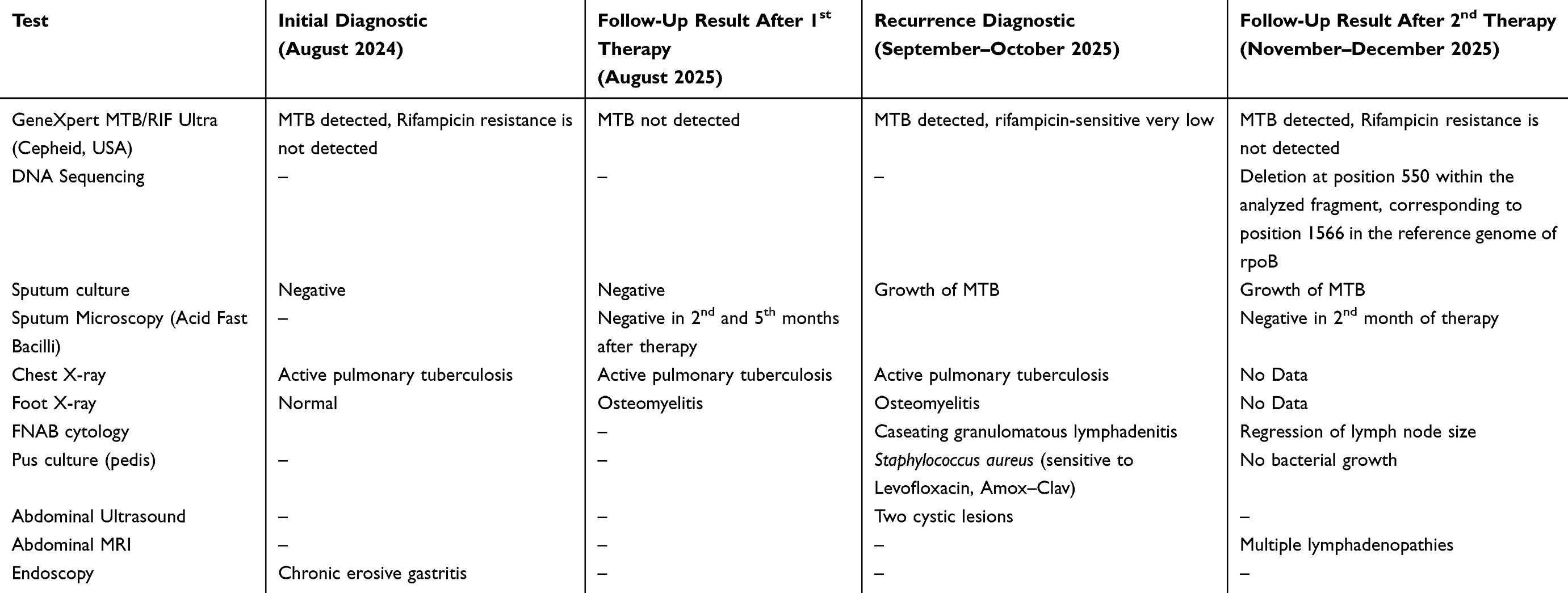 |
Table 2 The Specific Laboratory Results at Initial Diagnosis, Prior Follow-Up and Recurrence Diagnosis |
Chest X-ray posteroanterior view on 12 August 2024 (Figure 2A), showing features consistent with active pulmonary tuberculosis characterized by fibro-infiltrates and multiple cavities in the upper zone of the left lung, associated with superior retraction of the left hilum. Chest X-ray (posteroanterior view), after 1 year, 25 August 2025 (Figure 2B), demonstrating active pulmonary tuberculosis with partial radiologic improvement compared to Figure 2A. Chest X-ray (posteroanterior view) in September 2025 (Figure 2C), revealing active pulmonary tuberculosis with fibro-infiltrates and cavitary lesions in the upper and mid zones of the left lung, showing radiologic progression compared to Figure 2B.
 |
Figure 2 Chest X-ray. (A) Chest X-ray (posteroanterior view) 12 August 2024, showing features consistent with active pulmonary tuberculosis characterized by fibro-infiltrates and multiple cavities in the upper zone of the left lung, associated with superior retraction of the left hilum (red arrow). (B) Chest X-ray (posteroanterior view), after 1 year (25 August 2025), demonstrating active pulmonary tuberculosis with partial radiologic improvement compared to image A (red arrow). (C) Chest X-ray (posteroanterior view) in September 2025, revealing active pulmonary tuberculosis with fibro-infiltrates and cavitary lesions in the upper and mid zones of the left lung (red arrow), showing radiologic progression compared to image B. |
However, the patient continued to experience persistent abdominal pain during the initial evaluation period. Because of these symptoms, initiation of anti-tuberculosis therapy was temporarily postponed until further imaging evaluation was completed to determine the underlying cause. A follow-up abdominal ultrasound on October 31, 2025, revealed two multiloculated cystic lesions with internal debris and iso–to–hypoechoic characteristics involving the left iliopsoas and iliacus muscles, extending to both inguinal regions. The findings are suggestive of bilateral iliopsoas abscesses (Figure 3). Neck ultrasound revealed multiple hypoechoic masses with indistinct borders and central necrosis, compatible with tuberculous lymphadenitis.
 |
Figure 3 Abdominal Ultrasound. An abdominal Ultrasound conducted on October 31, 2025, shows two cystic lesions with internal debris and iso-to-hypoechoic characteristics (red arrows). (A) The lesions measured approximately 4.9×4.9 × 6.6 cm with an estimated volume of 43.7 mL on the right side, while (B) Those on the left side measured 5.2×5.2 × 6.7 cm, corresponding to an estimated volume of 50.2 m. No hepatic, splenic, or renal abnormalities were observed. |
During outpatient follow-up, she also reported the appearance of a palpable abdominal mass associated with worsening discomfort. Subsequent abdominal magnetic resonance imaging (MRI) on November 2025 revealed multiloculated abscess formation involving the iliopsoas and pelvic regions, consistent with tuberculous abscess formation associated with disseminated disease. (Figures 4 and 5). Based on the combination of microbiological findings, histopathological confirmation, and radiologic evidence of abscess formation, the patient was started on a modified anti-tuberculosis regimen consisting of rifampicin, isoniazid, and ethambutol (RHE) combined with levofloxacin
 |
Figure 4 Abdominal MRI of The Right Pubic Region. Abdominal MRI demonstrated a lesion with hypointense signal on T1WI+Contrast (A, red arrow), hyperintense signal on T2WI (B, red arrow), and restricted diffusion on DWI (C, red arrow) involving the right pubic bone and pubic symphysis. The lesion was continuous with a thick-walled complex cystic lesion containing internal debris, showing rim enhancement and measured approximately 3.8×10.0 × 3.7 cm (with an estimated volume ~17 cc) in the pelvic region. The lesion extended to involve the right obturator externus, adductor magnus, and adductor brevis muscles. |
 |
Figure 5 Abdominal MRI of The Left Pelvic Region. Abdominal MRI dated November 2025 shows a T1-weighted (T1/WI) hypointense (A), T1+Contrast (B), T2 -Stir (T2/WI) weighted hyperintense (C) and restricted diffusion of DWI images and RIM enhancement after contrast administration in the left pelvic region (D), involving the pelvic muscles and extending to the left anterior abdominal wall, impression more likely tuberculosis of the pubic symphysis with a left pelvic abscess (estimated volume 190 cc) (Red arrow). |
A radiograph of the left foot from a year ago shows normal findings with no sign of bony involvement (Figure 6A). The current radiograph (30 July 2025) revealed soft tissue swelling along with a destructive osteolytic lesion at the far end of the first metatarsal bone in the left foot, suggesting the presence of osteomyelitis (Figure 6B). After two months of standard treatment (9 October 2025), the soft tissue swelling and osteolytic lesion in the distal portion of the first metatarsal bone of the left foot remained persistent, but there was improvement in the degree of swelling (Figure 6C). Radiograph of the right humerus showed an osteolytic lesion that decreased in size and density following re-treatment.
 |
Figure 6 Left foot X-ray (anteroposterior and oblique views). (A) Radiograph of the left foot dated 16 August 2024 shows normal findings with no evidence of bony involvement. (B) Radiograph dated 30 July 2025 demonstrated soft tissue swelling accompanied by a destructive osteolytic lesion at the distal part of the first metatarsal bone of the left foot, indicating osteomyelitis (red arrow). (C) Radiograph dated 9 October 2025 reveals persistent soft tissue swelling and an osteolytic lesion at the distal first metatarsal of the left foot (red arrow), with interval improvement in the degree of swelling compared to image B. |
The patient continued on Rifampicin–Isoniazid–Ethambutol (RHE) regimen incorporating Levofloxacin daily for enhanced coverage against possible resistant M. tuberculosis strains and secondary bacterial pathogens. Following initiation of therapy, the patient showed progressive clinical improvement. The previously reported right-sided chest pain resolved. There was a marked relief of abdominal pain and the palpable abdominal mass was no longer detected during follow-up examination. Her general condition improved with better appetite and reduced inflammatory symptoms, with no new symptoms were reported during follow-up. These clinical findings support the diagnosis of recurrent multifocal tuberculosis with associated iliopsoas abscess.
Discussion
Recurrent multifocal tuberculosis remains a diagnostic and therapeutic challenge, particularly in resource-limited settings where molecular diagnostic tools are not yet widely available. This case illustrates the progression of disseminated tuberculosis affecting the pulmonary, lymphatic, skeletal, and muscular systems. The development of bilateral iliopsoas abscesses represents a rare but recognized manifestation of tuberculous myositis, most often resulting from hematogenous dissemination from a primary pulmonary or skeletal focus. The iliopsoas compartment provides an optimal environment for bacillary proliferation due to its relatively poor vascularity and proximity to spinal and retroperitoneal lymphatic drainage. Given the current findings, contrast-enhanced abdominal MRI was performed for better delineation and evaluation of potential extension to the adjacent pelvic structure.10,11 While Staphylococcus aureus remains the leading cause of primary psoas abscesses, secondary tuberculous psoas abscesses are frequently associated with spinal or vertebral tuberculosis. In this patient, the absence of spinal involvement on imaging and concurrent improvement in pulmonary and osseous lesions suggest hematogenous spread from prior extrapulmonary foci.12
In this case, recurrence occurred shortly after completion of a modified anti-tuberculosis regimen that excluded pyrazinamide, which may have reduced the sterilizing capacity of the regimen. In addition, intermittent corticosteroid use for arthritis may have contributed to partial immunosuppression, potentially facilitating reactivation of residual bacilli. The patient had a cough and weight loss, consistent with pulmonary tuberculosis, common symptoms. Negative culture sputum smears cannot exclude pulmonary tuberculosis. Discrepancies between the Xpert assay and culture have posed challenges in determining which individuals are infected with TB. Nevertheless, Xpert-Ultra remains recommended as the primary diagnostic tool in TB prevalence surveys, with culture used only for confirmatory purposes.13
Pulmonary TB with negative culture results likely represents an initial stage along the continuum between MTB infection and active disease and may progress to culture-positive disease in the absence of treatment. An approach that characterizes the clinical manifestations of culture-negative pulmonary tuberculosis may enhance clinician awareness, facilitate earlier diagnosis, and promote timely initiation of therapy, thereby potentially reducing the risk of progression to transmissible disease. The frequency and clinical manifestations of pulmonary TB with negative mycobacterial culture have not been thoroughly researched. Among the limited studies available, it was found that pulmonary TB with negative mycobacterial culture had fewer instances of hemoptysis and radiographic abnormalities compared to pulmonary TB with positive mycobacterial culture.14
From a laboratory standpoint, patient inflammatory markers (CRP 3.57 mg/dL) and hematologic profile (moderate anemia and neutrophil predominance) are consistent with subacute infection. The recurrence after a modified initial regimen excluding pyrazinamide may have contributed to partial sterilization, allowing latent bacilli to reactivate under suboptimal immune clearance.
Recurrent tuberculosis is generally classified into two types: endogenous reactivation and exogenous reinfection. Recurrence caused by the same strain as the initial episode is considered reactivation, whereas recurrence involving a different strain is regarded as reinfection. The risk factors for both forms of recurrent TB are varied and multifactorial. Anti-TB drug resistance, patient clinical characteristics, strain genotype, and HIV co-infection or diabetes are all considered factors that roughly account for the different types of recurrent TB.6,15 Prior to the advent of molecular genotyping techniques, it was difficult to ascertain the impact of reactivation and reinfection on the prevalence of recurrent TB, which could have adverse effects on public health. This highlights the importance of molecular diagnostics in understanding these dynamics.6
Given the above factors, molecular confirmation of drug susceptibility is essential. Common assays such as GeneXpert MTB/RIF are limited to rpoB gene mutations, which may lead to the missing of potential resistance to isoniazid, fluoroquinolones, or linezolid.16,17 The qPCR testing in this patient with multiple resistance targets associated to several loci (rpoB, katG, inhA, gyrA, and rplC), providing a comprehensive genotypic profile to guide individualized therapy. A previous study reported the development and analytical validation of a multiplex real-time PCR assay. This assay employs contact-quenching fluorescence detection, allele-specific primers, and 3′-blocked wild-type allele blockers to identify clinically relevant resistance-associated mutations in several genes, including the rpoB, katG, and inhA.18
Recent advances in molecular diagnostics have significantly improved the rapid detection of drug-resistant tuberculosis. The Xpert MTB/XDR assay can detect resistance to multiple anti-tuberculosis drugs, including isoniazid, fluoroquinolones, second-line injectable drugs, and ethionamide directly from clinical samples.19 In addition, line probe assays such as GenoType MTBDRplus and MTBDRsl are able to identify resistance to rifampicin, isoniazid, fluoroquinolones, and second-line injectable agents through the detection of specific genetic mutations.20 These molecular diagnostic tools are particularly valuable in settings where culture and phenotypic drug susceptibility testing are not readily available or require prolonged processing times. Early identification of drug resistance allows clinicians to initiate appropriate treatment regimens more rapidly, thereby improving patient outcomes and reducing transmission of resistant strains.
In the present case, GeneXpert MTB/RIF Ultra detected MTB with no rifampicin resistance. GeneXpert MTB/RIF Ultra has high sensitivity and specificity for the identification of MTB and drug-resistant MTB. However, its detection of rifampicin resistance is restricted to mutations in a conserved 81 base-pair (bp) rifampin resistance-determining region (RRDR) of the rpoB gene, potentially missing resistance associated with mutations outside this region, such as in this case,21 therefore Xpert results suggest the absence of rifampicin resistance, they do not definitively confirm susceptibility. Additionally, another obvious limitation of Xpert is its poor performance in extrapulmonary TB cases.22–24
Unexpectedly, in the present study, we analyzed mutations in the rpoB gene to investigate the genetic basis of rifampicin resistance. Sequence analysis of a 561 bp fragment of rpoB revealed a single nucleotide deletion when compared with the reference sequence (NCBI Reference Sequence: NC_000962.3). Specifically, the deletion was identified at position 550 within the analyzed fragment, corresponding to position 1566 in the reference genome.
Therapeutically, continuation with the RHE regimen plus levofloxacin. The use of drug class: fluoroquinolone aligns with WHO recommendations for suspected drug-sensitive or partially resistant TB reactivation.25 Levofloxacin is an important drug for MDR-TB and it used empirically for pre-XDR TB globally. In our case, based on specific clinical considerations, the patient had a history of relapse and concurrent secondary infections, and pyrazinamide could not be administered due to intolerance (drug-induced gastritis). Therefore, levofloxacin was introduced as part of a modified regimen to maintain adequate antimicrobial coverage, particularly addressing the possibility of secondary bacterial infection, while ensuring continuation of anti-TB therapy. The presence of relapse TB and clinical complexity contributed to a higher index of suspicion, and the decision was made in a controlled clinical setting with close monitoring, while awaiting further diagnostic confirmation. Levofloxacin administration to our patient offers improved penetration within cells and exhibits early bactericidal effects, which is especially advantageous for treating deep-seated or abscessed infections.26 The MRI performed with contrast has facilitated the identification of abscess extension and the assessment of treatment efficacy.
Taken together, these findings supported the diagnosis of drug-resistant recurrent TB and guided the selection of the treatment regimen in the next follow-up. We plan to use a six-month treatment regimen (BPaLM) composed of bedaquiline (B), pretomanid (P), linezolid (L), and moxifloxacin (M). According to the existing policy of the WHO, multiple treatment regimens are available for patients diagnosed with multidrug-resistant or rifampicin-resistant tuberculosis (MDR/RR-TB). The primary considerations that influence the selection of a treatment regimen encompass the patient’s drug-resistance profile, previous exposure to tuberculosis medications, the patient’s medical history, the drug-resistance profiles of close contacts, the age of the patient, the severity of pulmonary tuberculosis, and the location of any extrapulmonary tuberculosis lesions.27,28
This case reinforces the need for integrated diagnostic and advanced molecular approaches in recurrent TB, especially for multifocal disease in young adults without immunosuppression. The favorable clinical response to anti-tuberculosis therapy, including resolution of abdominal pain and disappearance of the abdominal mass, further supports the presumptive diagnosis of recurrent multifocal tuberculosis.
Conclusion
This case illustrates the progressive nature of recurrent multifocal tuberculosis with new muscular involvement in the form of bilateral iliopsoas abscesses following prior pulmonary, lymphatic, and skeletal disease. It highlights the limitations of conventional diagnostic methods and the potential value of PCR–based molecular testing for comprehensive resistance detection. Continued RHE plus Levofloxacin therapy and follow-up imaging, including contrast-enhanced MRI, are essential for monitoring disease evolution and treatment response. This case highlights the importance of combining molecular diagnostics with clinical and radiologic assessments to enhance personalized management of recurrent and multifocal TB in Indonesia. The integration of molecular diagnostics and phenotypic drug susceptibility testing plays an essential role in confirming drug susceptibility and guiding appropriate treatment strategies in recurrent tuberculosis.
Informed Consent and Ethics for Publication
Written consent for publication of case details was obtained from the patient. An ethical approval letter was obtained from Research and Innovation Ethics Committee Universitas Brawijaya No. 459/EC/KEPK-S1/11/2025.
Acknowledgments
The authors sincerely acknowledge the patient for her willingness and cooperation in supporting this investigation. We would also like to express our gratitude to dr. Eko Dian Syafira, dr. R.A. Rose Khasana Dewi, Sp.PA, and dr. Achmad Bayhaqi N.A., Sp.Rad (K), for their valuable contribution, expertise, and continuous support throughout the completion of this research.
Funding
This research received funding from the Directorate of Research and Community Service, Directorate General of Research and Development, Ministry of Education, Science and Technology (The Impactful Consortium Research Program grant number: 3088/2/UN6.3.1/PT.00/2025).
Disclosure
The authors declare that there are no conflicts of interest associated with this work.
References
1. WHO. Global tuberculosis report 2025 [Internet]. 2025. Available from: https://iris.who.int/server/api/core/bitstreams/e97dd6f4-b567-4396-8680-717bac6869a9/content.
2. Goldman R, Laube J. Latent tuberculosis infection vs. Active TB disease: what’s the difference? [Internet]. Everyday Health. 2023. Available from: https://www.everydayhealth.com/tuberculosis/guide/latent/#resources.
3. WHO. Tuberculosis [Internet]. World Health Organization. 2025. Available from: https://www.who.int/news-room/questions-and-answers/item/tuberculosis.
4. Price C, Nguyen A. Latent tuberculosis. In StatPearls [Internet]. Treasure Island (FL): StatPearls Publishing; 2024. https://www.ncbi.nlm.nih.gov/books/NBK599527/.
5. CDC. Clinical overview of tuberculosis disease [Internet]. Centers for Disease Control and Prevention. 2025. Available from: https://www.cdc.gov/tb/hcp/clinical-overview/tuberculosis-disease.html.
6. Qiu B, Wu Z, Tao B, et al. Risk factors for types of recurrent tuberculosis (reactivation versus reinfection): a global systematic review and meta-analysis. Int J Infect Dis. 2022;116:14–12. doi:10.1016/j.ijid.2021.12.344
7. GmbH HL. GenoType MTBDRplus Assay Documentation. Nehren, Germany; 2021.
8. Zumla A, Chakaya J, Centis R, et al. Tuberculosis treatment and management—an update on treatment regimens, trials, new drugs, and adjunct therapies. Lancet Respir Med. 2015;3(3):220–234. doi:10.1016/S2213-2600(15)00063-6
9. Gopalaswamy R, Dusthackeer VNA, Kannayan S, Subbian S. Extrapulmonary Tuberculosis—an update on the diagnosis, treatment and drug resistance. J Respir. 2021;1:141–164. doi:10.3390/jor1020015
10. Vasigh M, Karoobi M, Montazeri M, et al. Isolated psoas abscess caused by Mycobacterium tuberculosis: a rare case report. Clin Case Rep. 2022;10(5):e05823. doi:10.1002/ccr3.5823
11. Tone K, Hirano Y, Kuwano K. Iliopsoas gravity abscess secondary to a tuberculous empyema. Int J Mycobacteriol. 2021;10(3):335–337. doi:10.4103/ijmy.ijmy_129_21
12. Sari DG, Imanuela R, Resultanti R. Abses iliopsoas akibat tuberkulosis: sebuah laporan kasus. Jurnal Penyakit Dalam Indonesia. 2024;11. https://scholarhub.ui.ac.id/jpdi/vol11/iss3/7.
13. Floyd S, Klinkenberg E, de Haas P, et al. Optimising Xpert-Ultra and culture testing to reliably measure tuberculosis prevalence in the community: findings from surveys in Zambia and South Africa. BMJ Open. 2022;12(6):e058195. doi:10.1136/bmjopen-2021-058195
14. Nguyen M-VH, Levy NS, Ahuja SD, Trieu L, Proops DC, Achkar JM. Factors associated with sputum culture-negative vs culture-positive diagnosis of pulmonary tuberculosis. JAMA Network Open. 2019;2(2):e187617. doi:10.1001/jamanetworkopen.2018.7617
15. Vega V, Rodríguez S, Van der Stuyft P, Seas C, Otero L. Recurrent TB: a systematic review and meta-analysis of the incidence rates and the proportions of relapses and reinfections. Thorax. 2021;76(5):494–502. doi:10.1136/thoraxjnl-2020-215449
16. Xia H, Song Y, Zheng Y, et al. Detection of Mycobacterium tuberculosis rifampicin resistance conferred by borderline rpoB mutations: xpert mtb/rif is superior to phenotypic drug susceptibility testing. Infect Drug Resist. 2022;15:1345–1352. doi:10.2147/IDR.S358301
17. Kusumaningrum D, Mertaniasih NM, Soedarsono S. The performance of A rpob gene mutation linked to A resistant to rifampicin Mycobacterium tuberculosis isolate from an Indonesian referral hospital. Indian J Tuberc. 2025;72 Suppl 2:S47–S50. doi:10.1016/j.ijtb.2024.06.013
18. Goyani K, Patel D, Vaniawala S, Mukhopadhyaya PN. A novel multiplex qPCR platform for detection of Mycobacterium tuberculosis drug resistance using synthetic mutation standards and fluorescence quenching probes. medRxiv. 2025;11:2025–06. 10.1101/2025.06.10.25329335.
19. Pillay S, Steingart KR, Davies GR, et al. Xpert MTB/XDR for detection of pulmonary tuberculosis and resistance to isoniazid, fluoroquinolones, ethionamide, and amikacin. Cochrane Database Syst Rev. 2022;5(5):CD014841. doi:10.1002/14651858.CD014841.pub2
20. Seifert M, Georghiou SB, Catanzaro D, et al. MTBDR plus and MTBDR sl assays: absence of wild-type probe hybridization and implications for detection of drug-resistant tuberculosis. J Clin Microbiol. 2016;54(4):912–918. doi:10.1128/JCM.02505-15
21. Shinnick TM, Starks AM, Alexander HL, Castro KG. Evaluation of the Cepheid Xpert MTB/RIF assay. Expert Rev Mol Diagn. 2015;15(1):9–22. doi:10.1586/14737159.2015.976556
22. Haas AL, Ma A, Pham J, et al. Limitations of the MTB/RIF assay: an Xpert review of 4 clinical cases. Open Forum Infect Dis. 2025;12(4):ofaf132. doi:10.1093/ofid/ofaf132
23. Hopmeier D, Lampejo T, Rycroft J, Tiberi S, Melzer M. The limitations of the Cepheid GeneXpert® Mtb/Rif assay for the diagnosis and management of polyresistant pulmonary tuberculosis. Clin Infect Prac. 2020;7-8:100038. doi:10.1016/j.clinpr.2020.100038
24. Chen P, Sun W, He Y. Comparison of metagenomic next-generation sequencing technology, culture and GeneXpert MTB/RIF assay in the diagnosis of tuberculosis. J Thorac Dis. 2020;12(8):4014–4024. doi:10.21037/jtd-20-1232
25. World Health Organization. WHO consolidated guidelines on tuberculosis: module 4: treatment – drug-resistant tuberculosis treatment [Internet]. Geneva: World Health Organization; 2022. Available from: https://iris.who.int/bitstream/handle/10665/365309/9789240065352-eng.pdf.
26. Zeitlinger MA, Dehghanyar P, Mayer BX, et al. Relevance of soft-tissue penetration by levofloxacin for target site bacterial killing in patients with sepsis. Antimicrob Agents Chemother. 2003;47(11):3548–3553. doi:10.1128/AAC.47.11.3548-3553.2003
27. World Health Organization. Tuberculosis: multidrug-resistant (MDR-TB) or rifampicin-resistant TB (RR-TB) [Internet]. World Health Organization. 2024. Available from: https://www.who.int/news-room/questions-and-answers/item/tuberculosis-multidrug-resistant-tuberculosis-(mdr-tb).
28. CDC. Treatment for Drug-resistant tuberculosis disease [Internet]. Centers for Disease Control and Prevention. 2025. Available from: https://www.cdc.gov/tb/hcp/treatment/drug-resistant-tuberculosis-disease.html.
 © 2026 The Author(s). This work is published and licensed by Dove Medical Press Limited. The
full terms of this license are available at https://www.dovepress.com/terms
and incorporate the Creative Commons Attribution
- Non Commercial (unported, 4.0) License.
By accessing the work you hereby accept the Terms. Non-commercial uses of the work are permitted
without any further permission from Dove Medical Press Limited, provided the work is properly
attributed. For permission for commercial use of this work, please see paragraphs 4.2 and 5 of our Terms.
© 2026 The Author(s). This work is published and licensed by Dove Medical Press Limited. The
full terms of this license are available at https://www.dovepress.com/terms
and incorporate the Creative Commons Attribution
- Non Commercial (unported, 4.0) License.
By accessing the work you hereby accept the Terms. Non-commercial uses of the work are permitted
without any further permission from Dove Medical Press Limited, provided the work is properly
attributed. For permission for commercial use of this work, please see paragraphs 4.2 and 5 of our Terms.