Back to Journals » International Journal of Nanomedicine » Volume 9 » Issue 1
Amplified voltammetric detection of glycoproteins using 4-mercaptophenylboronic acid/biotin-modified multifunctional gold nanoparticles as labels
Authors Liu L, Xing Y, Zhang H, Liu R, Liu H, Xia N
Received 12 February 2014
Accepted for publication 23 March 2014
Published 27 May 2014 Volume 2014:9(1) Pages 2619—2626
DOI https://doi.org/10.2147/IJN.S62343
Checked for plagiarism Yes
Review by Single anonymous peer review
Peer reviewer comments 4
Lin Liu,1,2 Yun Xing,1 Hui Zhang,1 Ruili Liu,1 Huijing Liu,1 Ning Xia1,2
1College of Chemistry and Chemical Engineering, Anyang Normal University, Anyang, Henan, People’s Republic of China; 2College of Chemistry and Chemical Engineering, Central South University, Changsha, Hunan, People’s Republic of China
Abstract: Ultrasensitive detection of protein biomarkers is essential for early diagnosis and therapy of many diseases. Glycoproteins, differing from other types of proteins, contain carbohydrate moieties in the oligosaccharide chains. Boronic acid can form boronate ester covalent bonds with diol-containing species. Herein, we present a sensitive and cost-effective electrochemical method for glycoprotein detection using 4-mercaptophenylboronic acid (MBA)/biotin-modified gold nanoparticles (AuNPs) (MBA-biotin-AuNPs) as labels. To demonstrate the feasibility and sensitivity of this method, recombinant human erythropoietin (rHuEPO) was tested as a model analyte. Specifically, rHuEPO was captured by the anti-rHuEPO aptamer-covered electrode and then derivatized with MBA-biotin-AuNPs through the boronic acid–carbohydrate interaction. The MBA-biotin-AuNPs facilitated the attachment of streptavidin-conjugated alkaline phosphatase for the production of electroactive p-aminophenol from p-aminophenyl phosphate substrate. A detection limit of 8 fmol L-1 for rHuEPO detection was achieved. Other glycosylated and non-glycosylated proteins, such as horseradish peroxidase, prostate specific antigen, metallothionein, streptavidin, and thrombin showed no interference in the detection assay.
Keywords: electrochemical biosensor, boronic acid, signal amplification, alkaline phosphatase
Introduction
Ultrasensitive detection of protein biomarkers is essential for early diagnosis and therapy of many diseases. Glycoproteins are a type of protein that plays vital roles in a wide variety of biological processes, such as cellular adhesion, cell signaling, and immune response.1–3 Change in the level of glycoproteins has been associated with inflammation and cancers.1,4 Thus, highly sensitive methods for glycoprotein detection are in demand for clinical diagnostics and therapeutics.5 Analytical techniques including mass spectrometry, surface plasmon resonance, chromatography, and enzyme-linked immunosorbent assay have been devoted to achieving the sensitive detection of glycoproteins.6,7 These methods have adequate sensitivity but usually suffer from high cost, time-consuming procedure, and professional operation. Recently, electrochemical biosensors have raised considerable interest for protein detection in clinical applications due to their intrinsic advantages, such as high sensitivity, fast response time, simple instrumentation, and low cost.8–12
For ultrasensitive detection of analytes with electrochemical techniques, a popular approach is driving the enhancement of sensitivity with signal amplification. Over the past decades, nanotechnology brings new possibilities for the development of signal-amplified electrochemical biosensors. Nanomaterials such as carbon-based nanostructures and magnetic/metallic nanoparticles have been successfully applied as carriers/tracers to develop amplified electrochemical bioassays.13–16 Among these nanomaterials, gold nanoparticles (AuNPs) are one kind of nanomaterial employed in diagnostics and bioassays in view of their good biological compatibility, high surface-to-volume ratio, and excellent conducting capability.14,17–19 To realize the highly sensitive detection of proteins, AuNPs have been commonly coated with biorecognition units such as antibodies for the sandwich-type or competitive assays. These methods include the utilization of AuNPs as carriers for loading of probe/signal molecules or as indicators to induce the metal deposition for both optical and conductivity detection. However, the application of antibody-coated nanoparticles could be hindered, especially in resource-poor countries, by the relatively poor stability and high cost of antibodies.
Differing from other types of proteins, glycoproteins contain carbohydrate moieties in the oligosaccharide chains, which enable glycoproteins to be selectively recognized and separated from a biological matrix.20,21 Typically, lectin is one of the major available tools for glycoproteins recognition. However, lectin has a certain specificity for the overall topology and sugar compositions, thus limiting its application in high throughput analysis.22 Alternately, boronic acid can form boronate ester covalent bonds with diol-containing species.23,24 Based on the interaction, there has been particular attention in employing boronic acids as recognition units for the immobilization and separation of glycoproteins.21,25–27 For example, Li et al25 developed a universal and facile approach for imprinting and detection of five distinct glycoproteins using boronic acid-based ultraviolet (UV)-initiated polymerization and a photolithographic fabrication route. Xu et al27 prepared the boronic acid-functionalized Au-coated Si wafer as the matrix-assisted laser desorption/ionization for enrichment of glycopeptides. Herein, we report a signal-amplified electrochemical biosensor for the detection of glycoproteins using 4-mercaptophenylboronic acid (MBA)/biotin-modified multifunctional AuNPs (denoted MBA-biotin-AuNPs) as labels. Specifically, the captured glycoproteins were first recognized by MBA-biotin-AuNPs through the boronic acid–carbohydrate interaction. Then, streptavidin (SA)-conjugated alkaline phosphatase (ALP) (SA-ALP) was attached by MBA-biotin-AuNPs via the biotin–SA interaction, promoting the production of electrochemically active p-aminophenol (p-AP) from p-aminophenyl phosphate (p-APP) substrate. To demonstrate the feasibility and sensitivity of this method, the glycoprotein recombinant human erythropoietin (rHuEPO) was tested as a model analyte.
Materials and methods
Chemicals and reagents
MBA, tris(carboxyethyl)phosphine (TCEP), 6-mercapto-1-hexanol (MCH), SA-ALP, horseradish peroxidase, bovine serum albumin (BSA), thrombin, SA, trisodium citrate, tris-(hydroxymethyl)aminomethane hydrochloride (TrisHCl), serum, KH2PO4, and K2HPO4 were purchased from Sigma-Aldrich Co. (St Louis, MO, USA). p-Aminophenyl phosphate (p-APP) was from Enzo Life Sciences, Inc. (Farmingdale, NY, USA). rHuEPO was acquired from BioVision, Inc. (Milpitas, CA, USA). Metallothionein was purchased from Hunan Lugu Biotech Co., Ltd (Changsha, People’s Republic of China). Prostate-specific antigen (PSA) was purchased from Linc-Bio Science Co. Ltd. (Shanghai, People’s Republic of China). The peptide with a sequence of CALNNGK(biotin)G was synthesized and purified by China Peptides Co., Ltd. (Shanghai, People’s Republic of China). The thiolated single-stranded deoxyribonucleic acid (DNA) probe (5′-SH-(CH2)6-GATTGAAAGGTCTGTTTTTGGGGTTGGTTTGGGTCAATA) was purchased from Sangon Biotech. Co., Ltd (Shanghai, People’s Republic of China). The DNA was dissolved in pH 7.4 TE (Tris-EDTA) buffer solution consisting of 10 mmol L-1 TrisHCl and 1 mmol L-1 ethylenediaminetetraacetic acid (EDTA), and kept at -18°C. The electrolyte buffer (EB) for the enzymic catalytic reaction and the electrochemical experiment was 10 mmol L-1 Tris (pH 7.4) containing 1 mmol L-1 MgCl2 and 50 mmol L-1 Na2SO4.
Synthesis of MBA-biotin-AuNPs
AuNPs with a diameter of ~13 nm were synthesized by the trisodium citrate reduction method, as reported previously.17 Briefly, 5 mL of 38.8 mmol L-1 trisodium citrate was added into 50 mL of 1 mmol L-1 HAuCl4 boiling solution, and the resulting solution was then kept continuously boiling for 30 minutes and then cooled to room temperature. The resultant red solution was stored at 4°C. Modification of AuNPs was performed through the ligand-exchange reaction. A 5 mL volume of the as-prepared AuNPs was first mixed with 5 mL phosphate-buffered saline (PBS) (1 mmol L-1, pH 7.4) containing 50 μmol L-1 TCEP and 2 μmol L-1 CALNNGK(biotin)G for 2 hours, followed by addition of MBA (0.5 μmol L-1). After stirring for 2 hours again, the resultant suspension was thoroughly rinsed with PBS to remove the free MBA and CALNNGK(biotin)G. We found that the average loading number of CALNNGK(biotin)G per Au nanoparticle was around 580 by measuring the free peptide in solution with mass spectroscopy. The synthesized MBA-biotin-AuNPs were characterized by a Cary® 50 spectrophotometer (Agilent Technologies, Santa Clara, CA, USA). The morphology was observed by a Tecnai G2 T20 (FEI Company, Hillsboro, OR, USA) transmission electron microscope.
Procedure for rHuEPO detection
For the detection of rHuEPO, a cleaned gold electrode of 2 mm diameter was immersed in an anti-rHuEPO aptamer (1.0 μmol L-1) solution containing 50 μmol L-1 TCEP for 12 hours. The amount of probe immobilized on the gold electrode was chosen according to the well-established protocol.28 To block the unreacted gold surface, the electrode was soaked successively in a 0.1 mmol L-1 MCH solution for 5 minutes and in a 1% BSA solution for 30 minutes. Then, 10 μL of rHuEPO solution was cast onto the sensor surface for 30 minutes. This step was followed by washing the electrode thoroughly with water to remove non-specifically adsorbed substances. For the attachment of SA-ALP, the electrode was first exposed to the MBA-biotin-AuNP suspension for 30 minutes and then immersed in a 10 μL of 1 μmol L-1 SA-ALP solution for 10 minutes. After the electrode was rinsed with water again, the electrode was soaked in a solution containing 0.5 mmol L-1 p-APP for 30 minutes in a homemade plastic cell. Because p-AP is light-sensitive and not stable in air, in this sensing system the enzymatic reaction was conducted in an N2-saturated EB solution in the dark. Finally, voltammetric determination was carried out on a DY2013 electrochemical workstation (Digi-Ivy Inc., Austin, TX, USA). The auxiliary electrode and the reference electrode were a platinum wire and Ag/AgCl, respectively. Electrochemical impedance spectroscopy was collected on a CHI 660E (CH Instruments, Shanghai, People’s Republic of China) electrochemical workstation at the potential of 0.25 V in the frequency range of 0.01–500 kHz. The redox mediator was 1 mmol L-1 [Fe(CN)6]3-/4- (1:1) solution containing 0.1 mol L-1 KCl.
Results and discussion
Mechanism of the detection
Aptamers, small strands of DNA or RNA (ribonucleic acid), can specifically bind to targets with high affinity.29 In comparison with antibodies, aptamers possess distinctive several advantages such as small sizes, high stability, ease of synthesis, and a lack of immunogenicity. This allows aptamers to be the most valuable molecular acceptors for the capture and recognition of biomolecules. Furthermore, antibodies will be unsuitable for the present system since they contain carbohydrate moieties in the oligosaccharide chains. Thus, aptamers were used as the acceptors for the capture of glycoproteins in this work. rHuEPO is a glycoprotein hormone that has been used extensively for the treatment of several anemias associated with acute and chronic diseases, by stimulating red blood cell production. To demonstrate the feasibility of our method for glycoprotein detection, rHuEPO was tested. The principle of the method is presented in Figure 1. First, rHuEPO was captured by the anti-rHuEPO aptamer-covered gold electrode. Among the published anti-rHuEPO aptamers, the 39-base oligonucleotide (GATTGAAAGGTCTGTTTTTGGGGTTGGTTTGGGTCAATA) binds to rHuEPO with the highest affinity.30–33 The captured rHuEOP molecules were then derivatized with MBA-biotin-AuNPs through the boronic acid–carbohydrate interaction, which facilitated the attachment of SA-ALP. Finally, the p-AP produced from p-APP substrate was detected electrochemically.34 This method would be very sensitive, since one rHuEPO molecule can capture more than one MBA-biotin-AuNP, and each MBA-biotin-AuNP can load more than one SA-ALP molecule.
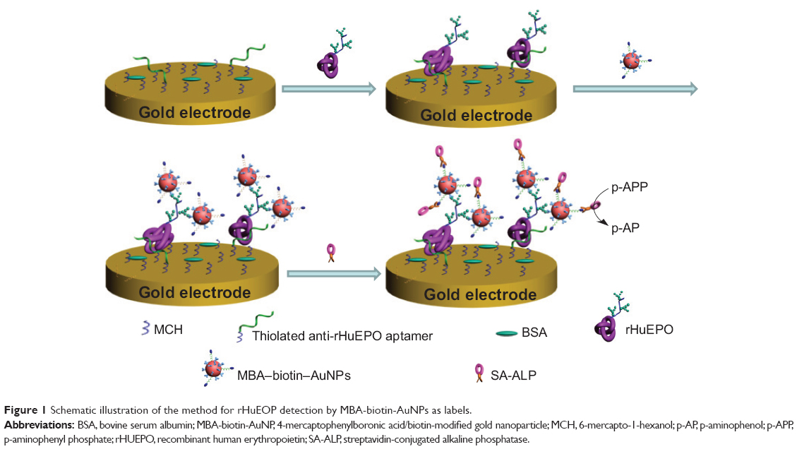 | Figure 1 Schematic illustration of the method for rHuEOP detection by MBA-biotin-AuNPs as labels. |
Characterization of MBA-biotin-AuNPs
Modification of AuNPs with multiple functionalities can provide more flexibility for multiplexing in bioanalytical application. For example, Kong et al35 reported the colorimetric dopamine detection using MBA-DSP-AuNPs (AuNPs modified with MBA and dithiobis[succinimidylpropionate]). Such bifunctional AuNPs can doubly recognize dopamine by reacting with the diol and amine groups. AuNPs modified with CALNN peptide are water-soluble and extremely stable.36 Wang et al37 demonstrated that multifunctional AuNPs capped with CALNNGK(biotin)G peptide and DNA targets are readily obtained in one step through the ligand-exchange reaction. Such functionality facilitates SA to be absorbed on the surface of AuNPs through the strong biotin–SA interaction. In the present work, the bifunctional AuNPs capped with MBA and CALNNGK(biotin)G groups were prepared by the formation of the Au–S covalent bond. The synthesized MBA-biotin-AuNPs were characterized by UV-visible spectroscopy. As shown in Figure 2, the MBA-biotin-AuNPs displayed a characteristic UV-visible absorption spectrum with a plasmon band at 520 nm (curve b). Such absorption was ascribed to the surface plasmon resonance of the AuNPs (curve a). This demonstrated that the synthesized MBA-biotin-AuNPs were stable and monodisperse. The result was also confirmed by transmission electron microscopy, as shown in the inset of Figure 2.
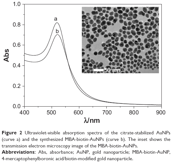 | Figure 2 Ultraviolet-visible absorption spectra of the citrate-stabilized AuNPs (curve a) and the synthesized MBA-biotin-AuNPs (curve b). The inset shows the transmission electron microscopy image of the MBA-biotin-AuNPs. |
Feasibility for rHuEPO detection
Electrochemical impedance spectroscopy has been used to examine the property of self-assembled monolayers, including surface coverage and monolayer composition, and the conduction ability of the modified electrode. Herein, the immobilization of the aptamers and the attachment of rHuEPO and modified gold nanoparticles were characterized by electrochemical impedance spectroscopy. A modified Randles equivalent circuit was used to fit the impedance spectra and to determine electrical parameters for each step. As shown in the inset of Figure 3A, the circuit included Rs (the electrolyte resistance between working and reference electrodes), Zw (the Warburg impedance), Q (a constant phase element representing the double layer capacitance for an unmodified electrode or the capacitance of the self-assembled monolayers for the modified electrodes) and Ret (the electron-transfer resistance). As shown in Figure 3A, the bare gold electrode showed a small electron transfer resistance (curve a), demonstrating a fast electron transfer process. The increase in the Ret implied the immobilization of anti-rHuEPO aptamers (curve b), and the result can be explained by the fact that the negatively charged aptamers repulsed the negatively charged [Fe(CN)6]3-/4- from the sensor surface. The modification of Au/aptamers with MCH and BSA resulted in an increase in the Ret (curve c). The result is understandable, since the block of unreacted gold surface by the MCH and BSA could retard the interfacial electron transfer kinetics of [Fe(CN)6]3-/4- anions. Interestingly, we found that incubation of the aptamer-modified electrode with rHuEPO solution led to a small increase in the Ret (curve d), indicating that rHuEPO was captured by the sensing electrode. In turn, we found that the charge-transfer impedance decreased with the attachment of MBA-biotin-AuNPs (curve e), which was attributed to the high conducting capability of AuNPs.
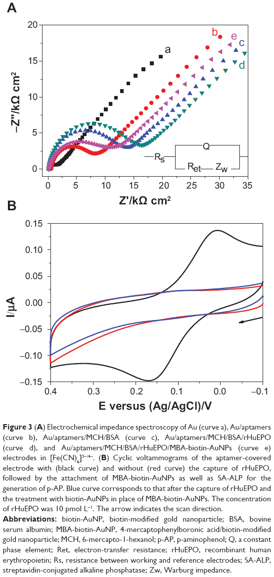 | Figure 3 (A) Electrochemical impedance spectroscopy of Au (curve a), Au/aptamers (curve b), Au/aptamers/MCH/BSA (curve c), Au/aptamers/MCH/BSA/rHuEPO (curve d), and Au/aptamers/MCH/BSA/rHuEPO/MBA-biotin-AuNPs (curve e) electrodes in [Fe(CN)6]3-/4-. (B) Cyclic voltammograms of the aptamer-covered electrode with (black curve) and without (red curve) the capture of rHuEPO, followed by the attachment of MBA-biotin-AuNPs as well as SA-ALP for the generation of p-AP. Blue curve corresponds to that after the capture of rHuEPO and the treatment with biotin-AuNPs in place of MBA-biotin-AuNPs. The concentration of rHuEPO was 10 pmol L-1. The arrow indicates the scan direction. |
Figure 3B shows the cyclic voltammograms of the aptamer-covered electrode with and without incubation with rHuEPO, followed by the attachment of MBA-biotin-AuNPs and the follow-up capture of SA-ALP for the production of p-AP. The voltammetric response in the black curve was caused by the oxidization of the generated p-AP. In the case of absence of the rHuEPO capture step, no redox peaks were observed (red curve). The result indicated that the attachment of MBA-biotin-AuNPs is dependent on the capture of rHuEPO. This is understandable since phenylboronic acid functionalized materials show no affinity to DAN and BSA.26,38 For the control, biotin-AuNPs were used in place of MBA-biotin-AuNPs. The absence of redox peaks in the blue curve confirmed that the attachment of AuNPs is strongly dependent upon the boronic acid-carbohydrate. Therefore, the method is feasible for the detection of rHuEPO. Moreover, we also investigated the influence of enzyme reaction time on the current, and found that the oxidation current increased with the reaction time and began to level off after 20 minutes. Thus, the sensor electrode was incubated in the p-APP solution for 20 minutes before electrochemical measurement.
Sensitivity and selectivity to rHuEPO
Differential pulse voltammetry is more sensitive than cyclic voltammetry, since it can decrease the background charging currents. Furthermore, we assessed the linear range, detection limit, and selectivity of this method with differential pulse voltammetry. The differential pulse voltammograms obtained at different rHuEPO concentrations are shown in Figure 4A. As a result, we found that the current increased linearly with the rHuEPO concentration in the range of 0.02–2.0 pmol L-1 and began to level off beyond 2 pmol L-1 (Figure 4B). All the relative standard deviations were found to be below 9.5%. The linear regression equation was expressed as Ipa (μA) =0.038+0.14 [rHuEPO] (pmol L-1) (R2=0.99). The limit of detection was estimated to be 8 fmol L-1, which is at least one order of magnitude lower than those achievable by mass spectrometry and spectroscopy (Table 1). The lower detection limit can be attributed to the multiplex amplification of AuNPs and the high turnover frequency of ALP.19 Moreover, this value is comparable to (or even lower than) those achievable using other electrochemical strategies for detection of glycoproteins including PSA (1.6 pg mL-1),17 sialylated glycoproteins fetuin (0.33 fM) and asialofetuin (0.54 fM),39 lactoferrin (145 pg mL-1),40 α-fetoprotein (4 pg mL-1),41 and ovalbumin (0.83 pg mL-1).42
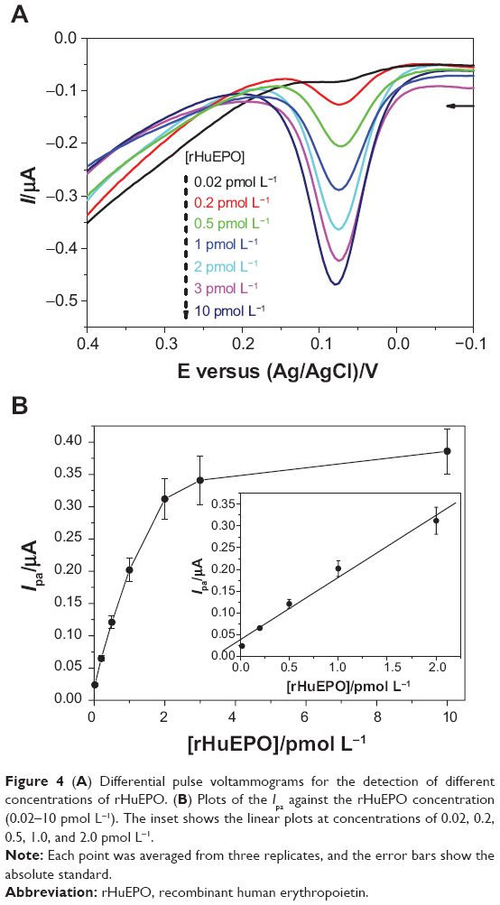 | Figure 4 (A) Differential pulse voltammograms for the detection of different concentrations of rHuEPO. (B) Plots of the Ipa against the rHuEPO concentration (0.02–10 pmol L-1). The inset shows the linear plots at concentrations of 0.02, 0.2, 0.5, 1.0, and 2.0 pmol L-1. |
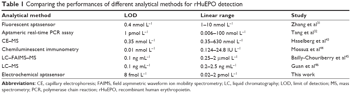 | Table 1 Comparing the performances of different analytical methods for rHuEPO detection |
To demonstrate the selectivity of this sensor, we tested other proteins (glycoproteins and nonglycoproteins), such as horseradish peroxidase, PSA, metallothionein, SA, and thrombin, at a concentration ten times higher than that of rHuEPO. As shown in Figure 5, the negligible currents in these cases are indicative of the high selectivity of the method to rHuEPO. Furthermore, to investigate the interference, the sensor electrode was incubated with the rHuEPO solution containing above tested five interfering proteins. As a result, no apparent difference in the current was observed in the cases of absence and presence of these interferences, further verifying that the aptamer was highly specific to rHuEPO. Moreover, in the spectrum analysis, the protein detection is usually disturbed by the strong light scattering effect of serum. To demonstrate the viability of the sensor in biomedical samples, we examined its performance in the solution of blood serum. As a result, the current in 10% serum was the same as in the buffer, demonstrating that a serum environment shows no interference in the detection assay. Since phenylboronic acid can react with various carbohydrates, phenylboronic acid-based materials have been widely used in the selective separation of glycoproteins such as lactoferrin, horseradish peroxidase, ribonuclease B, α-acid glycoprotein, α-fetoprotein, and ovalbumin.21 We believe that the strategy developed here would also be suitable for the sensitive and selective detection of these glycoproteins on electrodes covered with the matched capture probes. Moreover, compared with mass spectrometry and spectroscopy, our method is simple, selective, and cost-effective. For example, in the mass spectrometric assay, sample cleanup and preconcentration (eg, analyte extraction and enrichment by immunoaffinity separation, with antibodies and dual digestion by trypsin as well as peptide-N/O-glycosidase) is required considering the complex sample matrix and low concentrations of glycoproteins in biological fluids.46
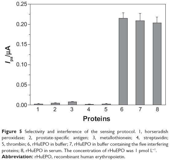 | Figure 5 Selectivity and interference of the sensing protocol. 1, horseradish peroxidase; 2, prostate-specific antigen; 3, metallothionein; 4, streptavidin; 5, thrombin; 6, rHuEPO in buffer; 7, rHuEPO in buffer containing the five interfering proteins; 8, rHuEPO in serum. The concentration of rHuEPO was 1 pmol L-1. |
Conclusion
In conclusion, we have reported a sensitive and selective electrochemical strategy for glycoprotein detection. MBA-biotin-AuNPs were used for the recognition of glycoproteins and the follow-up attachment of SA-ALP. To demonstrate the feasibility and sensitivity, rHuEPO was tested as the model. The detection limit of 8 fmol L-1 is at least one order of magnitude lower than those achievable by other techniques. Compared with other sandwich-type electrochemical biosensors, the presented approach obviated the use of expensive antibodies for the capture and recognition of targets. We believe that the strategy would find many detection applications for various glycoproteins by rationally designing the surface chemistry of electrodes.
Acknowledgments
Partial support of this work by the National Natural Science Foundation of China (Nos. 21205003, 21305004), the Joint Fund for Fostering Talents of National Natural Science Foundation of Henan Province (U1304205), and the Science and Technology Foundation of Henan Province (14A150042) is gratefully acknowledged.
Disclosure
The authors report no conflicts of interest in this work.
References
Spiro RG. Protein glycosylation: nature, distribution, enzymatic formation, and disease implications of glycopeptide bonds. Glycobiology. 2002;12:43R–56R. | ||
Hart GW, Copeland RJ. Glycomics hits the big time. Cell. 2010;143: 672–676. | ||
Park S, Kim W, Kim Y, et al. Array-based analysis of secreted glycoproteins for rapid selection of a single cell producing a glycoprotein with desired glycosylation. Anal Chem. 2010;82:5830–5837. | ||
Chaubard J-L, Krishnamurthy C, Yi W, Smith DF, Hsieh-Wilson LC. Chemoenzymatic probes for detecting and imaging fucose-α(1-2)-galactose glycan biomarkers. J Am Chem Soc. 2012;134:4489–4492. | ||
Imperiali B. The chemistry-glycobiology frontier. J Am Chem Soc. 2012;134:17835–17839. | ||
Zamfir A, Vakhrushev S, Sterling A, et al. Fully automated chip-based mass spectrometry for complex carbohydrate system analysis. Anal Chem. 2004;76:2046–2054. | ||
Haiber S, Herzog H, Burba P, Gosciniak B, Lambert J. Quantification of carbohydrate structures in size fractionated aquatic humic substances by two-dimensional nuclear magnetic resonance. Fresen J Anal Chem. 2001;369:457–460. | ||
Lu Y, Li XC, Zhang LM, et al. Aptamer-based electrochemical sensors with aptamer-complementary DNA oligonucleotides as probe. Anal Chem. 2008;80:1883–1890. | ||
Tang DP, Yuan R, Chal YQ. Ultrasensitive electrochemical immunosensor for clinical immunoassay using thionine-doped magnetic gold nanospheres as labels and horseradish peroxidase as enhancer. Anal Chem. 2008;80:1582–1588. | ||
Lai GS, Yan F, Wu J, Leng CA, Ju HX. Ultrasensitive multiplexed immunoassay with electrochemical stripping analysis of silver nanoparticles catalytically deposited by gold nanoparticles and enzymatic reaction. Anal Chem. 2011;83:2726–2732. | ||
Xiong P, Gan N, Cao Y, et al. An ultrasensitive electrochemical immunosensor for alpha-fetoprotein using an envision complex-antibody copolymer as a sensitive label. Materials. 2012;5:2757–2772. | ||
Song S, Xu H, Fan C. Potential diagnostic applications of biosensors: current and future directions. Int J Nanomed. 2006;1:433–440. | ||
Pruneanu S, Pogacean F, Biris AR, et al. Electro-catalytic properties of graphene composites containing gold or silver nanoparticles. Electrochim Acta. 2013;89:246–252. | ||
Saha K, Agasti SS, Kim C, Li XN, Rotello VM. Gold nanoparticles in chemical and biological sensing. Chem Rev. 2012;112:2739–2779. | ||
Zhou HK, Gan N, Li TH, et al. The sandwich-type electrochemiluminescence immunosensor for alpha-fetoprotein based on enrichment by Fe3O4-Au magnetic nano probes and signal amplification by CdS-Au composite nanoparticles labeled anti-AFP. Anal Chim Acta. 2012;746: 107–113. | ||
Chin YT, Liao EC, Wu CC, Wang GJ, Tsai JJ. Label-free detection of single-nucleotide polymorphisms associated with myeloid differentiation-2 using a nanostructured biosensor. Biosens Bioelectron. 2013;49:506–511. | ||
Xia N, Deng D, Zhang L, et al. Sandwich-type electrochemical biosensor for glycoproteins detection based on dual-amplification of boronic acid-gold nanoparticles and dopamine-gold nanoparticles. Biosens Bioelectron. 2013;43:155–159. | ||
Liu SN, Wu P, Li W, Zhang H, Cai CX. Ultrasensitive and selective electrochemical identification of hepatitis C virus genotype 1b based on specific endonuclease combined with gold nanoparticles signal amplification. Anal Chem. 2011;12:4752–4758. | ||
Nam EJ, Kim EJ, Wark AW, et al. Highly sensitive electrochemical detection of proteins using aptamer-coated gold nanoparticles and surface enzyme reactions. Analyst. 2012;137:2011–2016. | ||
Bertok T, Klukova L, Sediva A, et al. Ultrasensitive impedimetric lectin biosensors with efficient antifouling properties applied in glycoprofiling of human serum samples. Anal Chem. 2013;85:7324–7332. | ||
Wang X, Xia N, Liu L. Boronic acid-based approach for separation and immobilization of glycoproteins and its application in sensing. Int J Mol Sci. 2013;14:20890–20912. | ||
Jin S, Cheng Y, Reid S, Li M, Wang B. Carbohydrate recognition by boronolectins, small molecules, and lectins. Med Res Rev. 2010;30: 171–257. | ||
Siegel D. Applications of reversible covalent chemistry in analytical sample preparation. Analyst. 2012;137:5457–5482. | ||
Piest M, Engbersen JFJ. Role of boronic acid moieties in poly(amido amine)s for gene delivery. J Control Release. 2011;155:331–340. | ||
Li L, Lu Y, Bie Z, Chen H-Y, Liu Z. Photolithographic boronate affinity molecular imprinting: a general and facile approach for glycoprotein imprinting. Angew Chem Int Ed. 2013;52:7451–7454. | ||
Nie H, Chen Y, Lü C, Liu Z. Efficient selection of glycoprotein-binding DNA aptamers via boronate affinity monolithic capillary. Anal Chem. 2013;85:8277–8283. | ||
Xu Y, Zhang L, Lu H, Yang P. On-plate enrichment of glycopeptides by using boronic acid functionalized gold-coated Si wafer. Proteomics. 2010;10:1079–1086. | ||
Steel AB, Herne TM, Tarlov MJ. Electrochemical quantitation of DNA immobilized on gold. Anal Chem. 1998;70:4670–4677. | ||
Farjami E, Campos R, Nielsen JS, et al. RNA aptamer-based electrochemical biosensor for selective and label-free analysis of dopamine. Anal Chem. 2013;85:121–128. | ||
Zhang Z, Guo L, Guo A, et al. In vitro lectin-mediated selection and characterization of rHuEPO-α-binding ssDNA aptamers. Bioorg Med Chem. 2010;18:8016–8025. | ||
Zhang Z, Guo L, Tang J, Guo X, Xie J. An aptameric molecular beacon-based “Signal-on” approach for rapid determination of rHuEPO-α. Talanta. 2009;80:985–990. | ||
Tang J, Guo L, Shen R, et al. Quantification of rHuEPO-α by magnetic beads-based aptameric real-time PCR assay. Analyst. 2010;135:2924–2929. | ||
Sun J, Guo A, Zhang Z, Guo L, Xie J. A conjugated aptamer-gold nanoparticle fluorescent probe for highly sensitive detection of rHuEPO-α. Sensors. 2011;11:10490–10501. | ||
Arai T, Nishijo T, Matsumae Y, et al. Noninvasive measurement of alkaline phosphatase activity in embryoid bodies and coculture spheroids with scanning electrochemical microscopy. Anal Chem. 2013;85:9647–9654. | ||
Kong B, Zhu A, Luo Y, et al. Sensitive and selective colorimetric visualization of cerebral dopamine based on double molecular recognition. Angew Chem Int Ed. 2011;50:1837–1840. | ||
Lévy R, Thanh NTK, Christopher Doty R, et al. Rational and combinatorial design of peptide capping ligands for gold nanoparticles. J Am Chem Soc. 2004;126:10076–10084. | ||
Wang Z, Lévy R, Fernig DG, Brust M. The peptide route to multifunctional gold nanoparticles. Bioconjugate Chem. 2005;16:497–500. | ||
Sørensen MD, Martins R, Hindsgaul O. Assessing the terminal glycosylation of a glycoprotein by the naked eye. Angew Chem Int Ed. 2007;46:2403–2407. | ||
Bertok T, Gemeiner P, Mikula M, Gemeiner P, Tkac J. Ultrasensitive impedimetric lectin based biosensor for glycoproteins containing sialic acid. Microchim Acta. 2013;180:151–159. | ||
Pan Y, Sonn GA, Sin MLY, et al. Electrochemical immunosensor detection of urinary lactoferrin in clinical samples for urinary tract infection diagnosis. Biosens Bioelectron. 2010;26:649–654. | ||
Wang H, Li H, Zhang Y, et al. Label-free immunosensor based on Pd nanoplates for amperometric immunoassay of alpha-fetoprotein. Biosens Bioelectron. 2014;53:305–309. | ||
Eissa S, L’Hocine L, Siaj M, Zouro M. A graphene-based label-free voltammetric immunosensor for sensitive detection of the egg allergen ovalbumin. Analyst. 2013;138:4378–4384. | ||
Haselberg R, de Jong GJ, Somsen GW. Low-flow sheathless capillary electrophoresis-mass spectrometry for sensitive glycoform profiling of intact pharmaceutical proteins. Anal Chem. 2013;85:2289–2296. | ||
Mossuz P, Girodon F, Hermouet S, et al. Serum erythropoietin measured by chemiluminescent immunometric assay: an accurate diagnostic test for absolute erythrocytosis. Clin Chem. 2005;51:1018–1021. | ||
Bailly-Chouriberry L, Cormant F, Garcia P, et al. A new analytical method based on anti-EPO monolith column and LC-FAIMS-MS/MS for the detection of rHuEPOs in horse plasma and urine samples. Analyst. 2012;137:2445–2453. | ||
Guan F, Uboh CE, Soma LR, et al. LC-MS/MS method for confirmation of recombinant human erythropoietin and darbepoetin a in equine plasma. Anal Chem. 2007;79:4627–4635. |
 © 2014 The Author(s). This work is published and licensed by Dove Medical Press Limited. The
full terms of this license are available at https://www.dovepress.com/terms
and incorporate the Creative Commons Attribution
- Non Commercial (unported, 3.0) License.
By accessing the work you hereby accept the Terms. Non-commercial uses of the work are permitted
without any further permission from Dove Medical Press Limited, provided the work is properly
attributed. For permission for commercial use of this work, please see paragraphs 4.2 and 5 of our Terms.
© 2014 The Author(s). This work is published and licensed by Dove Medical Press Limited. The
full terms of this license are available at https://www.dovepress.com/terms
and incorporate the Creative Commons Attribution
- Non Commercial (unported, 3.0) License.
By accessing the work you hereby accept the Terms. Non-commercial uses of the work are permitted
without any further permission from Dove Medical Press Limited, provided the work is properly
attributed. For permission for commercial use of this work, please see paragraphs 4.2 and 5 of our Terms.