
Back to Journals » Clinical Ophthalmology » Volume 20
Amniotic Membrane Transplantation versus Tarsorrhaphy for the Treatment of Persistent Corneal Epithelial Defects: A Comparative Study in a Tertiary Eye Care Facility in Riyadh, Saudi Arabia
Authors Alasqah AA, AlRashedi M, AlDihan KA, AlSulaiman HM, Hawsawi R
Received 11 November 2025
Accepted for publication 11 February 2026
Published 27 March 2026 Volume 2026:20 580763
DOI https://doi.org/10.2147/OPTH.S580763
Checked for plagiarism Yes
Review by Single anonymous peer review
Peer reviewer comments 3
Editor who approved publication: Dr Scott Fraser
Abdulrahman Abdullah Alasqah,1 Musa AlRashedi,2 Khalid A AlDihan,3 Hamad M AlSulaiman,4 Rawan Hawsawi1
1Anterior Segment Department, King Khaled Eye Specialist Hospital, Riyadh, Saudi Arabia; 2Retina Division, King Khaled Eye Specialist Hospital, Riyadh, Saudi Arabia; 3Glaucoma Division, King Khaled Eye Specialist Hospital, Riyadh, Saudi Arabia; 4Oculoplastics division, King Khaled Eye Specialist Hospital, Riyadh, Saudi Arabia
Correspondence: Abdulrahman Abdullah Alasqah, Department of Anterior Segment, King Khaled Eye Specialist Hospital, Al Urubah Road, Umm Al Hamam Al Gharbi, Riyadh, 11462, Saudi Arabia, Email [email protected]
Background: To evaluate the effectiveness and safety of amniotic membrane transplantation (AMT) compared to tarsorrhaphy for treating persistent corneal epithelial defects (PED) resistant to medical therapy. The primary outcome was complete healing at 1, 2, 3, and 6 months. Secondary outcomes included healing time, recurrence rates, and postoperative complications.
Methods: This retrospective study was conducted at King Khaled Eye Specialist Hospital (KKESH), Riyadh, Saudi Arabia. We analyzed electronic medical records of patients treated from January 2018 to December 2021. Patients with PED (epithelial defect > 14 days) receiving either AMT or tarsorrhaphy were included. Data collected included demographics, etiology of PED, size and duration of the PED, type of intervention and postoperative outcomes. Complications assessed included recurrent PED, microbial keratitis, and corneal scarring.
Results: Of 67 eyes, 37 underwent AMT (median age 65) and 30 underwent tarsorrhaphy (median age 69). Baseline exposure keratopathy was more prevalent in the tarsorrhaphy group (40% vs. 2.7%; p< 0.001), while microbial keratitis was more common in the AMT group (57% vs. 20%; p=0.002). Healing rates at 1 month were 59% (22/37) for AMT and 50% (15/30) for tarsorrhaphy (p=0.43). By 6 months, healing rates were 86% (32/37) for AMT and 70% (21/30) for tarsorrhaphy (p=0.10). Complications included recurrent PED (2.7% for AMT vs. 6.7% for tarsorrhaphy; p=0.6) and microbial keratitis (2.7% in both groups; p=0.6). Corneal scarring was significantly more frequent in the tarsorrhaphy group (37% vs. 8.1%; p=0.004).
Conclusion: Both AMT and tarsorrhaphy successfully facilitated PED healing, with AMT demonstrating a numerically higher, though not statistically significant, healing rate (86% vs 70%). Tarsorrhaphy was associated with a higher incidence of corneal scarring, potentially linked to underlying exposure keratopathy. Future prospective investigations with standardized treatment assignments are warranted.
Keywords: persistent epithelial defect, amniotic membrane transplant, tarsorrhaphy, complications
Introduction
One of the commonest ophthalmic presentations is the corneal epithelial defect. Persistence of this defect for more than 14 days is labeled as persistent epithelial defect (PED).1,2 There are multiple etiologies to PED, including exposure keratopathy, limbal stem cell deficiency, infectious causes, keratoplasty, neurotrophic keratopathy, and severe dry eye syndrome.3–7 In the United States, the incidence of PED per year is estimated to be less than 200,000. Furthermore, the incidence of PED in patients who underwent keratoplasty and diabetic vitrectomy is estimated to be around 7500 and 5000 cases per year, respectively.2 If left untreated, PED can lead to potentially devastating complications, such as microbial keratitis, neovascularization, corneal scaring, and melting.1,2 PED is typically treated in a stepwise approach, that starts with medical treatment, including artificial tears autologous serum and whole-blood-derived products, anti-inflammatory medications, prophylactic antibiotics drops and bandage contact lenses.1,8 Surgical intervention is reserved for refractory cases, which includes debridement, amniotic membrane transplant (AMT) and tarsorrhaphy.9
Because of the noticeable lack of studies that compare the clinical outcome(s) of AMT and tarsorrhaphy, whether one surgical approach is superior to another is a question that remains to be answered. In this study, we aim to compare and study the clinical outcomes of AMT and tarsorrhaphy in the healing of PEDs.
Materials and Methods
Study Design and Settings
This retrospective comparative study is to compare and study the outcomes of AMT and tarsorrhaphy in the management of PED at King Khaled Eye Specialist Hospital (KKESH), Riyadh, Saudi Arabia between January 2018 and December 2021. Ethical approval obtained through the Institutional Review Board (IRB) at KKESH. The approval reference number is: RD/26001/IRB/0229-22. The requirement for informed patient consent was waived by the IRB due to the retrospective nature of the study. All patient data were kept confidential, and the study adhered to the tenets of the Declaration of Helsinki. The objective is to compare and study the clinical outcome between the two interventions in terms of PED healing rate.
Identification of Study Participants
We included both genders of Saudi patients aged 18 years and above who suffered from persistent epithelial defects after maximum medical therapy and were managed by AMT or tarsorrhaphy regardless of their past medical history or other surgeries. AMT is carried out by directly placing the cryopreserved graft over the defect, using sutures or fibrin glue while tarsorrhaphy is used to close the eyelids temporarily or permanently.
Data Collection Process
We collected data from the patient’s electronic medical records. The following variables were collected: age, gender, comorbid disorders (eg. diabetes and hypertension), previous ocular surgeries, the underlining etiology of PED, type of intervention (AMT, tarsorrhaphy). The following data were collected during the follow-up at 1,2,3 and 6 months: healed PED, unhealed PED and possible postoperative complications (microbial keratitis, or recurrent corneal epithelial defect). A corneal epithelial defect failing to re-epithelialize after 14 days of standard conservative therapy was labelled as PED. Standard conservative therapy consisted of conventional medical management (eg., preservative-free artificial tears, ointments, antibiotics, and autologous serum eye drops) administered at the discretion of the treating ophthalmologist. Healed PED was defined as complete epithelial healing determined using fluorescein staining.
Data Analysis
All data were analyzed using the R statistical software (version 4.3.0; R Foundation for Statistical Computing, Vienna, Austria). Descriptive statistics were calculated for all variables. Continuous variables were reported as medians with interquartile ranges (IQR), while categorical variables were reported as frequencies and percentages. The Mann–Whitney U-test was used to compare the medians between the amniotic membrane transplant (AMT) and temporary tarsorrhaphy (TT) groups. For categorical variables, the chi-square test or Fisher’s exact test were used, as appropriate, to evaluate the differences between the two groups. Multivariate regression models with backward elimination were employed to adjust for potential confounders associated with the study outcomes. A p-value of less than 0.05 was considered statistically significant. All p-values were two-sided.
Results
A total of 67 patients were included in this study. Of these, 37 underwent amniotic membrane transplantation (AMT), while 30 received tarsorrhaphy. The median age was 65 years (IQR: 54–74) in the AMT group and 69 years (IQR: 56–78) in the tarsorrhaphy group (p = 0.2). 23/37 (57%) and 24/30 (80%) were males in the AMT and tarsorrhaphy groups, respectively (p = 0.044). Regarding the etiology of PED, exposure keratopathy was significantly higher in the tarsorrhaphy group 12/30 (40%) compared to the amniotic membrane group 1/37 (2.7%) (p = < 0.001). Microbial Keratitis showed a significantly higher in the amniotic membrane group 21/37 (57%) compared to the tarsorrhaphy group 6/30 (20%) (p = 0.002). Other causes did not exhibit significant differences between the two groups. The median size of the corneal epithelial defect (PED) was 6 mm (IQR: 3–11) in the AMT group and 6 mm (IQR: 2–16) in the tarsorrhaphy group (p = 0.7) (Table 1). Most of the patients in the tarsorrhaphy group underwent a temporary procedure rather than a permanent one (80% vs. 20%). Healing of the epithelial defect is similar between the two groups with no statistical difference across the follow-up visits (p = > 0.05). Microbial keratitis occurred in 2.7% of patients in both the AMT and tarsorrhaphy groups. The rate of corneal scar formation was significantly higher in the tarsorrhaphy group (37%) compared to the AMT group (8.1%) (p = 0.004). No significant differences were observed in complications like microbial keratitis or recurrent PED (Table 2).
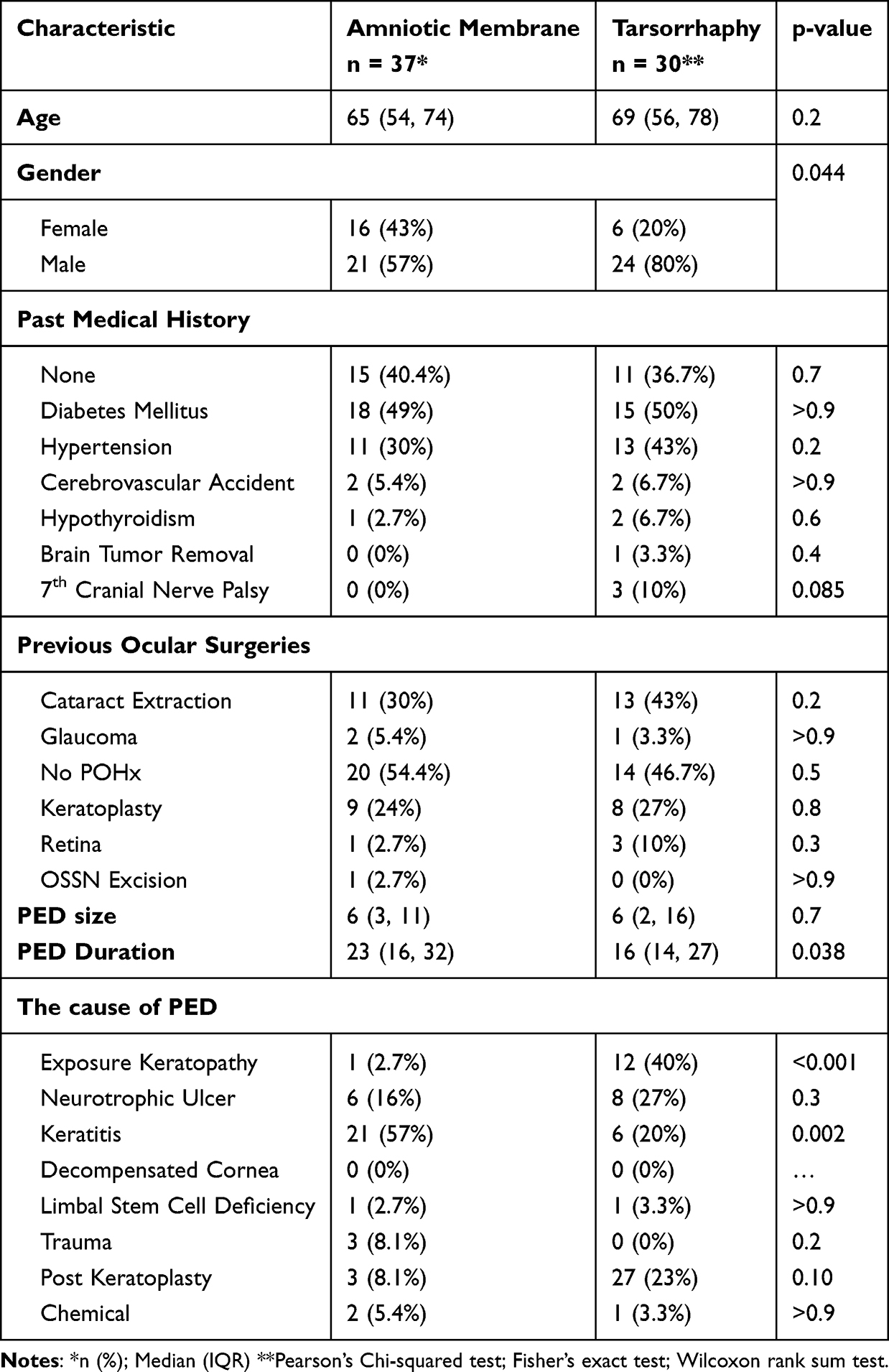 |
Table 1 Baseline Characteristics of and Amniotic Membrane Transplant and Tarsorrhaphy Groups (n=67) |
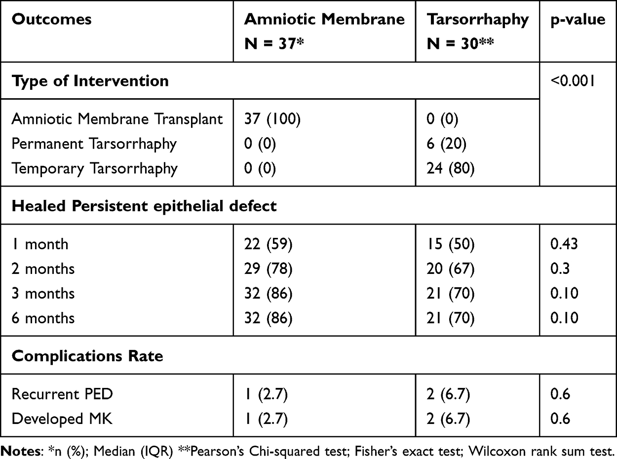 |
Table 2 Comparative Analysis for Eyes Undergoing Amniotic Membrane Transplant vs Tarsorrhaphy |
Multivariate analysis (Table 3) was performed to control for potential confounders, including age, gender, prior keratoplasty, PED size and duration, etiology (exposure keratopathy or keratitis), and surgical procedure (AMT vs tarsorrhaphy). Cox regression was utilized for the time-to-event outcome (healed PED) and binary logistic regression for the categorical outcomes of recurrent PED and MK development; all models were run with backward elimination. While none of the factors reached statistical significance at the p < 0.05 level, several factors demonstrated clinically relevant trends or borderline significance, suggesting potential associations with PED healing, recurrence, and MK development. Regarding PED healing, both PED size (p = 0.189) and surgical procedure (p = 0.194) emerged as clinically relevant factors. Healing occurred in 88.9% (32/36) of cases with a size ≤6.0mm compared to 67.7% (21/31) for those >6.0mm. Similarly, healing rates were 86.5% (32/37) for the AMT group versus 70.0% (21/30) for the tarsorrhaphy group. Kaplan–Meier analysis of the cumulative probability of PED healing is illustrated in Figure 1. The median time to healing was 1 month for AMT group compared to 2 months for the tarsorrhaphy group. Although AMT showed a shorter median recovery time, the difference in healing rates between the two procedures did not reach statistical significance over the follow-up period (log-rank p = 0.083). For PED recurrence, age approached statistical significance (p = 0.053), with recurrence noted in 6.1% (2/33) of patients >68.0 years compared to 2.9% (1/34) in those ≤67.0 years. Finally, prior keratoplasty showed a trend toward an association with MK development (p = 0.093), occurring in 11.8% (2/17) of patients with a history of keratoplasty versus 2.0% (1/50) in those without.
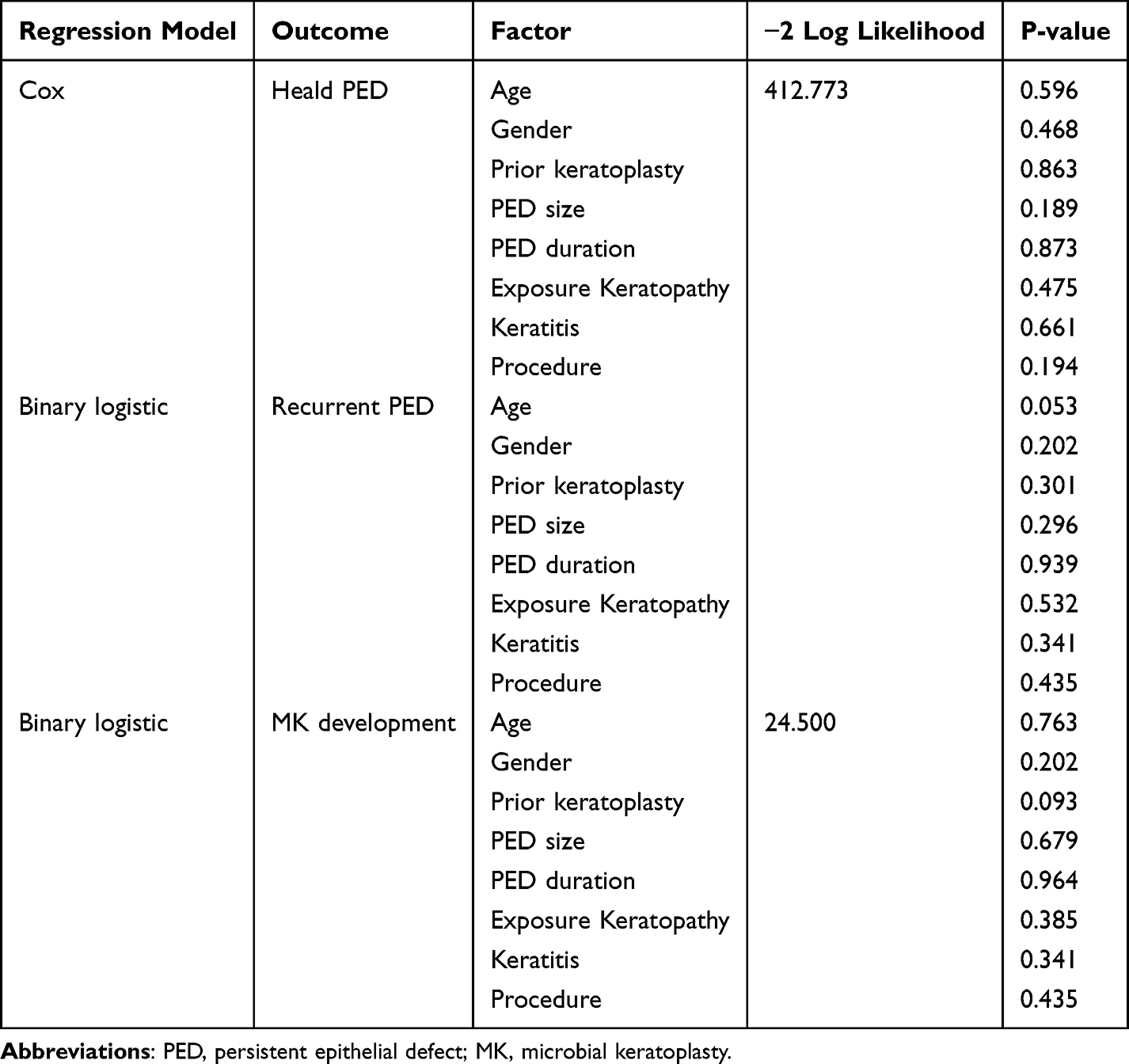 |
Table 3 Multivariate Regression Analysis of Factors for Persistent Epithelial Defect Healing, Recurrence, and Development of Microbial Keratitis |
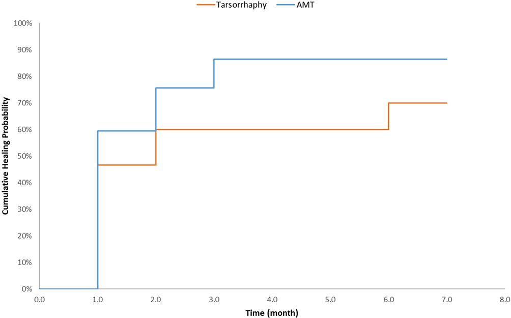 |
Figure 1 Cumulative healing rates of persistent epithelial defect over time by surgical intervention. |
Discussion
Under normal circumstances, the cornea resurfaces epithelial defects within approximately seven days. However, factors such as limbal stem cell deficiency, exposure keratopathy, and neurotrophic keratopathy can impede this process, resulting in persistent epithelial defects (PEDs) that fail to heal within two weeks despite conventional therapy.10,11 This study evaluates and compares the efficacy of two surgical interventions—amniotic membrane transplantation (AMT) and tarsorrhaphy—in managing PEDs, focusing on healing rates and associated outcomes.
Tarsorrhaphy, whether temporary or permanent, is a well-established intervention that minimizes corneal exposure, facilitating repair even in challenging conditions.11 Conversely, AMT is recognized for its role in ocular surface reconstruction, particularly in corneal pathologies like PED.1,11,12 This study aimed to compare the outcomes of these two approaches, with a sub-analysis of combined cases, to determine their relative effectiveness in promoting PED healing.
In our study, the median duration of PED was 23 days in the AMT group and 16 days in the tarsorrhaphy group. This aligns closely with findings by Dhillon HK et al, who reported a mean PED duration of 20.1 days.9 In contrast, Prabhasawat et al documented a longer duration of approximately 5.45 weeks, while Saleem T et al reported an even more extended mean of 7.67 weeks for AMT-treated patients.12,13 These variations may reflect differences in patient demographics, PED severity, or treatment protocols across studies.
The median PED size in our study was 6 mm for both groups, which is smaller than the 26 mm reported by Dhillon HK et al but larger than the 3 mm noted by Prabhasawat et al.9,12 This discrepancy suggests variability in the baseline characteristics of PEDs across cohorts, potentially influencing healing outcomes. Etiologically, our study identified exposure keratopathy (13 eyes), microbial keratitis (27 eyes), and neurotrophic ulcers (14 eyes) as predominant causes. Comparatively, Dhillon HK et al reported exposure keratopathy in 15 eyes, post-keratoplasty defects in 32, trauma in 5, and idiopathic cases in 5, while Saleem T et al found bacterial keratitis (17.2%) and vegetative trauma (13.8%) as leading causes. These differences highlight the multifactorial nature of PED and its context-specific etiologies.
At the six-month follow-up, healing rates were 86% (32/37) in the AMT group and 70% (21/30) in the tarsorrhaphy group. Although this difference appears clinically meaningful, it did not reach statistical significance, likely due to inadequate statistical power given the study’s sample size. Dhillon HK et al reported higher healing rates at one month—76.7% for AMT and 90% for tarsorrhaphy—whereas Saleem T et al observed a 91.4% success rate with AMT.9,13 Mimouni M et al, in a smaller cohort of nine eyes treated with dehydrated AMT, achieved an 89% resolution rate14 Additionally, Ozcan AA et al reported complete healing in all cases using tarsorrhaphy combined with sutureless AMT, and Shuja D et al noted an 85% resolution rate with tarsorrhaphy alone by the fourth week.15,16 Notably, our study’s extended follow-up period (six months) contrasts with the one-month endpoint in these prior studies, potentially accounting for differences in reported outcomes.
Multivariate analysis provided further insight into factors influencing these outcomes. Notably, larger PED size (>6 mm) showed a trend toward reduced healing rates, consistent with the biological understanding that extensive defects require longer migration times for epithelial coverage.17,18 Additionally, our analysis indicated that AMT may offer a faster recovery trajectory, with a median healing time of one month compared to two months for tarsorrhaphy. Although the overall healing difference did not reach statistical significance (log-rank p = 0.083), this acceleration in epithelial closure is clinically relevant for reducing patient morbidity. We also observed a trend associating older age with PED recurrence and prior keratoplasty with microbial keratitis; these findings align with the age-related decline in corneal regenerative capacity and the chronically compromised ocular surface in transplant recipients.18,19,20
Complication rates in our study showed no significant difference between groups, with microbial keratitis occurring in 2.2% of patients in both the AMT and tarsorrhaphy cohorts. This contrasts with Mimouni M et al, who reported no recurrences or complications with dehydrated AMT.14 Shuja D et al noted that three of 21 tarsorrhaphy-treated patients failed to heal, requiring subsequent AMT.16 Regarding long-term sequelae, a higher rate of corneal scarring was observed in the tarsorrhaphy group; however, this finding must be interpreted with caution. Tarsorrhaphy is frequently the primary intervention for exposure keratopathy, a condition inherently prone to stromal damage and scarring due to chronic desiccation.17–22 Therefore, the increased scarring likely reflects the severity of the underlying etiology rather than a direct consequence of the surgical technique.
Limitation
The retrospective design limits causal inference, though it enabled a larger sample size. Variability in PED etiology, patient demographics, and medication compliance may have influenced outcomes, suggesting a need for prospective, controlled studies. Furthermore, patient selection based on individual clinician judgment may have introduced selection bias, potentially influencing the study results.
Conclusion
Both AMT and tarsorrhaphy are effective for PED, with AMT showing a clinically meaningful, though not statistically significant, trend toward higher healing rates (86% vs. 70%). Multivariate analysis indicates that larger defect size hinders healing, while AMT may provide faster recovery. The higher scarring rate in the tarsorrhaphy group likely reflects the underlying severity of exposure keratopathy rather than the procedure itself. Given the variability in PED characteristics across studies, future research requires standardized protocols. Ultimately, treatment selection should be tailored to the specific etiology and patient needs. Our findings highlight the value of long-term follow-up, but larger prospective studies are needed to confirm the optimal approach.
Funding
This research was funded by King Khaled Eye Specialist Hospital & Research Center (KKESH&RC), Riyadh, Saudi Arabia.
Disclosure
The authors report no conflicts of interest in this work.
References
1. Vaidyanathan U, Hopping GC, Liu HY, et al. Persistent corneal epithelial defects: a review article. Med Hypothesis Discov Innov Ophthalmol. 2019;8(3):163.
2. Wilson SE, Medeiros CS, Santhiago MR. Pathophysiology of corneal scarring in persistent epithelial defects after PRK and other corneal injuries. J Refract Surg. 2018;34(1):59–8. doi:10.3928/1081597X-20171128-01
3. Wirostko B, Rafii M, Sullivan DA, Morelli J, Ding J. Novel therapy to treat corneal epithelial defects: a hypothesis with growth hormone. Ocul Surf. 2015;13(3):204–212.e1. doi:10.1016/j.jtos.2014.12.005
4. Ahmad S. Concise review: limbal stem cell deficiency, dysfunction, and distress. Stem Cells Transl Med. 2012;1(2):110–115. doi:10.5966/sctm.2011-0037
5. Shaikh S, Ta CN. Evaluation and management of herpes zoster ophthalmicus. Am Fam Physician. 2002;66(9):1723–1730.
6. Bonini S, Rama P, Olzi D, Lambiase A. Neurotrophic keratitis. Eye. 2003;17(8):989–995. doi:10.1038/sj.eye.6700616
7. Rumelt S, Bersudsky V, Blum-Hareuveni T, Rehany U. Persistent epithelial defects and ulcers in repeated corneal transplantation: incidence, causative agents, predisposing factors and treatment outcomes. Graefes Arch Clin Exp Ophthalmol. 2008;246(8):1139–1145. doi:10.1007/s00417-008-0797-4
8. Young AL, Cheng ACO, Ng HK, Cheng LL, Leung GYS, Lam DSC. The use of autologous serum tears in persistent corneal epithelial defects. Eye. 2004;18(6):609–614. doi:10.1038/sj.eye.6700721
9. Dhillon H, Bahadur H, Raj A. A comparative study of tarsorrhaphy and amniotic membrane transplantation in the healing of persistent corneal epithelial defects. Indian J Ophthalmol. 2020;68(1):29. doi:10.4103/ijo.IJO_617_19
10. Moin M, Qayyum I, Ahmad AU, Hussain M. Role of temporary tarsorrhaphy using super glue in the management of corneal disorders. Pak J Ophthalmol. 2009;25(3):139–144.
11. Katzman LR, Jeng BH. Management strategies for persistent epithelial defects of the cornea. Saudi J Ophthalmol. 2014;28(3):168–172. doi:10.1016/J.SJOPT.2014.06.011
12. Prabhasawat P, Tesavibul N, Komolsuradej W. Single and multilayer amniotic membrane transplantation for persistent corneal epithelial defect with and without stromal thinning and perforation. Br J Ophthalmol. 2001;85(12):1455–1463. doi:10.1136/BJO.85.12.1455
13. Saleem T, Bokhari SA. Outcome of amniotic membrane transplant in persistent corneal epithelial defects. Pak J Ophthalmol. 2022;38(1):52–57. doi:10.36351/PJO.V38I1.1340
14. Mimouni M, Trinh T, Sorkin N, et al. Sutureless dehydrated amniotic membrane for persistent epithelial defects. Eur J Ophthalmol. 2021;32(2):875–879. doi:10.1177/11206721211011354
15. Ozcan AA, Ulas B, Ciloglu E. Tarsorrhaphy with sutureless amnion transplantation: practical management of ocular surface pathologies. Beyoglu Eye J. 2022;7(4):313–319. doi:10.14744/BEJ.2022.10327
16. Shuja D, Rana J, Dwivedi K, Garg M. Role of tarsorrhaphy in healing of persistent epithelial defects in today’s era. IP Int J Ocular Oncol Oculoplasty. 2025;6(3):192–195. doi:10.18231/J.IJOOO.2020.043
17. Crosson CE, Klyce SD, Beuerman RW Epithelial wound closure in the rabbit cornea. A biphasic process. Invest Ophthalmol Vis Sci. 1986;27(4):464–473.
18. V. LA, Saghizadeh M Progress in corneal wound healing. Prog Retin Eye Res. 2015;49:17–45. doi:10.1016/J.PRETEYERES.2015.07.002
19. Sun JP, Chen WL, Huang JY, Hou YC, Wang IJ, Hu FR Microbial keratitis after penetrating keratoplasty. Am J Ophthalmol. 2017;178:150–156. doi:10.1016/J.AJO.2017.03.022
20. Vajpayee RB, Sharma N, Sinha R, Agarwal T, Singhvi A Infectious keratitis following keratoplasty. Surv Ophthalmol. 2007;52(1):1–12. doi:10.1016/J.SURVOPHTHAL.2006.10.001
21. Kousha O, Kousha Z, Paddle JJ Exposure keratopathy in critically ill adult patients: incidence and risk factors. Eye (Lond). 2018;32(5):928–932.
22. Kuruvilla S, Peter J, David S, et al. Incidence and risk factor evaluation of exposure keratopathy in critically ill patients: a cohort study. J. Crit. Care. 2015;30(2):400–404. doi:10.1016/J.JCRC.2014.10.009
 © 2026 The Author(s). This work is published and licensed by Dove Medical Press Limited. The
full terms of this license are available at https://www.dovepress.com/terms
and incorporate the Creative Commons Attribution
- Non Commercial (unported, 4.0) License.
By accessing the work you hereby accept the Terms. Non-commercial uses of the work are permitted
without any further permission from Dove Medical Press Limited, provided the work is properly
attributed. For permission for commercial use of this work, please see paragraphs 4.2 and 5 of our Terms.
© 2026 The Author(s). This work is published and licensed by Dove Medical Press Limited. The
full terms of this license are available at https://www.dovepress.com/terms
and incorporate the Creative Commons Attribution
- Non Commercial (unported, 4.0) License.
By accessing the work you hereby accept the Terms. Non-commercial uses of the work are permitted
without any further permission from Dove Medical Press Limited, provided the work is properly
attributed. For permission for commercial use of this work, please see paragraphs 4.2 and 5 of our Terms.