Back to Journals » International Journal of Women's Health » Volume 17
Aerobic Vaginitis Caused By Streptococcus &Bgr;- Hemolyticus and Staphylococcus Epidermidis in a 26-Year-Old Woman with a History of Frequent Antibiotics Administration
Authors Achdiat PA ![]() , Saraswati AL
, Saraswati AL ![]() , Handayani Y
, Handayani Y ![]() , Rowawi R, Maharani RH
, Rowawi R, Maharani RH ![]()
Received 17 January 2025
Accepted for publication 13 April 2025
Published 19 May 2025 Volume 2025:17 Pages 1437—1444
DOI https://doi.org/10.2147/IJWH.S516616
Checked for plagiarism Yes
Review by Single anonymous peer review
Peer reviewer comments 3
Editor who approved publication: Dr Vinay Kumar
Pati Aji Achdiat, Amalia Laksmi Saraswati, Yulisa Handayani, Rasmia Rowawi, Retno Hesty Maharani
Department of Dermatology and Venereology, Faculty of Medicine Universitas Padjadjaran – Dr. Hasan Sadikin Hospital, Bandung, West Java, Indonesia
Correspondence: Pati Aji Achdiat, Department of Dermatology and Venereology, Faculty of Medicine, Universitas Padjadjaran – Dr. Hasan Sadikin Hospital, Jl. Pasteur 38, Bandung, West Java, Indonesia, 40161, Email [email protected]
Abstract: Aerobic vaginitis (AV) is a form of vaginitis caused by the presence of aerobic bacteria, primarily group B Streptococcus, Escherichia coli, Enterococcus faecalis, Streptococcus viridans, and Staphylococcus epidermidis. This condition is characterized by clinical signs of acute inflammation, including vaginal redness, burning, increased vaginal secretions with a pH of 5, sticky vaginal discharge, leukocyte infiltration, and vulvar pruritus, which is defined as itching and irritation, and commonly occurs in individuals between the age of 15– 30 years and 31– 45 years. The report presents a case of AV caused by Streptococcus β-hemolyticus and Staphylococcus epidermidis in a 26-year-old female patient at Hasan Sadikin Hospital, Bandung. The treatment process includes the administration of amoxicillin 500 mg three times daily for 14 days. The patient was unmarried, routinely performed vaginal douching with betel soap, and frequently used a panty liner. Subsequently, a history of frequent antibiotic use was reported, however, the specific names of previously administered antibiotics remain unknown, except for clindamycin, which is the only agent specifically recalled. The patient also had a history of high exposure to stressors and underwent a bacterial culture examination taken from the vaginal introitus, which showed the presence of Streptococcus β-hemolyticus and Staphylococcus epidermidis. This case highlights the significance of AV in the differential diagnosis of vaginal infections, especially in atypical patient populations, and emphasizes the need for appropriate diagnostic measures such as bacterial culture to guide effective treatment strategies.
Keywords: aerobic vaginitis, streptococcus β-hemolyticus, staphylococcus epidermidis, aerobic vaginitis
Introduction
Aerobic vaginitis (AV) is a type of vaginitis related to aerobic bacteria, primarily including group B Streptococcus, Escherichia coli, Enterococcus faecalis, Streptococcus viridans, and Staphylococcus epidermidis. This condition is characterized by clinical symptoms of acute inflammation, such as vaginal redness, burning sensations, increased vaginal secretions with a pH of 5.5–6.5,1 sticky discharge, leukocyte infiltration, and vulvar itching as well as irritation. Subsequently, it commonly occurs in individuals between the age of 15–45 years.2–5 Vaginitis is a term used to describe a range of diseases presenting symptoms affecting the vulva and vagina. It is a leading cause of healthcare visits among women, accounting for an estimated 6 to 10 million consultations annually in the United States.6 This condition is not life-threatening, but it can reduce quality of life due to discomfort, pain, decreased sexual function, and self-confidence.7,8 AV is an important clinical problem, often misdiagnosed as bacterial vaginosis (BV), leading to inappropriate treatment with metronidazole, which is typically ineffective and may result in chronic infection. The condition is associated with various gynecological and obstetric complications, including pelvic inflammatory disease and an increased susceptibility to sexually transmitted infections. Moreover, AV is a well-documented cause of adverse pregnancy outcomes, such as preterm birth.9
The three primary infectious causes of vaginitis are BV, vulvovaginal candidiasis, and trichomoniasis.7,10 However, approximately 7–72% of women with vaginitis caused by abnormal vaginal flora other than BV are undiagnosed, and this phenomenon is often referred to as transitional flora.6 In 1999, Donders11 introduced the term “aerobic vaginitis (AV)” to describe this newly identified condition. It has been reported that AV occurs in 5% of all cases in specialized vaginal clinics.10 The incidence of this condition increases with age, onset of sexual activity, and parity.12 According to the study by Khan et al the disease commonly affects individuals within the age range of 15–30 years and 31–45 years. The cause of AV is aerobic bacteria, primarily Gram-positive bacteria (71%) including group β-hemolytic streptococci, staphylococci, and enterococci, as well as Gram-negative bacteria (29%), such as Enterobacteriaceae, particularly Escherichia coli.2,3 The following factors have been identified as independent risk factors for AV, such as the use of intrauterine devices, the presence of external hemorrhoids, prolonged antibiotic use, and frequent vaginal douching. Studies have also shown that stress induces vaginal dysbiosis, resulting in a reduction in the dominance of Lactobacillus species.13,14 A case of AV caused by Streptococcus β-hemolyticus and Staphylococcus epidermidis in a 26-year-old female patient who had never had sexual intercourse was reported.
Case Report
In May 2024, a 26-year-old unmarried woman visited a venereology clinic in a top referral hospital, Dr. Hasan Sadikin Hospital, Bandung, West Java, Indonesia, due to vaginal discharge persisting for one year. The discharge had a sour odor, a yellowish-white color, and a viscous consistency. Additionally, the patient experienced pruritic and burning sensations, which worsened in humid conditions. Notably, she frequently used panty liners and practiced vaginal douching with betel soap. Given these symptoms, the patient was referred from a General Hospital with a diagnosis of recurrent bacterial vaginosis (BV), while vulvovaginal candidiasis and trichomoniasis were considered differential diagnoses. An oral ketoconazole 200 mg was prescribed to be taken twice daily for seven days, along with a single 150 mg dose of oral fluconazole. Previously, treatment was carried out using Clindamycin 300 mg, taken twice daily for seven days. However, the symptoms did not improve, and the patient had previously undergone treatment for symptoms at a local clinic for one year. During this period, multiple antibiotic therapies were frequently administered, though the specific medications could not be identified, and no improvement was observed. There was no history of marriage, romantic relationships, or sexual activity. Before the onset of symptoms, significant stress was reported. While studying chemical engineering at a university, considerable academic difficulties were encountered, leading to psychiatric assistance. For the past six months, psychiatric medications have been administered, including 250 mg of divalproex sodium once daily and 20 mg of fluoxetine once daily, following a diagnosis of borderline personality disorder. Medical history also revealed an allergy to metronidazole.
During physical examination, the patient’s vital signs and overall condition were normal, and a dermatological examination showed no skin abnormalities. A venereological examination showed external genitalia within normal limit (Figure 1) but purulent discharge with a pH of 5 was observed at the vaginal and vulvar introitus, with a Nugent score of 5, and 3 of Amsel criteria. A Gram stain examination showed polymorphonuclear leukocytes (PMNs), Gram-positive rod-shaped bacteria, and Gram-positive cocci-shaped bacteria (Figure 2a and b). The patient was given clindamycin 300 mg, taken orally twice a day for seven days. Inspeculo examination could not be performed since the patient never had sexual intercourse, thereby bacterial culture of vaginal and vulval introitus swabs was performed. The sample of vaginal secretion was taken 2–3 cm from the vaginal introitus, and put into the swab transfer medium, and were examined in the microbiology laboratory. Furthermore, bacterial culture, fungal culture, Neisseria gonorrhoeae culture, microscopy, Christie–Atkins–Munch-Petersen test, and resistance test were carried out respectively. The culture was carried out on chocolate agar with a conclusion negative for Neisseria gonorrhoea, and blood agar medium which showed growth of three colonies such as Staphylococcus epidermidis, Lactobacillus sp., and β-hemolyticus Streptococcus (Figure 3). Staphylococcus epidermidis was identified using a conventional biochemical test. A catalase test was performed using a 3% H202 reagent, with a positive result for Staphylococcus species. To determine the species of Staphylococcus, a novobiocin test was conducted to differentiate between the epidermis and saprophytic species. The result was sensitive and an epidermis was obtained. Subsequent bacterial resistance testing for β-hemolyticus Streptococcus showed sensitivity to amoxicillin (33.3 mm), erythromycin (26.3 mm), clindamycin (22.9 mm), moxifloxacin (22.9 mm), and ciprofloxacin (21.7 mm) (Figure 4).
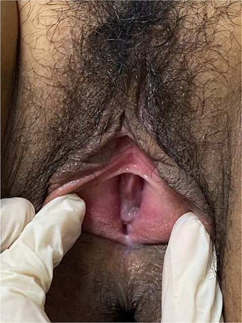 |
Figure 1 External genitalia showed no sign of inflammation. |
 |
Figure 2 (a) Gram staining from vaginal smears shows polymorphonuclear cells and bacteria, (b) Highlighting scant Gram-positive rod-shaped bacteria (yellow box), and numerous Gram-positive cocci-shaped bacteria (blue box). |
 |
Figure 3 Microscopic examination form bacterial culture specimen found (a) Staphylococcus aureus and β-hemolyticus Streptococcus (b) Lactobacillus sp. |
 |
Figure 4 Examination of antibiotic resistance in cultures of β-hemolyticus Streptococcus showed sensitivity to a) amoxicillin, b) clindamycin, and c) ciprofloxacin. |
Amoxicillin 500 mg was prescribed to be taken orally three times a day, for two weeks. On the 52nd day of observation after taking amoxicillin therapy, the patient reported that the vaginal discharge had disappeared and symptoms had improved.
Discussion
The term AV was first introduced by Donders et al15 in 2002 as a clinical entity distinct from BV. In AV, the transition from a healthy lactobacillus-dominated microbiota to vaginal dysbiosis is characterized by inflammatory symptoms, including vaginal redness, stinging and burning sensations, the presence of thick yellow vaginal discharge, and pain during intercourse. The vaginal discharge is uniform and pus-like, with a yellow or yellow-green appearance. It is accompanied by a rotting, malodorous smell, opposite to the BV with a fishy odor.4,16 Patient in this case report, complaining of yellowish-white discharge, sour smell, with pruritus and burning sensation. The signs of inflammation in the external genitalia have decreased, likely as a result of the prior prolonged use of antibiotics.
AV is defined as a disorder of the vaginal microflora (dysbiosis) involving aerobic bacteria, enteric bacteria, vaginal inflammation, and incomplete epithelial maturation.4 AV is a type of vaginitis linked to aerobic microorganisms, primarily group B streptococci and Escherichia coli. Furthermore, other studies have identified additional bacteria as contributors to AV, such as Enterococcus faecalis, Streptococcus viridans, and Staphylococcus epidermidis, which are frequently discovered in patients with AV. All of these microorganisms are aerobic bacteria.17 In this patient, Gram stain examination showed PMNs, Gram-positive rod-shaped bacterial rods, and Gram-positive bacterial cocci. The patient was then followed for microbiological culture examination of a smear taken from the vaginal introitus, which showed the presence of Lactobacillus sp., Streptococcus β-hemolyticus, and Staphylococcus epidermidis. The prevalence of Gram-positive bacteria is much higher than the number of Lactobacillus in this patient, coupled with complaints of vaginal discharge and changes in pH to alkaline, indicating abnormalities in the condition of the vagina. The Nugent test result also suggested BV, but the culture failed to find the causative bacteria, which indicated that the patient had AV.
The pathogenesis of AV consists interrelated pathogenic processes which involve: (1) A depletion of lactobacillus-dominated microflora, facilitating the adhesion and colonization of various urogenital pathogens, including group B Streptococcus and Staphylococcus aureus; (2) Dysregulation of local immune responses, characterized by a pronounced elevation in interleukin-1β (IL-1β) levels (178.8 pg/mL) compared to BV (71.2 pg/mL); (3) A marked increase in pro-inflammatory cytokines, such as interleukin-6 (IL-6) and interleukin-8 (IL-8), which is not typically observed in BV, and (4) Estrogen deficiency, inducing a shift in exfoliating epithelial cells from superficial towards intermediate cells, and in more severe cases, towards parabasal cells, as observed in AV.4,9
The following have been identified as distinct risk factors for AV, such as the use of intrauterine devices, external hemorrhoids, prolonged antibiotic use, regular vaginal douching, and stress. Stress, in particular, can contribute to vaginal dysbiosis by disrupting the dominance of Lactobacillus species.13,14 Stress exposure triggers the release of cortisol and norepinephrine through the hypothalamic-pituitary-adrenal and sympathetic-adrenal-medullary pathways. Cortisol suppresses the growth of vaginal epithelium, which is linked to estrogen maturation and glycogen buildup. This results in reduced levels of vaginal free glycogen and Lactobacilli, leading to a decrease in the production of lactic acid and hydrogen peroxide (H2O2), as well as a change in pH. Consequently, the dysbiotic environment promotes the growth of pathogenic bacteria.13 The risk factors associated with this case emerged during the patient’s academic studies. A significant increase in stress was reported, which coincided with the onset of vaginal discharge, suggesting a potential trigger for the initial symptoms. Additionally, a diagnosis of borderline personality disorder was made, and several psychiatric medications were prescribed.
Antibiotic treatment disrupts the microbial balance of the vaginal mucosa. In a healthy vaginal ecosystem, low bacterial diversity, primarily dominated by Lactobacillus species, is indicative of normal vaginal flora. In a study by Oh JE et al antibiotic treatment led to a notable reduction in bacterial count, primarily decreasing Gram-positive bacteria, in the vaginal mucosa.3 Douching disrupts the balance of vaginal microflora or triggers inflammation due to physical or chemical irritation, making women more susceptible to infections. Although most commercial douching products consist of fragrance, acetic acid, and water, some also contain surfactants such as oxtoxynol-9 or cetyl pyridinium chloride.18 Brotman et al reported that microbiological studies have demonstrated douching’s ability to change the composition of vaginal flora, but there are only a few reports, and they are limited to very short observation periods.12 Onderdonk et al, reported by Brotman et al found that douching with saline or acetic acid resulted in changes to the vaginal microflora within 10 minutes and that normal flora took up to 72 hours to return to pre-douching levels. Exposure to the bactericidal agent povidone-iodine in women resulted in significant short- and long-term alterations in the vaginal microflora. These changes facilitated the overgrowth of pathogenic organisms with a faster growth rate than Lactobacillus species.12 In contrast, Monif et al, reported by Brotman et al found that normal levels of vaginal flora were restored four hours after iodine spraying. Pavlova and Tao, reported by Brotman et al noted that in broth culture, three douche products containing vinegar selectively suppressed vaginal pathogens linked to BV, group B streptococcal vaginitis, and candidiasis, while at the same time not affecting Lactobacillus.12 The patient had a history of long-term antibiotic use due to complaints of vaginal discharge that did not improve from the previous clinic, and also often performs vaginal douching using betel soap which can increase the risk of AV.
AV represents a shift from a healthy, lactobacillus-dominant vaginal environment to disrupted vaginal flora, marked by clinical symptoms such as inflammation, vaginal redness, burning, and thick and tacky discharge (pH 5.0–8.0 vs normal pH 3.8–4.5). In clinical terms, the condition presents with uniform or pus-like, yellow to yellow-green, and accompanied by an unpleasant, foul smell, but negative on the KOH test.2 Bleeding and erosion of the vaginal mucosa may occur. This patient complained of a yellowish vaginal discharge with a slight odor and itching with a pH of 5. The patient was not examined with a speculum due to the absence of a history of sexual intercourse. Instead, a pH test was conducted by applying litmus paper to the labium minus and the vaginal introitus. Samples for Gram staining and bacterial culture were also collected from these areas. The area was initially sanitized to prevent bacterial contamination. A 2-centimeter ose was subsequently inserted into the vagina, avoiding contact with the hymen and other regions of the labia.
To establish the diagnosis of AV, bacteriologic criteria are used. This approach allows a more detailed and comprehensive categorization of the vaginal microbiota, thereby avoiding undefined and vague categories. The vaginal microbiota should be classified into normal and abnormal forms, based on bacteriological criteria. The abnormal vaginal microbiota may be primarily composed of aerobic microorganisms, excessive growth of anaerobic bacteria, or a combination of both. It is crucial to differentiate AV from trichomoniasis, mucopurulent cervicitis, and endometritis. In addition, AV in general is often confused with BV. These conditions may present with similar symptoms, including increased vaginal discharge, a lack of Lactobacillus, and elevated pH levels.2,16 In BV, a diverse range of anaerobic bacteria, including Gardnerella vaginalis, Atopobium vaginae, Ureaplasma urealyticum, Mycoplasma hominis, Mobiluncus, are present.19 In comparison, AV is primarily characterized by a predominance of enteric bacteria, particularly Escherichia coli and group B streptococci.2,16 The bacterial culture examination did not identify any bacteria associated with the differential diagnoses. However, Lactobacillus sp., β-hemolyticus, Streptococcus, and Staphylococcus epidermidis were detected. Consequently, the differential diagnoses can be ruled out.
In 2002, Donders et al cited by Han C. et al suggested a microscopic method for diagnosing AV. In 2004, Tempera et al cited by Han C. et al recommended using a combination of clinical features and wet smear microscopy, which includes abnormal yellowish vaginal discharge, increased vaginal pH, and foul odor, but with a negative KOH test and the presence of a high number of leukocytes observed through microscopic examination (magnification 400x) and Lactobacillus grade (LBG) IIa, IIb, or III values, according to Donders et al16 viz: LBG I represents normal microflora, primarily consisting of lactobacilli, with little to no cocci bacteria present. However, caution should be exercised to avoid confusing cellular cytoplasmic debris from lysed epithelial cells (epithelolysis) with cocci and empty nuclei from lysed epithelial cells with leukocytes. LBG II indicates a reduced number of lactobacilli, mixed with other bacteria. It is further classified into two subtypes based on the balance between lactobacilli and other bacteria: slightly disturbed but relatively normal microflora (LBG IIa) and mildly disturbed, moderately abnormal microflora (LBG IIb). Lastly, LBG III represents highly abnormal microflora, dominated by various bacteria and lacking lactobacilli.4 In AV patients a score of 5 signifies moderate AV.
Management of patients with AV with targeted antibiotic administration or targeted probiotic therapy can have an advantageous influence on the management of AV. Therefore, the management of AV should involve a comprehensive approach that considers multiple strategies, rather than relying on a single antibiotic treatment, taking into account hormonal adjustment in certain cases.9 In the study of Mumtaz et al, significant growth of aerobic pathogens was observed in 76.6% of patients with AV. Staphylococcus aureus was the most prevalent vaginal pathogen in patients aged 31–40 years, followed by enteric gram-negative bacilli and other gram-positive cocci. Several antibiotics, including third-generation cephalosporins, penicillin, quinolones, sulfonamides, and tetracyclines, are highly effective (over 80%) against common aerobic vaginal pathogens.6 Antibiotics should only be used at the beginning of the infection and for a short time to manage symptoms in complex and severe cases, such as staphylococcal or macular streptococcal vaginitis, which are rare and specific forms of severe AV. According to Donders et al,9 the use of metronidazole is unlikely to be effective in treating AV, as its microbiota primarily consists of a smaller number of aerobic enteric bacteria, in contrast to the large quantities of diverse anaerobic bacterial subtypes found in BV. Therefore, when metronidazole treatment fails in women with symptomatic vulvovaginitis, it is often a key indicator to consider AV as the underlying cause. On the other hand, using clindamycin as the first-line treatment for AV may have limitations. Infection control may be temporary, it may not target all species involved in AV, and, most concerning, it is susceptible to the development of resistance in patients with recurrent infections, particularly with methicillin-resistant S. aureus (MRSA) and group B streptococci. Shifting the focus to Gram-negative bacilli instead of Gram-positive cocci, Tempera et al investigated the local use of 51 mg meclocycline and 100 mg kanamycin to treat AV. These drugs are not absorbed and, like quinolones, help preserve vaginal lactobacilli. After 6 days of treatment, 80% of those treated with meclocycline and 100% of those treated with kanamycin were cured by day 7. However, after 13–16 days, only women treated with kanamycin remained in remission with normal vaginal pH and LBG1 lactobacilli. More recently, excellent treatment outcomes have been reported using vaginal products containing probiotics (107 viable Lactobacillus acidophilus) combined with a very low dose of 30 μg estriol (E3) for treating severe vaginal atrophy in menopausal women.2 This patient was previously treated with clindamycin 300 mg twice a day for 7 days, but there was no clinical improvement. We performed a bacterial culture and bacterial resistance examination, so the drug administration was based on the results that showed the sensitivity of bacteria to amoxicillin was still good. The patient received a single course of amoxicillin (500 mg) three times daily for 14 days. Following antibiotic treatment, clinical and microscopic improvements were observed. Although probiotics have not been routinely administered to patients with AV, their potential role in future management strategies warrants consideration.
Conclusion
In conclusion, this case emphasized considering aerobic vaginitis as a possible cause of vaginal infections other than BV, vulvovaginal candidiasis, and trichomoniasis, in patients who do not improve despite repeated antibiotics for other suspected vaginitis. In addition, this case also emphasized the importance of proper diagnostic procedures, such as bacterial culture, to determine the appropriate treatment.
Ethical Statement
The publication of images was included in patients’ consent for the publication of the case. Institutional approval was obtained to publish the case details from Dr. Hasan Sadikin Hospital Ethical Committee with ethical approval number DP.04.03/D.XIV.6.5/64/2025.
Acknowledgments
The authors would like to express their gratitude to the staff of the Departments of Dermatology and Venereology at the Faculty of Medicine, Universitas Padjadjaran - Dr. Hasan Sadikin General Hospital.
Funding
There is no funding to report.
Disclosure
The authors report no conflicts of interest in this work.
References
1. Raheem ZK, Said LA. Incidence of symptomatic aerobic vaginitis among some Iraqi women in Baghdad city. Revis Bionatura. 2022;7(4):59. doi:10.21931/RB/2022.07.04.59
2. Tempera G, Furneri PM. Management of aerobic vaginitis. Gynecol Obstet Invest. 2010;70(4):244–249. doi:10.1159/000314013
3. Oh JE, Kim BC, Chang DH, et al. Dysbiosis-induced IL-33 contributes to impaired antiviral immunity in the genital mucosa. Proc Natl Acad Sci U S A. 2016;113(6):62–71. doi:10.1073/pnas.1518589113
4. Donders GGG, Bellen G, Grinceviciene S, Ruban K, Vieira-Baptista P. Aerobic vaginitis: no longer a stranger. Res Microbiol. 2017;168(9– 10):845–858. doi:10.1016/j.resmic.2017.04.004
5. Jahic M. Aerobic vaginitis caused by enterococcus faecalis - clinical features and treatment. Mater Sociomed. 2022;34(4):291–295. doi:10.5455/msm.2022.34.291-295
6. Mumtaz S, Ahmad M, Aftab I, et al. Aerobic vaginal pathogens and their sensitivity pattern. J Ayub Med Coll Abbottabad. 2008;20(1):113–117.
7. Eckert LO. Acute vulvovaginitis. N Engl J Med. 2006;355:1244–1252. doi:10.1056/NEJMcp053720
8. Sobel JD. Vaginitis. N Engl J Med. 1997;337(26):1896–1903. doi:10.1056/NEJM199712253372607
9. Donders GGG, Ruban K, Bellen G. Selecting anti-microbial treatment of aerobic vaginitis. Curr Infect Dis Rep. 2015;17(5):24. doi:10.1007/s11908-015-0477-6
10. French L, Horton J, Matousek M. Abnormal vaginal discharge: using office diagnostic testing more effectively. J Fam Pract. 2004;53(10):805–814.
11. Donders GGG. Microscopy of the bacterial flora on fresh vaginal smears. J Infect Dis Obstet Gynecol. 1999;7:177–179.
12. Brotman RM, Klebanoff MA, Nansel TR, et al. A longitudinal study of vaginal douching and bacterial vaginosis—a marginal structural modeling analysis. Am J Epidemiol. 2008;168(2):188–196. doi:10.1093/aje/kwn103
13. Amabebe E, Anumba DOC. Psychosocial stress, cortisol levels, and maintenance of vaginal health. Front Endocrinol. 2018;9:568. doi:10.3389/fendo.2018.00568
14. Li N, Yue Y, Chen Q. Pathogen profile and risk factors of aerobic vaginitis in pregnant women: a retrospective cohort study. Ann Palliat Med. 2021;10(8):8881–8888. doi:10.21037/apm-21-1710
15. Donders GGG, Vereecken A, Bosmans E, et al. Definition of a type of abnormal vaginal flora that is distinct from bacterial vaginosis; aerobic vaginitis. Brit J Obstet Gynecol. 2002;109:34–43. doi:10.1111/j.1471-0528.2002.00432.x
16. Han C, Wu W, Fan A, et al. Diagnostic and therapeutic advancements for aerobic vaginitis. Arch Gynecol Obstet. 2015;291(2):251–257. doi:10.1007/s00404-014-3525-9
17. Fan A, Yue Y, Geng N, Zhang H, Wang Y, Xue F. Aerobic vaginitis and mixed infections: comparison of clinical and laboratory findings. Arch Gynecol Obstet. 2013;287(2):329–335. doi:10.1007/s00404-012-2571-4
18. Pavlova SI, Tao L. In vitro inhibition of commercial douche products against vaginal microflora. Infect Dis Obstet Gynecol. 2000;8:99–104. doi:10.1002/(SICI)1098-0997(2000)8:2<99::AID-IDOG7>3.0.CO;2-N
19. Abou Chacra L, Fenollar F, Diop K. Bacterial vaginosis: what do we currently know? Front Cell Infect Microbiol. 2022;11:672429. PMID: 35118003; PMCID: PMC8805710. doi:10.3389/fcimb.2021.672429
 © 2025 The Author(s). This work is published and licensed by Dove Medical Press Limited. The
full terms of this license are available at https://www.dovepress.com/terms
and incorporate the Creative Commons Attribution
- Non Commercial (unported, 4.0) License.
By accessing the work you hereby accept the Terms. Non-commercial uses of the work are permitted
without any further permission from Dove Medical Press Limited, provided the work is properly
attributed. For permission for commercial use of this work, please see paragraphs 4.2 and 5 of our Terms.
© 2025 The Author(s). This work is published and licensed by Dove Medical Press Limited. The
full terms of this license are available at https://www.dovepress.com/terms
and incorporate the Creative Commons Attribution
- Non Commercial (unported, 4.0) License.
By accessing the work you hereby accept the Terms. Non-commercial uses of the work are permitted
without any further permission from Dove Medical Press Limited, provided the work is properly
attributed. For permission for commercial use of this work, please see paragraphs 4.2 and 5 of our Terms.