
Back to Journals » International Journal of Nanomedicine » Volume 20
Advances in Lipid-Based Nanomedicine: Pathway Specific siRNA Therapy and Optimizing Delivery for Hepatocellular Carcinoma
Authors Thalij KM  , You HW, Aher KB, Bhavar GB, Kumbhar ST , Habeeb M
Received 28 April 2025
Accepted for publication 14 August 2025
Published 30 August 2025 Volume 2025:20 Pages 10541—10566
DOI https://doi.org/10.2147/IJN.S532246
Checked for plagiarism Yes
Review by Single anonymous peer review
Peer reviewer comments 5
Editor who approved publication: Professor Eng San Thian
Karkaz M Thalij,1 Huay Woon You,2,3 Kiran Balasaheb Aher,4,* Girija Balasaheb Bhavar,5,* Smita Tukaram Kumbhar,6 Mohammad Habeeb7,*
1Department of Food Science, College of Agriculture, Tikrit University, Tikrit, Iraq; 2Department of Mathematical Sciences, Faculty of Science and Technology, Universiti Kebangsaan Malaysia, Bangi, Selangor, Malaysia; 3Pusat PERMATA@Pintar Negara, Universiti Kebangsaan Malaysia, Bangi, Selangor, Malaysia; 4Department of Pharmaceutical Quality Assurance SVKM NMIMS Global University, Dhule, Maharashtra, India; 5Department of Pharmaceutical Chemistry, SVKM NMIMS Global University, Dhule, Maharashtra, India; 6Department of Pharmaceutical Chemistry, Sanjivani College of Pharmaceutical Education and Research, Kopargaon, Maharashtra, India; 7Department of Pharmaceutics, Crescent School of Pharmacy, B.S. Abdur Rahman Crescent Institute of Science and Technology, Chennai, Tamilnadu, India
*These authors contributed equally to this work
Correspondence: Mohammad Habeeb, Email [email protected] Kiran Balasaheb Aher, Email [email protected]
Abstract: Hepatocellular carcinoma (HCC) is a major global health issue, ranking as the sixth most common cancer and a leading cause of cancer-related deaths worldwide. Risk factors for HCC include chronic hepatitis B and C, obesity, alcohol abuse, diabetes, and metabolic disorders. Current treatments, such as surgery, transplantation, and chemotherapy, are often ineffective in advanced stages due to tumor resistance and the inability to target key oncogenic pathways. Recent advances in small interfering RNA (siRNA) therapy offer a promising solution to silence these pathways and hinder tumor progression. Nanoparticles, especially lipid-based nanoparticles (LNPs) like liposomes, solid lipid nanoparticles, exosomes etc. have emerged as an effective platform for siRNA delivery. LNPs provide critical advantages, including protection of siRNA from enzymatic degradation, improved cellular uptake, and precise tumor targeting through functionalization strategies. Compared to polymeric and metallic nanocarriers, LNPs demonstrate superior biocompatibility, biodegradability, and safety profiles. Furthermore, their ability to exploit natural mechanisms, such as apolipoprotein E (ApoE)-mediated uptake via low-density lipoprotein receptors on hepatocytes, enhances liver-specific delivery. This review explores advancements in siRNA therapeutics for HCC, highlighting nanoparticle-based delivery, cell signaling targets, and synthesis strategies. It also examines AI’s role in optimizing siRNA design, formulation, and personalized treatment. These innovations enhance pathway-specific therapies, advancing clinical translation and improving HCC outcomes.
Keywords: hepatocellular carcinoma, cell signalling, targeted delivery, lipid nanomedicine, siRNA
Graphical Abstract:

Introduction
Liver cancer ranks among the most frequently diagnosed cancers globally and remains a major cause of cancer-related mortality.1 In 2022, an estimated 866,136 new cases of liver cancer were reported, along with 758,725 associated deaths worldwide.2 The World Health Organization (WHO) estimates that liver cancer will result in over one million deaths by 2030. Hepatocellular carcinoma (HCC), the most common form of liver cancer, is associated with several well-established risk factors, including obesity, chronic infections with hepatitis B (HBV) and hepatitis C (HCV) viruses, excessive alcohol consumption, autoimmune hepatitis, diabetes, and metabolic disorders. Current clinical treatments for HCC include surgical resection, ablation, liver transplantation, radiotherapy, transarterial chemoembolization (TACE), combination therapies, and chemotherapy. The choice of treatment depends on the clinical stage and specific diagnosis of the disease.3 Early-stage patients often achieve the best outcomes with surgical resection or liver transplantation. However, as the disease progresses, therapies like radiotherapy or combination treatments become more common, though their effectiveness is frequently limited by resistance mechanisms. HCC is characterized by the aberrant activation of several oncogenic signaling pathways, including Wnt/β-catenin, PI3K/AKT/mTOR, and MAPK/ERK.4 These pathways contribute to tumor growth, angiogenesis, and resistance to apoptosis, complicating treatment efforts. Conventional chemotherapy often lacks the specificity to target these pathways simultaneously, leading to incomplete suppression of tumor activity and the emergence of drug resistance. Nanotechnology offers a promising alternative for addressing these challenges by enabling targeted therapies through the design of nanostructures.5 Researchers are increasingly focusing on combining therapeutic agents with nanocarriers, targeting ligands, and stimuli-responsive components to enhance treatment efficacy. The development of effective nanocarriers requires an intricate understanding of nanomaterial properties, biological interactions, and tissue-specific characteristics.6 Among the various nanocarrier platforms, lipid nanoparticles (LNPs) loaded with small interfering RNA (siRNA) have emerged as a particularly promising strategy. This approach enables the silencing of key oncogenic genes within HCC-associated pathways, effectively disrupting tumor progression. LNPs exhibit several advantages over other nanocarrier types.7 They efficiently encapsulate siRNA, protecting it from enzymatic degradation, and facilitate efficient cellular uptake due to their natural compatibility with cell membranes.8 Compared to polymeric nanoparticles, which can release potentially toxic degradation products, and metallic nanoparticles, which pose risks of long-term accumulation and systemic toxicity, LNPs offer superior biocompatibility, biodegradability, and safety in clinical applications.9 Furthermore, LNPs demonstrate exceptional efficiency in delivering siRNA due to their ability to escape endosomes and their functional adaptability for tumor-specific targeting. Leveraging the enhanced permeability and retention (EPR) effect, LNPs can be functionalized with targeting moieties for precise delivery to hepatocellular carcinoma cells. Recent advances in siRNA delivery systems have addressed key challenges in vivo, with LNPs showing great potential for liver-specific applications. The liver’s unique characteristics, including its high vascular perfusion and fenestrated endothelium, facilitate the delivery of LNPs to hepatocytes.10 Moreover, LNPs interact with serum proteins during circulation, acquiring components that guide their targeting.11 A critical mechanism involves LNPs binding to apolipoprotein E (ApoE) on their surface, which enhances their uptake by hepatoma cells and primary hepatocytes. ApoE binds to the low-density lipoprotein receptor (LDLR), highly expressed on hepatocyte membranes, ensuring targeted delivery.7 In circulation, LNPs behave as neutral liposomes, acquiring ApoE and exploiting this interaction to specifically deliver siRNA to liver cells.12 These advancements in LNP technology have been validated in clinical settings, such as mRNA vaccine development, underscoring their safety and effectiveness.13 By overcoming the limitations of conventional chemotherapy and other nanocarrier systems, siRNA-loaded LNPs offer a pathway-specific and highly targeted therapeutic approach for treating HCC. These innovations hold significant potential for clinical application, combining safety, precision, and efficacy to address the unmet needs in liver cancer therapy.14
Cell Signaling
The progression of hepatocellular carcinoma (HCC) is driven by dysregulation in critical signaling pathways, including RTKs, RAS/RAF/MEK/ERK, PI3K/AKT/mTOR, Wnt/β-catenin, JAK/STAT, Hedgehog (Hh), and Hippo. RTKs and their downstream pathways, such as RAS/RAF/MEK/ERK and PI3K/AKT/mTOR, promote tumor growth, metastasis, and therapy resistance.15 The Wnt/β-catenin pathway supports migration, invasion, and stem cell maintenance, while JAK/STAT signaling regulates tumor proliferation, angiogenesis, and immune modulation, offering potential for combination therapies.16 Significantly, the progression of hepatocellular carcinoma (HCC) is not driven by isolated signaling events, but rather by an intricate network of cross-talking pathways that collectively influence tumor proliferation, angiogenesis, immune evasion, and therapy resistance. For example, EGFR signaling interacts with the PI3K/AKT/mTOR and RAS/RAF/MEK/ERK pathways, amplifying oncogenic signals that drive uncontrolled growth and survival. Similarly, VEGF-mediated angiogenesis is modulated by upstream regulators such as PDGF, FGF, and HGF, which coordinate vascular remodeling and endothelial cell proliferation. Furthermore, Wnt/β-catenin and JAK/STAT pathways can converge on shared transcriptional targets, supporting tumor immune evasion and stemness. This dynamic interplay allows tumors to bypass inhibition of a single pathway through compensatory activation of others, contributing to therapeutic escape. Therefore, understanding these inter-pathway interactions is critical for designing siRNA-based combination strategies that target multiple signaling nodes simultaneously. Such approaches hold promise for overcoming resistance and enhancing the efficacy of nanomedicine-based interventions in HCC.
Overactivation of Hh signaling drives metastasis and chemotherapy resistance, and the Hippo pathway controls cell proliferation and apoptosis. Targeting these pathways offers promising strategies for improved HCC treatment shown in (Figure 1).17
 |
Figure 1 Illustrates the lipid nanoparticles loaded siRNA blocking the oncogenic pathways in liver. Created in BioRender. Habeeb, M. (2025) https:// BioRender.com/12awtfv. |
VEGF Signaling Pathway
Angiogenesis is a critical process in the growth and metastasis of hepatocellular carcinoma (HCC), and vascular endothelial growth factor (VEGF) is a key regulator of this process. VEGF-A, the primary angiogenic driver, interacts with receptors VEGFR1, VEGFR2, and VEGFR3 to mediate vascular permeability, endothelial cell proliferation, and migration. VEGFR2, predominantly expressed in endothelial cells, is the principal receptor responsible for angiogenesis.18 The hypoxic microenvironment of HCC further upregulates VEGF expression, promoting tumor progression. Targeting VEGF signaling with siRNA has demonstrated significant potential in inhibiting tumor angiogenesis. VEGF-specific siRNA reduces VEGF-A expression, effectively impairing vascular permeability and angiogenesis. Delivery systems, including lipid nanoparticles (LNPs), enhance the stability and tumor-targeting capabilities of VEGF siRNA, leading to notable tumor size reduction in preclinical models. Similarly, siRNA targeting VEGFR2 suppresses endothelial cell proliferation and migration, proving effective in achieving specific VEGFR2 knockdown.19 Combination therapies integrating VEGF or VEGFR2 siRNA with systemic treatments like sorafenib show synergistic effects, reducing tumor growth and invasion more effectively than monotherapies.20
EGFR Signaling Pathway
The epidermal growth factor receptor (EGFR) plays a central role in HCC progression by regulating epithelial cell proliferation, survival, and motility.17 Overexpression of EGFR correlates with aggressive tumor growth and poor prognosis. Ligands such as HB-EGF promote tumor cell proliferation, invasion, and angiogenesis. siRNA therapeutics targeting EGFR provide a precise approach to disrupt this pathway. Silencing EGFR with siRNA reduces receptor expression and downstream signaling, impairing critical pathways like RAS/RAF/MEK/ERK and PI3K/AKT depicted in (Figure 1).21 Lipid nanoparticles carrying EGFR siRNA have shown effective tumor suppression in preclinical studies. Targeting specific ligands like HB-EGF with siRNA also reduces ligand-induced EGFR activation, suppressing angiogenesis and tumor cell migration. Advanced delivery platforms, including exosomes and biodegradable micelles, enhance the tumor-specific delivery of EGFR siRNA, minimizing off-target effects and systemic toxicity.22
FGF/FGFR Signaling Pathway
Fibroblast growth factor (FGF) and its receptors (FGFRs) are pivotal in HCC tumor growth, angiogenesis, and therapy resistance.23 FGF2 and FGF19 are overexpressed in HCC, with the FGF19-FGFR4 axis particularly implicated in tumor progression.24 FGF signaling also interacts with pathways like Wnt/β-catenin and STAT3, amplifying oncogenic effects. siRNA targeting the FGF signaling axis has shown promising results.25 Silencing FGF19 inhibits tumor proliferation and angiogenesis, with lipid-based siRNA formulations demonstrating substantial efficacy in preclinical HCC models. siRNA targeting FGFR4 disrupts the FGF19-FGFR4 axis, impairing downstream signaling pathways and overcoming therapy resistance.15 Furthermore, combination approaches using FGFR-specific siRNA with inhibitors of complementary pathways enhance therapeutic outcomes, providing a robust strategy against aggressive HCC phenotypes.26,27
HGF/c-Met Pathway
The HGF/c-Met axis drives tumor proliferation, angiogenesis, and metabolic adaptation in HCC.28 Overexpression of c-Met is associated with poor prognosis and resistance to therapies such as sorafenib. siRNA targeting HGF or c-Met offers a precise strategy to disrupt this pathway. Silencing c-Met with siRNA downregulates receptor expression, reducing tumor cell survival and invasion.29 Similarly, HGF-targeted siRNA blocks ligand-induced c-Met activation, disrupting downstream signaling pathways such as PI3K/AKT and STAT3.30 These siRNA therapeutics are often encapsulated in lipid to enhance stability and delivery efficiency.31 Combining c-Met siRNA with agents like defactinib, a PTK2 inhibitor, further amplifies antitumor effects, overcoming drug resistance and improving therapeutic outcomes.32
IGF Signaling Pathway
Insulin-like growth factors (IGFs) regulate tumor neovascularization, proliferation, and metastasis.33 IGF-1R overexpression and abnormal IGF-2 secretion are commonly observed in HCC, contributing to aggressive tumor behavior. siRNA-based strategies effectively target IGF signaling in HCC.34 Silencing IGF-1R with siRNA impairs receptor-mediated autophagy and tumor growth. Encapsulation of IGF-1R siRNA in nanoparticles enhances delivery efficiency and tumor penetration. Targeting IGF-2 with siRNA disrupts autocrine signaling loops, reducing tumor proliferation and metastasis.15 Combination strategies that pair IGF-specific siRNA with anti-inflammatory agents or chemotherapy further inhibit tumor progression, providing a robust approach to manage therapy-resistant HCC.16
PDGFR Signaling Pathway
Platelet-derived growth factor receptors (PDGFRα and PDGFRβ) contribute to epithelial-mesenchymal transition, angiogenesis, and metastasis in HCC.35 PDGF-induced signaling enhances VEGF production, amplifying tumor vascularization. siRNA therapeutics targeting PDGFR pathways effectively suppress tumor progression.16 Silencing PDGFRα with siRNA inhibits angiogenesis and cell proliferation by disrupting VEGF-mediated pathways15 Nanoparticle-based delivery systems for PDGFR siRNA have demonstrated efficacy in reducing VEGF production and tumor growth.36 These therapies also show potential in combination with other inhibitors targeting angiogenesis pathways, offering a comprehensive strategy to combat HCC.37
RAS/RAF/MEK/ERK Pathway
The RAS/RAF/MEK/ERK pathway, a core component of the MAPK cascade, is frequently dysregulated in HCC, promoting cell proliferation, survival, and metastasis.38 Aberrant activation of this pathway is associated with poor prognosis and therapeutic resistance. siRNA targeting critical components of this pathway, such as RAS or MEK, effectively inhibits oncogenic signalling.39 MEK-specific siRNA downregulates ERK phosphorylation, impairing tumor cell proliferation and inducing apoptosis. Delivery via lipid nanoparticles enhances the stability and cellular uptake of these siRNAs.40 Combining MEK-targeted siRNA with sorafenib synergistically inhibits HCC progression, offering a promising approach for therapy-resistant cases illustrated in (Figure 1).41
PI3K/AKT/mTOR Pathway
The PI3K/AKT/mTOR pathway is highly dysregulated in hepatocellular carcinoma (HCC), contributing to tumor growth, survival, and resistance to therapies.42 Activation of PI3Ks leads to phosphorylation of AKT, which in turn activates mTOR complexes, driving cell proliferation and inhibiting apoptosis. This pathway also promotes lipid biosynthesis and autophagy, critical for tumor metabolism.43 Dysregulation, including overexpression of mTOR and aberrant PDK1 activity, is observed in nearly 50% of HCC cases. Targeting this pathway with siRNA offers a precise strategy to suppress tumor-promoting signals. siRNA silencing PI3K or AKT effectively reduces downstream signaling, impairing cell proliferation and promoting apoptosis.44 For example, pan-PI3K siRNA reduces the activation of all isoforms, while specific siRNAs target key subunits such as p110α for greater specificity. mTOR-specific siRNA, delivered via advanced lipid-based nanoparticles, downregulates mTOR activity and impairs tumor growth. Additionally, combining siRNA targeting mTOR with inhibitors of autophagy or angiogenesis further enhances antitumor effects. Third-generation inhibitors targeting both mTORC1 and mTORC2 pathways show promise when paired with siRNA to overcome therapy resistance.45
Wnt/β-Catenin Pathway
The Wnt/β-catenin pathway plays a central role in hepatocellular carcinoma by promoting cell survival, proliferation, and invasion.46 Dysregulation is often driven by mutations in CTNNB1, encoding β-catenin, resulting in its nuclear accumulation and transcriptional activation of oncogenic genes.47 This pathway also interacts with EGFR and matrix metalloproteinases (MMPs), further enhancing tumor progression. siRNA strategies effectively target aberrant Wnt signaling. β-catenin-specific siRNA suppresses its expression, preventing its accumulation and nuclear translocation.48 This reduces the activation of pro-survival genes and sensitizes tumor cells to chemotherapies. Delivery systems such as micelles or liposomes enhance the stability and specificity of β-catenin siRNA. Furthermore, siRNA targeting co-receptors such as LRP5/6 interrupts upstream Wnt signaling, impairing β-catenin activation. Preclinical studies show that siRNA targeting β-catenin reduces tumor growth, migration, and invasion, highlighting its therapeutic potential for Wnt-dysregulated HCC displayed in (Figure 1).49
JAK–STAT Pathway
The JAK–STAT pathway is vital for regulating inflammation, immune responses, and tumor cell survival in HCC.50 Activation of this pathway, especially through STAT3, is associated with poor prognosis, metastasis, and resistance to therapies.51 STAT3 drives the expression of anti-apoptotic genes such as Bcl-2 and Survivin, promoting tumor cell survival.52 siRNA therapeutics targeting STAT3 have shown significant potential. STAT3-specific siRNA reduces its phosphorylation and transcriptional activity, impairing tumor progression. Lipid nanoparticles encapsulating STAT3 siRNA enable efficient delivery to tumor sites, minimizing systemic toxicity. Additionally, siRNA targeting JAK2 disrupts upstream activation of STAT proteins, further reducing pro-tumorigenic signaling. Combining STAT3-targeted siRNA with immune checkpoint inhibitors or chemotherapy enhances therapeutic outcomes, providing a synergistic approach for combating therapy-resistant HCC.
Hedgehog (Hh) Signaling Pathway
The Hedgehog (Hh) signaling pathway is crucial for embryonic development and tissue repair but is aberrantly activated in HCC, promoting tumor growth, angiogenesis, and resistance to therapies.53 Hh ligands bind to Ptch receptors, releasing inhibition on Smoothened (Smo), which activates Gli transcription factors.54 Dysregulation of this pathway supports invasion, metastasis, and recurrence.55 Targeting the Hh pathway with siRNA provides an effective strategy to inhibit its oncogenic effects. siRNA silencing Smo disrupts downstream activation, reducing Gli-mediated transcription of pro-tumorigenic genes. Gli1-specific siRNA further impairs nuclear translocation and transcriptional activity, suppressing cell proliferation and survival. Delivery via lipid nanoparticles ensures tumor-specific siRNA delivery, enhancing therapeutic efficacy.56 Combining Hh-targeted siRNA with small-molecule inhibitors such as vismodegib has shown synergistic antitumor effects, particularly in reducing metastasis and improving sensitivity to radiotherapy in HCC models.
Hippo Signaling Pathway
The Hippo signaling pathway regulates organ size, cell proliferation, and apoptosis.57 Dysregulation of this pathway, particularly overactivation of YAP/TAZ, drives HCC progression, metastasis, and resistance to chemotherapy.58 Elevated nuclear YAP levels correlate with poor prognosis and aggressive tumor behavior. siRNA targeting the Hippo pathway focuses on silencing YAP or its downstream targets. YAP-specific siRNA reduces its nuclear activity, impairing the transcription of genes linked to cell survival and tumor growth. Nanoparticle-based delivery systems enhance the stability and specificity of YAP siRNA, ensuring efficient tumor targeting. Combining YAP-targeted siRNA with chemotherapeutic agents like doxorubicin restores drug sensitivity and improves overall treatment outcomes. Verteporfin-loaded nanoparticles, which disrupt YAP-TEAD interactions, show enhanced antitumor effects when paired with YAP-specific siRNA, providing a promising approach for treating advanced HCC.59
Fabrication of siRNA Loaded Nanomedicines for Blocking the HCC
The preparation of siRNA-loaded lipid nanoparticles (LNPs) encompasses several methodologies, each tailored to enhance stability, encapsulation efficiency, and controlled release. Among these, microfluidic mixing has emerged as a prominent technique. This method involves the rapid and controlled mixing of lipid solutions (comprising components like ionizable lipids, phospholipids, cholesterol, and PEGylated lipids) dissolved in ethanol with aqueous siRNA solutions, typically in a microfluidic device. The process facilitates the spontaneous self-assembly of LNPs, offering advantages such as scalability, reproducibility, and high encapsulation efficiency, often exceeding 90%. Notably, this technique has been instrumental in the development of mRNA vaccines, including those targeting COVID-1960 Another approach is the ethanol injection method, where lipid components dissolved in ethanol are injected into an aqueous siRNA solution under stirring, leading to nanoparticle formation through rapid solvent exchange. This method is appreciated for its simplicity and the lack of requirement for specialized equipment; however, it poses challenges in achieving precise size control and may exhibit batch-to-batch variability.61 The reverse-phase evaporation technique involves creating a water-in-oil emulsion by mixing lipids dissolved in organic solvents (such as chloroform or isopropanol) with an aqueous siRNA phase, followed by sonication. Subsequent removal of the organic solvent under reduced pressure induces nanoparticle formation. While this method can achieve high encapsulation efficiency, concerns regarding residual solvent toxicity and complex purification processes persist.62 The thin-film hydration method entails dissolving lipids in organic solvents to form a thin film, which is then hydrated with an aqueous siRNA solution.63 The mixture is subjected to vortexing or sonication to facilitate nanoparticle formation, with size control achieved through extrusion. This well-established method is suitable for laboratory-scale studies but may result in low siRNA encapsulation efficiency and variability in particle size.64 High-pressure homogenization utilizes high shear forces to mix lipid and siRNA solutions, leading to nanoparticle formation. This scalable method produces uniform nanoparticles; however, it is equipment-intensive and poses a risk of siRNA degradation due to shear stress.61 Lastly, calcium or magnesium-induced nanoprecipitation leverages ionic interactions between cationic lipids and siRNA to promote nanoparticle formation. While this method can achieve high encapsulation efficiency, potential issues include stability concerns and cytotoxicity arising from excess ions.65 The fabrication of NPC/Bmi1siR nanocapsules begins with the preparation of two separate reverse microemulsions—one containing an aqueous solution of KCl (potassium chloride) and the other containing a highly soluble platinum complex, cis-diaminedihydroplatinum (II) (often abbreviated as CDDP). When these microemulsions are mixed, the confined water nanodroplets act as individual reactors, where the KCl and platinum complex react at the droplet interfaces to form a uniformly sized nanoparticle core (NPC) shown in (Figure 2). Concurrently, the core is loaded with Bmi1siR, which stands for B lymphoma Mo-MLV insertion region 1 small interfering RNA, targeting the Bmi1 gene implicated in cancer progression and stem cell regulation. Following core formation and siRNA incorporation, the nanoparticles are coated with a cationic lipid layer that not only stabilizes the nanocapsules in biological environments but also enhances cellular uptake by promoting electrostatic interactions with negatively charged cell membranes. This comprehensive approach results in a robust NPC/Bmi1siR formulation designed for efficient siRNA delivery and gene silencing in targeted cells.66 Jayesh et al, describe the proposed mechanism of lipid nanoparticle (LNP) formation with and without siRNA. At pH 4, rapid mixing of lipid dispersions leads to the formation of small unilamellar vesicles. In the absence of siRNA (Figure 3A), as the pH increases via PBS dialysis (pH 7.4), ionizable lipids become neutral, reducing electrostatic repulsion and facilitating vesicle fusion. During this process, PEG-lipid, DSPC, and cholesterol migrate to the outer monolayer, while neutral KC2 localizes to the core, forming an internal oil droplet. Fusion continues until the PEG-lipid concentration stabilizes the structure, preventing further aggregation. In the presence of siRNA (Figure 3B), initial vesicles form with siRNA enclosed between lipid monolayers. As the pH rises, ionizable lipid neutralization promotes fusion, but PEG-lipid and DSPC/cholesterol segregate to the surface, restricting excessive aggregation. Ethanol (≥25% v/v) enhances lipid exchange—except for ionizable lipids complexed with siRNA—enabling rapid formation of stable LNP structures. DSPC and cholesterol contribute to the stabilization of smaller LNPs at acidic pH, while further pH elevation drives additional fusion, ultimately leading to the encapsulation of siRNA within the LNP core.67 The fabrication of C-siRNA/Macrophage membrane combined with TSL-cRGD (C-siRNA/MTSLR) is depicted in (Figure 4A), while its proposed mechanism is illustrated in (Figure 4B). Following injection, C-siRNA/MTSLR evades rapid clearance by the reticuloendothelial system (RES) and accumulates near the tumor site, facilitated by macrophage membrane proteins and cRGD targeting. Upon the application of thermotherapy to the tumor region, the C-siRNA/MTSLR undergoes phase transition, leading to the controlled release of C-siRNA in the tumor vicinity. The released C-siRNA then enters tumor cells, where it is degraded by cytoplasmic enzymes, ultimately releasing free siRNA to exert its therapeutic effect.68 Cruz W and they team fabricated the high-density lipoprotein (HDL)-mimicking peptide phospholipid scaffold (HPPS) nanoparticles involves creating a lipid film using a mixture of DMPC and cholesteryl oleate, followed by hydration with phosphate-buffered saline (PBS) and sonication shown in (Figure 5). An apolipoprotein A-1 mimetic peptide is added to the rehydrated solution and incubated overnight to form HPPS particles. Cholesterol-modified small interfering RNA (siRNA) targeting SALL4 is loaded onto HPPS by mixing at optimized molar ratios, followed by incubation at room temperature. The integrity of the particles is assessed using agarose gel electrophoresis, and the final formulation is purified using fast protein liquid chromatography (FPLC). The resulting nanoparticles are characterized for size (approximately 13.8 nm), morphology, and functional properties, demonstrating effective delivery of siRNA into hepatocellular carcinoma cells via scavenger receptor class B type 1 (SR-BI), with no systemic immunogenicity observed.69
 |
Figure 2 Fabrication and characterization of NPC/Bmi1siR nanocapsules. (A) Schematic illustration of NPC/Bmi1siR nanocapsules preparation. The NPC/ Bmi1siR was fabricated by mixing two reverse microemulsions containing KCl solution and a highly soluble cis-diaminedihydroplatinum (II), coated with a cationic lipid layer. Reproduced from Yang T, Chen Y, Zhao P et al. Enhancing the therapeutic effect via elimination of hepatocellular carcinoma stem cells using Bmi1 siRNA delivered by cationic cisplatin nanocapsules. Nanomedicine. 2018;14(7):2009–2021. Copyright (2018), with permission from Elsevier.70 |
 |
Figure 3 Mechanism of lipid NPs Formation (A) without siRNA and (B) with siRNA.Reproduced from Kulkarni JA, Darjuan MM, Mercer JE et al. On the Formation and Morphology of Lipid Nanoparticles Containing Ionizable Cationic Lipids and siRNA. ACS Nano. 2018;12(5):4787–4795.Copyright © 2018, American Chemical Society.67 |
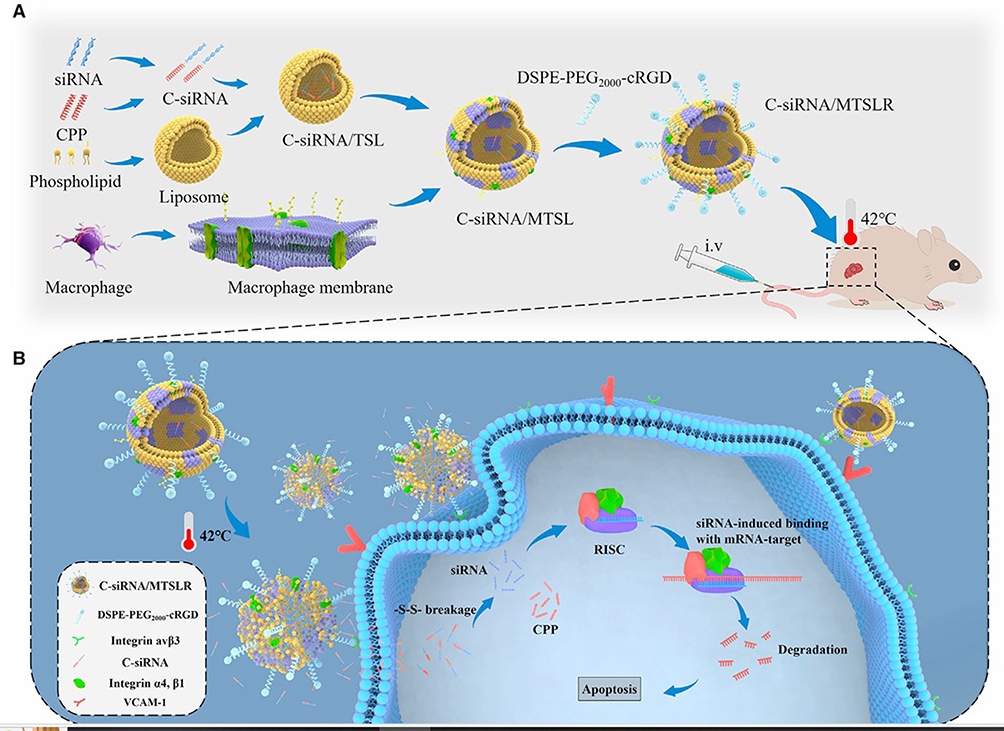 |
Figure 4 Synthesis and Conceptual Representation of C-siRNA/MTSLR (A) Diagram depicting the preparation process of C-siRNA/MTSLR. (B) Conceptual schematic illustrating the proposed mechanism of C-siRNA/MTSLR. Reproduced from Nai J, Zhang J, Li J et al. Macrophage membrane- and cRGD-functionalized thermosensitive liposomes combined with CPP to realize precise siRNA delivery into tumor cells. Mol Ther Nucleic Acids. 2022;27:349–362. CC BY-NC-ND 4.0.68 |
 |
Figure 5 Schematic of HPPS-siRNA and its synthesis process. (A) Diagram illustrating HPPS encapsulating siRNA targeting SALL4. (B) Stepwise synthesis of HPPS, beginning with the lipid mixture. Abbreviations: CO, cholesteryl oleate; DMPC, 1,2-dimyristoyl-sn-glycero-3-phosphocholine. Reproduced from Cruz W, Huang H, Barber B et al. Lipoprotein-Like Nanoparticle Carrying Small Interfering RNA Against Spalt-Like Transcription Factor 4 Effectively Targets Hepatocellular Carcinoma Cells and Decreases Tumor Burden. Hepatol Commun. 2020;4(5):769–782. Copyright 2020 Wolters Kluwer Health, Inc.69 |
Mechanism of Lipid Nanomedicines Loaded with siRNA Acting on the HCC
Nanoparticles (NPs) offer significant potential to enhance siRNA delivery by overcoming biological barriers, protecting siRNA from degradation, and improving its pharmacokinetics.71 Lack of nanomaterial protection siRNA faces challenges such as short half-life, rapid clearance, and insufficient target delivery. By encapsulating siRNA or attaching it to their surface via covalent or non-covalent interactions, NPs enhance stability and cellular uptake.72 These carriers extend siRNA circulation time, shield it from enzymatic degradation, and facilitate precise delivery, making them valuable tools for advancing cancer therapeutics through nanomedicine applications. Lipid nanoparticles (LNPs) have emerged as promising carriers for siRNA delivery targeting hepatocellular carcinoma (HCC).73 Lipid-based siRNA delivery systems encompass a diverse range of nanocarrier platforms, which differ in structure, composition, and functional performance. It is important to clarify that liposomes and solid lipid nanoparticles (SLNs) are not parallel categories but instead represent different levels within the same classification hierarchy. Liposomes serve as a broad class of lipid-based vesicular carriers characterized by bilayered phospholipid membranes, while SLNs are a distinct subtype comprising solid lipid cores stabilized by surfactants. Furthermore, certain hybrid systems—such as nanostructured lipid carriers (NLCs), thermosensitive liposomes, and cationic lipid-coated nanoparticles—integrate features from both classes, leading to cross-cutting designs. To enhance conceptual clarity, this section presents lipid-based nanomedicines as an overarching category, under which various subtypes such as conventional liposomes, SLNs, HDL-like particles, and multifunctional lipid nanocarriers are described according to their unique structural and functional attributes.These nanoparticles can encapsulate siRNA or attach it to their surface through covalent or non-covalent interactions, effectively protecting siRNA from degradation in biological fluids (Figure 6). LNPs enhance cellular uptake via ligand-mediated endocytosis or macropinocytosis, enabling efficient siRNA delivery to target tissues.74 To overcome challenges like endosomal entrapment, mechanisms such as bilayer disruption and the proton sponge effect facilitate siRNA escape into the cytoplasm, enhancing gene silencing efficacy. Conjugating ligands like RGD peptides or transferrin further improves target specificity, allowing precise siRNA delivery to HCC cells. LNPs also extend siRNA circulation time, enhance pharmacokinetics, and improve stability, making them a valuable tool in advancing HCC therapeutics.69
 |
Figure 6 Mechanisms of siRNA loaded lipid nanoparticles in hepatocellular carcinoma therapy. Created in BioRender. Habeeb, M. (2025) https://BioRender.com/ a0ag8ln. |
Different Lipid Loaded siRNA Nanomedicine for Targeting the HCC
Lipid-based siRNA nanomedicines offer a promising approach for hepatocellular carcinoma (HCC) treatment, ensuring stable, targeted, and efficient gene silencing while minimizing off-target effects. Advances in lipid formulations are enhancing precision and therapeutic outcomes. Below are some siRNA-loaded nanoparticles designed for HCC targeting tabulated in (Table 1).
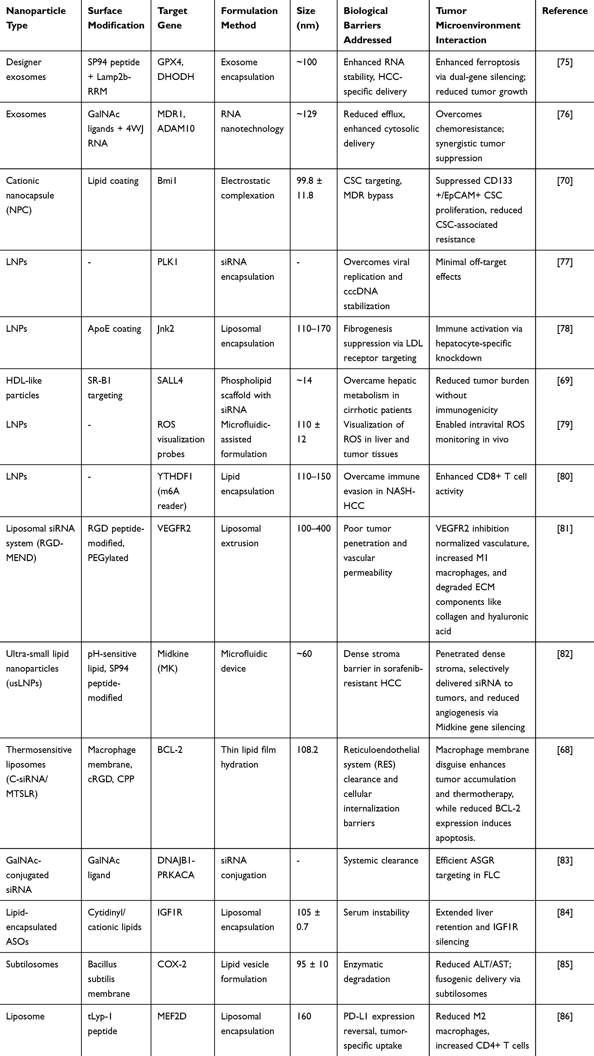 |
Table 1 Emerging Nanotechnologies for siRNA Drug Delivery and Liver Malignancy Suppression |
Solid Lipids Nanomedicines
Solid lipid nanomedicines loaded with siRNA provide a robust and efficient system for delivering gene therapy to hepatocellular carcinoma (HCC), ensuring enhanced stability and controlled release. These lipid-based nanoparticles improve tumor-targeted delivery, reducing off-target effects while maximizing therapeutic impact. Innovations in formulation techniques continue to refine their effectiveness for precision HCC treatment.73,78 Foca et al (2020) investigated lipid nanoparticles (LNPs) encapsulating PLK1-targeting siRNA for hepatitis B virus (HBV)-induced hepatocellular carcinoma (HCC). PLK1, a key player in HBV replication and HCC progression, was effectively silenced using RNA interference. LNPs exhibited significant HBV replication inhibition, reduced viral particles and antigen secretion, and minimal cytotoxicity in primary human hepatocytes (PHHs) and HepaRG cells (Table 1). The siRNA also decreased HBV RNA accumulation, suggesting transcriptional or post-transcriptional modulation, indicating a dual therapeutic role in HBV control and HCC prevention.77 Yang and they team explored cationic nanocapsules (NPCs) co-delivering cisplatin and Bmi1 siRNA to target cancer stem cells (CSCs) in HCC. Bmi1 siRNA effectively reduced CD133+ and EpCAM+ CSC populations, enhancing tumor suppression. The combined therapy showed synergistic efficacy, improved cytotoxicity, and induced cell cycle arrest (LC50: 2.05 μM). Lipid-coated NPCs ensured tumor-specific delivery, minimizing off-target effects. This strategy overcame multidrug resistance (MDR), enhancing therapeutic outcomes.70 A similar study was done by utilized ApoE-coated LNPs to selectively deliver siRNA targeting Jnk2 to hepatocytes via LDL receptor-mediated uptake. Jnk2 silencing exhibited antifibrotic and antitumoral effects, reducing liver fibrosis and suppressing tumor initiation in chronic liver disease (CLD) models. The treatment also activated immune responses, suggesting a dual role in mitigating liver damage and preventing HCC progression.78 In another study developed HDL-mimicking nanoparticles (HPPS) for delivering siRNA against SALL4, an oncogene associated with aggressive HCC. Leveraging SR-B1 receptor-mediated uptake, HPPS-SALL4 significantly reduced tumor burden and inhibited cancer cell proliferation. The formulation avoided hepatic metabolism and showed no systemic immunogenicity, demonstrating clinical potential for HCC therapy.69 Ishiguro and they team engineered milk-derived nanovesicles (MNVs) modified with EpCAM-specific aptamers for targeted siRNA delivery against β-catenin in liver cancer stem cells (LCSCs). EpCAM aptamers facilitated specific uptake, silencing β-catenin and impairing LCSC proliferation. This strategy significantly suppressed tumor growth in xenograft models, highlighting RNA-based nanotechnology for addressing aggressive HCC subtypes.87 Shashkovskaya in 2023 designed LNPs encapsulating ROS-sensitive fluorescent probes and plasmid-based ROS sensors (HyPer7) for real-time visualization of oxidative stress in HCC. Optimized for liver-specific delivery, these LNPs enabled intravital imaging of ROS dynamics, offering a novel diagnostic tool for early detection and therapeutic monitoring of liver diseases (Table 1).79 Wang et al (2023) explored YTHDF1’s role in immunosuppression in NASH-HCC, showing its involvement in the EZH2-IL-6 axis to recruit MDSCs. LNP-delivered siRNA targeting YTHDF1 reduced tumor burden and enhanced CD8+ T cell activity. YTHDF1 silencing synergized with anti-PD1 therapy, improving immunotherapy outcomes. This study highlights LNP-siRNA as a promising strategy for NASH-HCC treatment. LNP-siRNA therapy was explored for targeting YTHDF1 due to the absence of available inhibitors. An FDA-approved LNP formulation delivered siRNA against Ythdf1, achieving effective knockdown in vitro (Figure 7A). They designed siRNA effectively silenced Ythdf1 in vitro (Figure 7A), and to enhance in vivo stability, all pyrimidine bases were modified with 2’-O-Methyl (2’-OMe). LNP-siYthdf1 efficiently targeted Ythdf1 in RIL-175 cells, confirmed by immunostaining and flow cytometry (Figure 7B and C), and accumulated significantly in the mouse liver following tail vein injection (Figure 7A). Using an orthotopic NASH-HCC model with RIL-175 cells, mice were randomized seven days post-implantation and treated with LNP-siYthdf1, anti-PD-1, or both (Figure 7D). LNP-siYthdf1 significantly reduced YTHDF1 protein levels in tumors (Figure 7E) and, when combined with anti-PD-1, synergistically decreased tumor burden and proliferation while increasing apoptosis (Figure 7F and G). In contrast, anti-PD-1 alone had no effect (Figure 7F and G). Notably, treatments did not significantly impact body, liver, or spleen weight, indicating good tolerability and safety (Figure 7F). 80
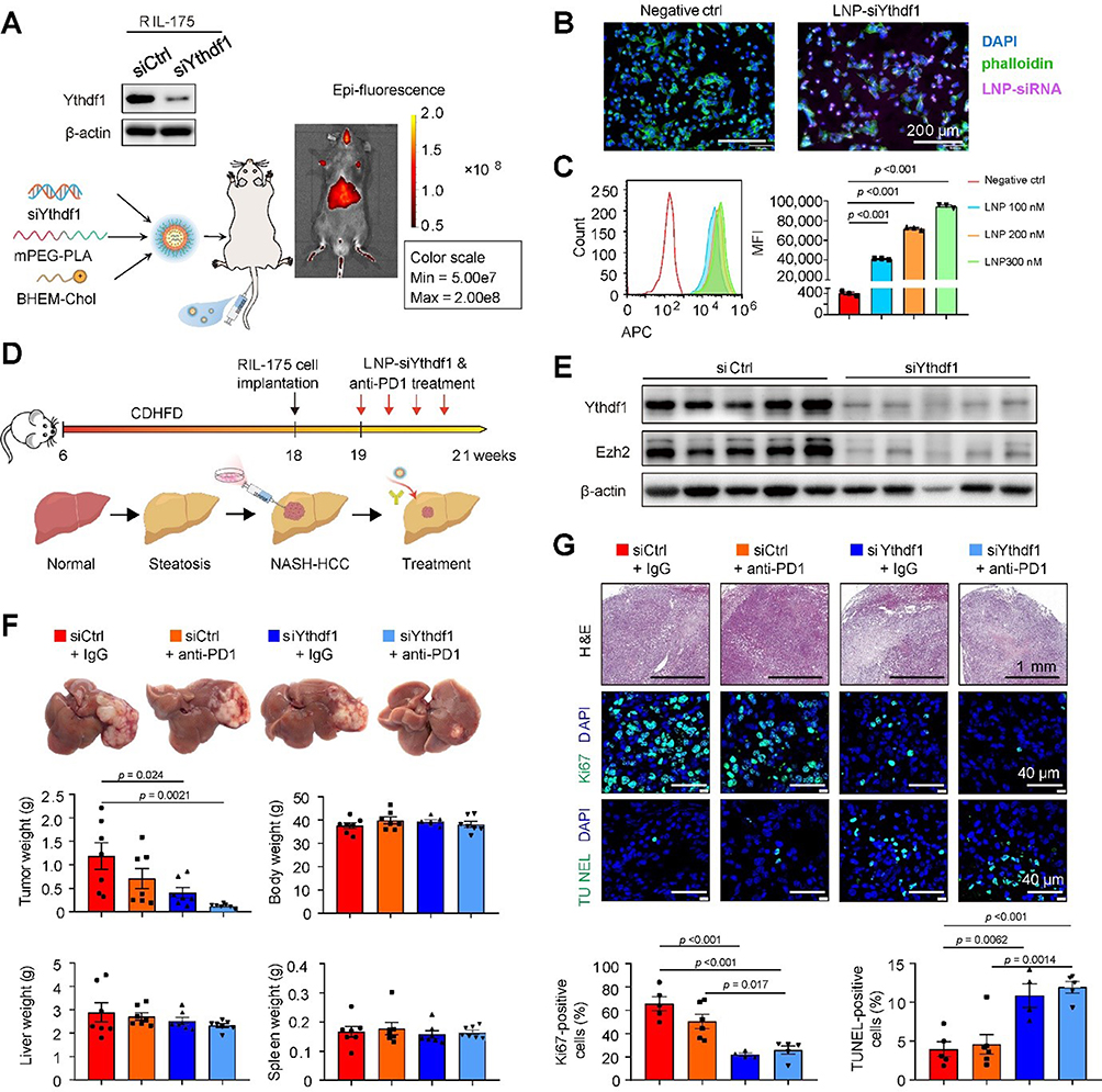 |
Figure 7 Illustrates LNP-siYthdf1 construction, knockdown validation, and in vivo imaging at 16h post-injection (A). Immunostaining and flow cytometry confirmed LNP-siYthdf1 uptake in RIL-175 cells (B and C). Mice with RIL-175 tumors were treated with LNP-siYthdf1 ± anti-PD-1 (D). Western blot confirmed YTHDF1 suppression in NASH-HCC tissues (E). Tumor burden reduction and stable body, liver, and spleen weights were observed (F). H&E staining showed decreased proliferation (Ki-67) and increased apoptosis (TUNEL) (G), highlighting LNP-siYthdf1’s efficacy in enhancing anti-PD-1 therapy. Reproduced from Wang L, Zhu L, Liang C et al. Targeting N6-methyladenosine reader YTHDF1 with siRNA boosts antitumor immunity in NASH-HCC by inhibiting EZH2-IL-6 axis. J Hepatol. 2023;79(5):1185–1200.Copyright (2023), with permission from Elsevier.80 |
Liposomes
siRNA-loaded nanoliposomes enhance targeted gene silencing in HCC by improving stability, tumor-specific delivery, and therapeutic efficacy tabulated in (Table 1). Advanced lipid-based formulations minimize off-target effects, offering a promising strategy for HCC treatment.88,89 Yamamoto et al (2017) developed the RGD-MEND system, a liposomal siRNA delivery platform targeting VEGFR2 in hyper vascularized HCC. This system, incorporating RGD peptides, PEG, and a pH-sensitive lipid (YSK05), enhanced siRNA efficacy by facilitating endosomal escape. VEGFR2 silencing normalized tumor vasculature, promoted M1 macrophage infiltration, and degraded ECM components, improving nanoparticle penetration. Its effectiveness was notable in tumor vessel (TV) type vasculature but limited in stroma vessel (SV) types.81 Correspondingly a research was done by utilizing an ultra-small lipid nanoparticles (usLNPs) co-encapsulating sorafenib and Midkine-siRNA for sorafenib-resistant HCC. Modified with a pH-sensitive lipid and SP94 peptide, these ~60 nm usLNPs overcame stromal barriers, ensuring deep tumor penetration. In vivo, a low sorafenib dose (2.5 mg/kg) significantly reduced toxicity, while Midkine-siRNA (IC50 ~0.1 mg/kg) achieved 85% tumor eradication, demonstrating potent synergy between chemotherapy and gene therapy.82 Nai they team in 2022 developed C-siRNA/MTSLR, a multifunctional liposomal system targeting BCL-2 in HCC. It incorporated TSLs, macrophage membrane camouflage, cRGD peptides, and CPPs for precise tumor targeting and cytoplasmic delivery. The system achieved ~86.5% siRNA loading efficiency and, with mild thermotherapy, enabled targeted BCL-2 silencing, enhancing apoptosis and tumor regression. Macrophage membrane coating minimized off-target effects, ensuring precise siRNA delivery. This approach presents a promising therapeutic strategy for HCC treatment. Nanoparticle uptake by HepG2 cells was analyzed using CLSM and FCM (Figure 8A and B). The highest fluorescence was observed in preheated C-siRNA/MTSLR-H and free C-siRNA, followed by unheated formulations (C-siRNA/MTSLR, C-siRNA/MTSL, C-siRNA/TSL, and siRNA/TSL). Free siRNA showed minimal uptake. Flow cytometry confirmed these trends, with significant differences among formulations. In RAW 264.7 macrophages, CLSM and FCM (Figure 8C and D) showed weaker fluorescence for C-siRNA/MTSL and C-siRNA/MTSLR compared to C-siRNA/TSL. FCM analysis revealed a 2-fold higher uptake of C-siRNA/TSL in macrophages, indicating reduced phagocytosis due to macrophage membrane modifications. The cytotoxicity of the nanoparticles on HepG2 cells was assessed using the CCK-8 assay. Except for the control, C-si.NC/MTSLR, and free siRNA groups, all treatments showed time-dependent inhibition of cell proliferation (Figure 8E). The greatest inhibition was observed in the C-siRNA and C-siRNA/MTSLR-H treated groups over time. Minimal apoptosis was observed in the control, free siRNA, and C-si.NC/MTSLR groups (Figure 8F). The highest apoptosis occurred with free C-siRNA and C-siRNA/MTSLR-H, decreasing in the order: C-siRNA/MTSLR-H > C-siRNA/MTSLR > C-siRNA/MTSL > C-siRNA/TSL > siRNA/TSL. Bcl-2 mRNA suppression was negligible in free siRNA and C-si.NC/MTSLR-H groups (Figure 8G). Other formulations showed significant Bcl-2 silencing, with C-siRNA/MTSLR-H being the most effective. Bcl-2 protein downregulation mirrored these results (Figure 8H).68 Neumayer et al in 2024 developed GalNAc-conjugated siRNA targeting the DNAJB1-PRKACA fusion in FLC. This enabled efficient liver-specific delivery via ASGR, achieving potent gene silencing (IC50: 1 pM). The siRNA significantly reduced tumor growth (~60%) in PDX models. It selectively targeted the fusion oncogene without affecting native genes. No toxicity was observed in vivo. These findings highlight the potential of liver-targeted siRNA therapy for FLC.83 Pan they et al in (2023) designed ASO-loaded lipid nanoparticles (DCP) to target IGF1R in HCC. Modified ASOs (CT102MOE5) improved serum stability and reduced dosing frequency. Encapsulation enhanced hepatic accumulation and intracellular delivery. The system achieved ~80% IGF1R silencing and ~57% apoptosis induction. In vivo, it significantly reduced tumor size. This approach enhances liver-targeted drug uptake and therapeutic efficacy.84 In another study subtilosomes was developed to deliver COX-2-targeting siRNA for DEN-induced HCC. These vesicles ensured high stability and sustained siRNA release (72 hours). The system reduced liver enzymes (ALT, AST) and suppressed COX-2 expression. Apoptosis induction (~18.53%) was superior to conventional liposomes or free siRNA. Subtilosomes improved membrane fusion and specific siRNA delivery. This novel carrier enhances HCC treatment efficiency.85 Du and they et al in 2025 developed tLyp-1-modified liposomes co-delivering SN38 and MEF2D-siRNA. SN38 activated the STING pathway, while MEF2D-siRNA reduced PD-L1 expression. tLyp-1 enhanced tumor-targeting via neuropilin-1 (NRP-1) binding. The system suppressed tumor growth and improved immune responses. It increased dendritic cell maturation and reduced immunosuppressive MDSCs and M2 macrophages. This dual-targeted therapy showed superior antitumor efficacy (Table 1).86
 |
Figure 8 Cellular Evaluation of Various Formulations. (A) CLSM imaging showed nanoparticle uptake in HepG2 cells, with Hoechst 33258 (blue) for nuclei and Cy5 (red) for fluorescence. (B) FCM analysis quantified uptake differences, showing significant variation among formulations. (C-D) RAW 264.7 cell uptake was visualized by CLSM and quantified by FCM, with C-siRNA/MTSLR showing reduced uptake. (E) Cytotoxicity assays revealed time-dependent effects, with C-siRNA/MTSLR exhibiting enhanced toxicity. (F) Apoptosis analysis demonstrated increased apoptotic and necrotic cell populations. (G) Bcl-2 mRNA expression was significantly reduced by C-siRNA/MTSLR-H. (H) Western blot confirmed decreased Bcl-2 protein levels, supporting effective gene silencing Reproduced from Nai J, Zhang J, Li J et al. Macrophage membrane- and cRGD-functionalized thermosensitive liposomes combined with CPP to realize precise siRNA delivery into tumor cells. Mol Ther Nucleic Acids. 2022;27:349–362. CC BY-NC-ND 4.0.68 |
Exosomes
Exosomes loaded with siRNA provide a biocompatible and effective strategy for delivering gene therapy to HCC, utilizing their innate tumor-targeting properties.90 These nanoscale carriers safeguard siRNA from degradation, ensuring enhanced stability and efficient gene silencing.91 Whether naturally derived or engineered, siRNA-loaded exosomes hold great potential for precise and targeted HCC treatment. Li et al, in 2022 developed ExoSP94-Lamp2b-RRM, designer exosomes engineered for targeted ferroptosis-mediated therapy in HCC. Functionalized with SP94 peptide for tumor targeting and Lamp2b-RRM for multi-siRNA delivery, they silenced GPX4 and DHODH, key ferroptosis suppressors (Table 1). This enhanced sorafenib-induced ferroptosis, increasing ROS production and lipid peroxidation, overcoming drug resistance. The strategy sensitized tumors to ferroptosis, inhibiting HCC progression. While IC50 values were not reported, significant tumor reduction and improved survival in vivo highlight its therapeutic potential. The therapeutic effect of SP94-Lamp2b-RRM-functionalized exosomes combined with sorafenib was evaluated in vivo. Tumors in mice treated with sorafenib + ExoSP94-Lamp2b-RRM containing multi-siRNA#1 showed significant reduction after 21 days, unlike those receiving scramble multi-siRNA (Figure 9A–C). However, the addition of ferroptosis inhibitor (ferrostatin-1) diminished this effect. Mice in the multi-siRNA#1 cotreatment group had longer survival compared to the scramble siRNA group (Figure 9D). Immunohistochemistry confirmed that multi-siRNA#1 significantly suppressed GPX4 and DHODH expression in tumors (Figure 9E). These findings suggest that ExoSP94-Lamp2b-RRM-multi-siRNA#1 enhances sorafenib efficacy by silencing ferroptosis suppressor genes, overcoming sorafenib resistance in HCC.75 Ellipilli et al, (2023) introduced ligand-displaying exosomes (Gal/Exo/miR122/PTX) leveraging RNA nanotechnology for targeted and synergistic drug delivery in liver cancer treatment (Table 1). These exosomes were functionalized with N-acetylgalactosamine (GalNAc) ligands for liver-specific targeting and loaded with a four-way junction (4WJ) RNA nanoparticle encapsulating Paclitaxel (PTX) and miRNA122 (miR122). MiR122 plays a dual role by targeting the oncogenic protein ADAM10 and the drug efflux protein MDR1, enhancing drug retention and sensitivity while simultaneously suppressing tumor growth. The integration of GalNAc ligands further optimized RNA stability and facilitated effective cytosolic delivery. Although specific IC50 values were not disclosed, the exosomes demonstrated significant therapeutic efficacy, including substantial tumor volume reduction and improved survival rates in murine xenograft models, highlighting their potential for liver cancer regression.76
 |
Figure 9 Therapeutic Effects of SP94-Lamp2b-RRM Functionalized Exosomes and Sorafenib in an HCC Mouse Model. Sorafenib-resistant HepG2 cells were orthotopically injected into the left liver lobes of nude mice, which were then divided into six treatment groups: Control (saline), Sora (sorafenib alone), Sora + ExoSP94 (sorafenib + SP94-Lamp2b-RRM exosomes containing multi-siRNA#1), Sora + ExoSP94-scramble (sorafenib + exosomes containing scramble siRNA), Sora + ExoSP94 + Vehicle (sorafenib + exosomes with multi-siRNA#1 in vehicle), and Sora + ExoSP94 + Fer-1 (sorafenib + exosomes with multi-siRNA#1 combined with ferrostatin-1). Tumor progression was assessed using IVIS imaging on days 1 and 21 (A), and normalized photon flux was analyzed on day 21 (B). Representative images of resected tumors (C) and survival rates of mice in different treatment groups (D) were evaluated. Immunohistochemical staining for GPX4 and DHODH in HCC tissues (E) demonstrated effective knockdown by multi-siRNA#1. Statistical analysis was performed using ANOVA with Dunnett’s test for (B) and the Log rank test for (D) (***p < 0.001). Reproduced from Li X, Yu Q, Zhao R et al. Designer Exosomes for Targeted Delivery of a Novel Therapeutic Cargo to Enhance Sorafenib-Mediated Ferroptosis in Hepatocellular Carcinoma. Front Oncol. 2022;12:898156. Copyright © 2022 Li, Yu, Zhao, Guo, Liu, Zhang, Zhang, Liu, Yu, Wang, Hao, Li, Zhang, Li, Zhang, Zhang and Gao. This is an open-access article distributed under the terms of the Creative Commons Attribution License (CC BY).75 |
Evaluation of Cost, Efficacy, and Scalability of Lipid Nanomedicine-Based siRNA Systems
Different lipid nanocarriers exhibit varied performance in terms of preparation cost, therapeutic efficacy, scalability, and cost-effectiveness for siRNA delivery in hepatocellular carcinoma (HCC). Lipid nanoparticles (LNPs), especially those prepared via microfluidic mixing, offer high encapsulation efficiency and strong therapeutic outcomes due to effective liver targeting.92 However, the method requires specialized equipment, making the initial production cost high, though it is highly scalable and has existing clinical approval precedent. HDL-mimicking nanoparticles show good biocompatibility and receptor-mediated uptake but involve complex synthesis steps, leading to moderate cost and scalability challenges tabulated in (Table 2). Exosome-based systems provide excellent targeting and immune compatibility, yet they are costly, difficult to scale, and exhibit batch-to-batch variability, limiting their industrial feasibility.93 In contrast, solid lipid nanoparticles (SLNs) are simpler and more economical to prepare, with high scalability, though their therapeutic performance may be lower than advanced LNP systems. Overall, LNPs strike the best balance between efficacy and clinical potential, while SLNs offer a cost-effective alternative suitable for broader implementation.
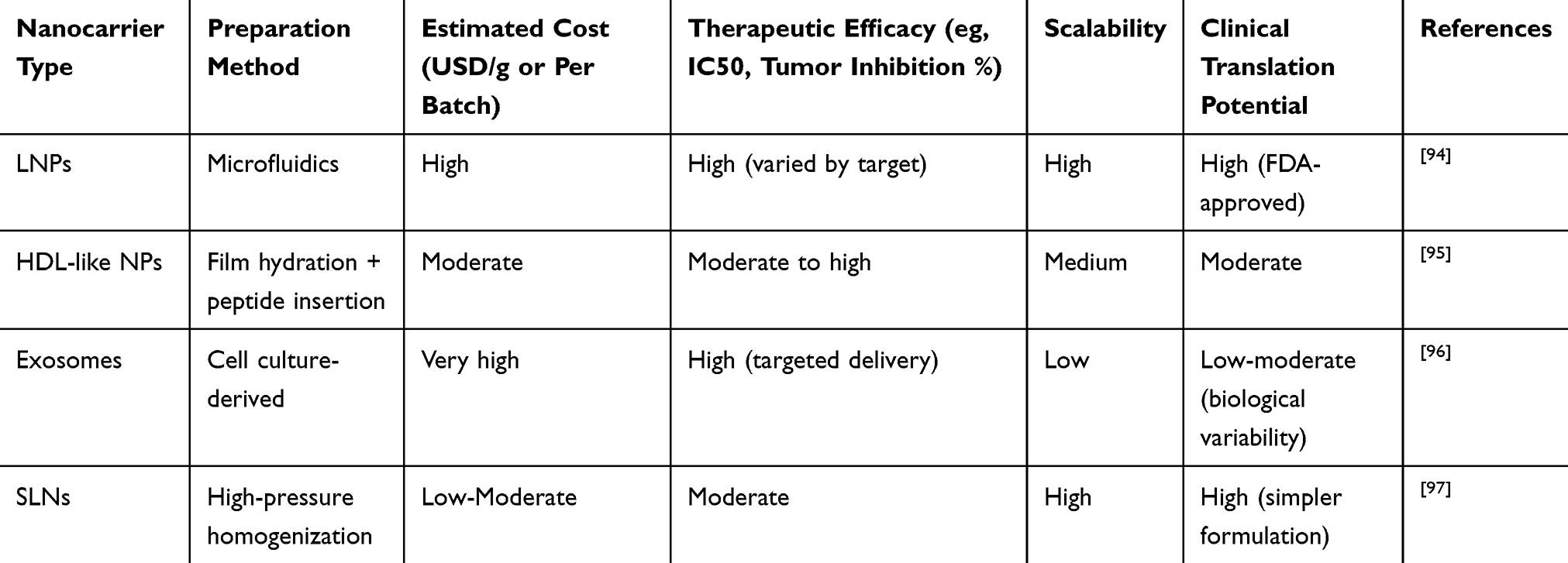 |
Table 2 Comparison of siRNA Lipid Nanomedicines by Cost, Efficacy, and Scalability |
Clinical Trails
Several siRNA-loaded lipid nanoparticle (LNP) formulations have been investigated in clinical trials targeting hepatocellular carcinoma (HCC), reflecting the growing interest in RNA interference-based therapies for liver-associated malignancies. One of the prominent candidates is TKM-080301, which delivers siRNA against the polo-like kinase 1 (PLK1) gene, a key regulator of cell cycle progression frequently overexpressed in various cancers including HCC.98 This formulation was evaluated in a Phase I/II clinical trial (NCT02191878) involving patients with advanced HCC and was administered intravenously.99 The treatment demonstrated a favorable safety and toxicity profile, indicating good tolerability in humans. However, no significant improvement in overall survival was observed, suggesting limited therapeutic efficacy as a monotherapy in advanced-stage disease.100 In parallel, an intratumoral administration approach of TKM-080301 was assessed in another Phase I trial (NCT01437007) for both primary and secondary liver cancers, though no results have been disclosed to date, leaving the effectiveness of local delivery strategies uncertain.101
Another siRNA-LNP formulation studied in the context of HCC is DCR-MYC, designed to silence the MYC oncogene, which plays a central role in driving cancer cell proliferation and metabolism. While DCR-MYC showed encouraging clinical and metabolic responses in a Phase I trial (NCT02110563) involving patients with solid tumors, multiple myeloma, or lymphoma, its performance in HCC was less promising. In a Phase Ib/II trial (NCT02314052) specifically targeting HCC, the results did not meet the sponsor’s expectations, leading to termination of the trial. This outcome underscores the complexity of translating siRNA therapeutics from general solid tumor models to the more biologically and genetically diverse setting of HCC.102 Overall, while early-phase trials confirmed the safety and feasibility of siRNA-LNPs in HCC patients, the therapeutic outcomes have been modest, with limited clinical benefit observed thus far. These studies highlight the need for enhanced targeting strategies, optimized delivery systems, and combination approaches to fully unlock the potential of siRNA nanotherapeutics in the treatment of HCC.73 Although the clinical progression of siRNA-based nanotherapeutics has been gradual, several siRNA-loaded lipid nanoparticles (LNPs) have entered early-stage clinical trials, particularly for treating solid tumors involving the liver. These trials are predominantly in Phase I and are primarily assessing safety profiles, with limited data available on their antitumor efficacy. Notably, the siRNA-LNP formulations tested so far lack active targeting mechanisms, and with a single exception, all have been delivered through intravenous infusion. Moreover, most of these therapies have been evaluated as standalone treatments.64
Role of AI in siRNA for Targeting HCC
Artificial intelligence (AI) enhances siRNA-based therapies for hepatocellular carcinoma (HCC) by optimizing target selection, delivery system design, and efficacy prediction. Machine learning models improve siRNA stability, minimize off-target effects, and personalize treatment strategies. AI-driven approaches accelerate the development of precision nanocarriers, making siRNA therapy more efficient and patient-specific. Chou et al developed an AI-assisted physiologically based pharmacokinetic (PBPK) model to predict nanoparticle (NP) transport to tumors in mice. As illustrated in (Figure 10), they constructed a quantitative structure–activity relationship (QSAR) model using machine learning techniques, including random forest, which outperformed other models in predictive accuracy. Notably, this AI-driven model was integrated into a PBPK framework to simulate tumor-targeted NP delivery efficiency and biodistribution. The optimized PBPK model, incorporating cellular uptake kinetics, successfully predicted peak delivery efficiencies at various time points post-intravenous NP administration, with high determination coefficients. Importantly, the AI-PBPK model’s predictions showed a strong correlation with experimental pharmacokinetic data on NP accumulation in tumors.103
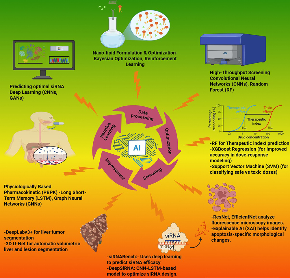 |
Figure 10 Artificial intelligence driven siRNA nanomedicine therapeutics enhancing drug design, screening, and pharmacokinetics. Created in BioRender. Habeeb, M. (2025) https://BioRender.com/ myyriym. |
Artificial intelligence (AI) has significantly advanced the field of siRNA therapeutics by enabling data-driven optimization of siRNA sequences, delivery systems, and pharmacokinetic models. While tools such as siRNABench and DeepSiRNA utilize machine learning models like convolutional neural networks (CNNs) and long short-term memory (LSTM) architectures to predict siRNA efficacy and minimize off-target effects, it is important to note that these tools are designed for general-purpose applications.104 These platforms evaluate sequence features such as thermodynamic stability, GC content, mRNA secondary structure accessibility, and potential off-target binding, thus guiding the initial design of effective siRNA candidates.105 Once optimized, these siRNA sequences can be further tailored for hepatocellular carcinoma (HCC) through disease-specific delivery systems, including lipid nanoparticles (LNPs), hepatocyte-targeting ligands, and liver-specific PBPK models. Moreover, AI-assisted physiologically based pharmacokinetic (PBPK) modeling and nanoparticle design can be customized to enhance liver-specific uptake and reduce systemic toxicity. Therefore, while the core AI tools may be generalized, their integration with HCC-focused delivery strategies and modeling frameworks provides a robust pathway for developing personalized and highly targeted siRNA therapeutics for liver cancer.106
Artificial intelligence (AI) is transforming siRNA-based therapeutics for liver cancer by enhancing drug design, delivery, and treatment monitoring. Deep learning models such as Convolutional Neural Networks (CNNs) and Generative Adversarial Networks (GANs) assist in predicting optimal siRNA sequences that effectively silence oncogenes associated with hepatocellular carcinoma (HCC), including MYC, VEGF, and MET.107,108 AI-powered tools like siRNABench and DeepSiRNA (CNN-LSTM models) further refine siRNA design to maximize efficacy while minimizing off-target effects.109 In addition to sequence optimization, AI-driven physiologically based pharmacokinetic (PBPK) modeling, incorporating Graph Neural Networks (GNNs) and Long Short-Term Memory (LSTM) networks, helps predict siRNA distribution and metabolism in the liver.110 These models aid in optimizing nanoparticle-based siRNA carriers, ensuring improved hepatic uptake while minimizing systemic toxicity. AI also plays a crucial role in formulating nanocarriers, with Bayesian Optimization and Reinforcement Learning fine-tuning lipid nanoparticles (LNPs) and polymeric carriers to enhance siRNA stability, bioavailability, and liver-specific targeting.111 Furthermore, AI-driven high-throughput screening (HTS) using CNNs and Random Forest (RF) models accelerates the identification of effective siRNA constructs. Machine learning algorithms such as XGBoost Regression and Support Vector Machines (SVMs) improve dose-response modeling and toxicity classification, ensuring a safe and effective therapeutic index.104 AI also contributes significantly to liver cancer imaging and treatment monitoring. DeepLabv3+ and 3D U-Net facilitate automatic segmentation of liver tumors in medical images, enabling more precise treatment planning. Additionally, deep learning architectures like ResNet and EfficientNet analyze fluorescence microscopy images to detect apoptosis-specific morphological changes induced by siRNA therapy. Explainable AI (XAI) further enhances interpretability, providing insights into siRNA-induced cellular responses.112 By integrating AI into every stage of siRNA-based liver cancer therapy, from molecular design to clinical evaluation, researchers can develop more effective, targeted, and personalized treatments. These AI-driven advancements significantly improve therapeutic outcomes while reducing off-target effects, ultimately revolutionizing precision medicine for liver cancer.
Conclusion
Hepatocellular carcinoma remains a formidable global health challenge due to its complex molecular landscape, late-stage diagnosis, and limited treatment options. siRNA offers a transformative approach to target oncogenic drivers at the genetic level; however, its clinical utility relies heavily on effective delivery systems. Recent advances in lipid-based nanomedicine, particularly lipid nanoparticles (LNPs), have significantly improved siRNA stability, biocompatibility, and liver-specific targeting. The development of multifunctional nanocarriers—such as thermosensitive liposomes, membrane-camouflaged systems, and ligand-functionalized particles—has further enhanced selective uptake, immune modulation, and tumor penetration.
Artificial intelligence has begun to play a vital role in optimizing siRNA therapeutics by enabling predictive design of siRNA sequences, enhancing delivery system personalization, and guiding physiologically based pharmacokinetic (PBPK) modeling for liver-specific applications. These AI-driven tools, when combined with precision nanocarrier engineering, contribute to the rational development of safer and more effective siRNA therapeutics.
Future research should focus on refining multi-pathway targeting strategies to prevent compensatory resistance, improving tumor microenvironment-responsive delivery systems, integrating siRNA with immune checkpoint blockade, and advancing real-time, image-guided siRNA delivery platforms. Additionally, AI-guided adaptive trial designs and systems biology models hold promise in accelerating clinical translation and personalizing treatment regimens. By integrating nanotechnology, RNA therapeutics, and computational tools, siRNA-based strategies have the potential to redefine precision medicine in HCC.
Acknowledgments
All authors contributed equally to this manuscript.They sincerely thank the Management of Crescent School of Pharmacy, BSACIST, Vandalur, Chennai, for their support, and gratefully acknowledge BioRender for facilitating the creation of high-quality scientific illustrations. The authors, Dr. Kiran Balasaheb Aher and Dr. Girija Balasaheb Bhavar, express their sincere gratitude to the SVKM NMIMS Global University, Dhule, Maharashtra, India and Dean, School of Pharmacy and Technology Management, Dhule for their invaluable support, which greatly contributed to the successful completion of this work.
Author Contributions
All authors contributed to data analysis, drafting or revising the article, have agreed on the journal to which the article will be submitted, gave final approval of the version to be published, and agree to be accountable for all aspects of the work.
Disclosure
The author(s) report no conflicts of interest in this work and the manuscript is approved by all authors for publication.
References
1. Nie Q, Jiang Y, Li M, et al. Global burden and international disparities in NASH-associated liver Cancer: mortality trends (1990–2021) and future projections to 2045. Front Public Health. 2025;13:1527328. doi:10.3389/fpubh.2025.1527328
2. Li Q, Ding C, Cao M, et al. Global epidemiology of liver cancer 2022: an emphasis on geographic disparities. Chin Med J (Engl). 2024;137(19):2334. doi:10.1097/CM9.0000000000003264
3. Bray F, Laversanne M, Sung H, et al. Global cancer statistics 2022: GLOBOCAN estimates of incidence and mortality worldwide for 36 cancers in 185 countries. CA Cancer J Clin. 2024;74(3):229–263. doi:10.3322/CAAC.21834
4. Habeeb M, Sugumaran A. Strategies of cell signaling and critical focus on etiology of hepatocellular carcinoma. 2022;12:5187–5198. doi:10.33263/BRIAC124.51875198
5. Mi C, Liu S. Targeting ZCJ of D, 2025 undefined. redefining hepatocellular carcinoma treatment: nanotechnology meets tumor immune microenvironment. Taylor & Francis. doi:10.1080/1061186X.2025.2479757
6. Bhange M, Oncology DTD. 2025 undefined. Convergence of nanotechnology and artificial intelligence in the fight against liver cancer: a comprehensive review. Springer. 2025;16(1):77. doi:10.1007/s12672-025-01821-y
7. Dogheim G, Chinnam S, Delivery MACD. 2025 undefined. lipid nanoparticles as a platform for miRNA and siRNA delivery in hepatocellular carcinoma. Ingentaconnect.com.
8. Chang Y, Lv Y, Wei P, et al. Multifunctional glyco-nanofibers: sirna induced supermolecular assembly for codelivery in vivo. Adv Funct Mater. 2017;27(44):1703083. doi:10.1002/ADFM.201703083
9. Weiskirchen R, Weiskirchen S, Grassi C, et al. Recent advances in optimizing siRNA delivery to hepatocellular carcinoma cells. Expert Opin Drug Deliv. doi:10.1080/17425247.2025.2484287
10. Zhang M, Guo R, Yuan Z, Challenges HWG. 2025 undefined. lipid nanoparticle (LNP)‐A vector suitable for evolving therapies for advanced hepatocellular carcinoma (HCC). Wiley Online Lib. doi:10.1002/gch2.202400217
11. Fan J, Xiao Z, Dong Y, Ye F. Undefined. nanocarrier‐mediated RNA delivery platform as a frontier strategy for hepatic disease treatment: challenges and opportunities. Wiley Online Lib.
12. Chang Y, Yang K, Wei P, et al. Cationic vesicles based on amphiphilic pillar[5]arene capped with ferrocenium: a redox-responsive system for drug/siRNA Co-delivery. Angewandte Chemie International Edition. 2014;53(48):13126–13130. doi:10.1002/ANIE.201407272
13. Xu J, Liu Y. Nanomaterials for liver cancer targeting: research progress and future prospects. Front Immunol. 2025;16. doi:10.3389/FIMMU.2025.1496498/FULL
14. Ma K, Chen Z, Liang K, Pei Y, Pei Z. A near-infrared light-driven Janus nanomotor for deep tumor penetration and enhanced tumor immunotherapy. Chem. Commun. 2024;60(71):9550–9553. doi:10.1039/D4CC03445G
15. Luo X, He X, Zhang X, et al. Hepatocellular carcinoma: signaling pathways, targeted therapy, and immunotherapy. MedComm. 2024;5(2). doi:10.1002/MCO2.474
16. Xue Y, Ruan Y, Wang Y, Xiao P, Xu J. Signaling pathways in liver cancer: pathogenesis and targeted therapy. Mol Biomed. 2024;5(1). doi:10.1186/S43556-024-00184-0
17. Zheng J, Wang S, Xia L, et al. Hepatocellular carcinoma: signaling pathways and therapeutic advances. Signal Transduct Target Ther. 2025;10(1):35. doi:10.1038/S41392-024-02075-W
18. Whittaker S, Marais R, Zhu AX. The role of signaling pathways in the development and treatment of hepatocellular carcinoma. Oncogene. 2010;29(36):4989–5005. doi:10.1038/ONC.2010.236
19. Raskopf E, Vogt A, Sauerbruch T, Schmitz V. siRNA targeting VEGF inhibits hepatocellular carcinoma growth and tumor angiogenesis in vivo. J Hepatol. 2008;49(6):977–984. doi:10.1016/j.jhep.2008.07.022
20. Simons M, Gordon E, Claesson-Welsh L. Mechanisms and regulation of endothelial VEGF receptor signalling. Nat Rev Mol Cell Biol. 2016;17(10):611–625. doi:10.1038/NRM.2016.87
21. Hu L, Shi W, Liu K, et al. EGFR bypass activation mediates acquired resistance to regorafenib in hepatocellular carcinoma. Front Med Lausanne. 2024;11:1464610. doi:10.3389/FMED.2024.1464610/BIBTEX
22. Halder S, Basu S, Lall SP, Ganti AK, Batra SK, Seshacharyulu P. Targeting the EGFR signaling pathway in cancer therapy: what’s new in 2023? Expert Opin Ther Targets. 2023;27(4–5):305. doi:10.1080/14728222.2023.2218613
23. Suzuki H, Iwamoto H, Tanaka T, et al. Fibroblast growth factor inhibition by molecular-targeted agents mitigates immunosuppressive tissue microenvironment in hepatocellular carcinoma. Hepatol Int. 2024;18(2):610–622. doi:10.1007/S12072-023-10603-Z
24. Liu Q, Huang J, Yan W, Liu Z, Liu S, Fang W. FGFR families: biological functions and therapeutic interventions in tumors. MedComm. 2023;4(5):e367. doi:10.1002/MCO2.367
25. Peng M, Deng J, Li X. Clinical advances and challenges in targeting FGF/FGFR signaling in lung cancer. Mol Cancer. 2024;23(1). doi:10.1186/S12943-024-02167-9
26. Zhang K, Barry AE, Lamm R, Patel K, Schafer M, Dang H. The role of RNA binding proteins in hepatocellular carcinoma. Adv Drug Deliv Rev. 2022;182. doi:10.1016/j.addr.2022.114114
27. Wang Y, Liu D, Zhang T, Xia L. FGF/FGFR signaling in hepatocellular carcinoma: from carcinogenesis to recent therapeutic intervention. Cancers. 2021;13(6):1360. doi:10.3390/CANCERS13061360
28. Moosavi F, Giovannetti E, Saso L, Firuzi O. HGF/MET pathway aberrations as diagnostic, prognostic, and predictive biomarkers in human cancers. Crit Rev Clin Lab Sci. 2019;56(8):533–566. doi:10.1080/10408363.2019.1653821
29. Wang H, Rao B, Lou J, et al. The Function of the HGF/c-met axis in hepatocellular carcinoma. Front Cell Dev Biol. 2020;8:55. doi:10.3389/FCELL.2020.00055
30. Xia Y, Huang C, Zhong M, et al. Targeting HGF/c-MET signaling to regulate the tumor microenvironment: implications for counteracting tumor immune evasion. Cell Commun Signaling. 2025;23(1):1–18. doi:10.1186/S12964-025-02033-1
31. Charbe NB, Amnerkar ND, Ramesh B, et al. Small interfering RNA for cancer treatment: overcoming hurdles in delivery. Acta Pharm Sin B. 2020;10(11):2075–2109. doi:10.1016/j.apsb.2020.10.005
32. Zhang T, Wang Y, Xie M, et al. HGF-mediated elevation of ETV1 facilitates hepatocellular carcinoma metastasis through upregulating PTK2 and c-MET. J Exp Clin Cancer Res. 2022;41(1). doi:10.1186/S13046-022-02475-2
33. Nwabo Kamdje AH, Seke Etet PF, Kipanyula MJ, et al. Insulin-like growth factor-1 signaling in the tumor microenvironment: carcinogenesis, cancer drug resistance, and therapeutic potential. Front Endocrinol (Lausanne). 2022:13.doi:10.3389/FENDO.2022.927390/FULL.
34. Cai W, Ma Y, Song L, et al. IGF-1R down regulates the sensitivity of hepatocellular carcinoma to sorafenib through the PI3K / akt and RAS / raf / ERK signaling pathways. BMC Cancer. 2023;23(1). doi:10.1186/S12885-023-10561-7
35. Guérit E, Arts F, Dachy G, Boulouadnine B, Demoulin JB. PDGF receptor mutations in human diseases. Cell. Mol. Life Sci. 2021;78(8):3867–3881. doi:10.1007/S00018-020-03753-Y
36. Bergé M, Bonnin P, Sulpice E, et al. Small interfering RNAs induce target-independent inhibition of tumor growth and vasculature remodeling in a mouse model of hepatocellular carcinoma. Am. J. Pathol. 2010;177(6):3192–3201. doi:10.2353/ajpath.2010.100157
37. Alawyia B, Constantinou C. Hepatocellular carcinoma: a narrative review on current knowledge and future prospects. Curr Treat Options Oncol. 2023;24(7):711–724. doi:10.1007/S11864-023-01098-9
38. Yang S, Liu G. Targeting the Ras/Raf/MEK/ERK pathway in hepatocellular carcinoma. Oncol Lett. 2017;13(3):1041. doi:10.3892/OL.2017.5557
39. Moon H, Ro SW. MAPK/ERK signaling pathway in hepatocellular carcinoma. Cancers. 2021;13(12):3026. doi:10.3390/CANCERS13123026
40. Song Y, Bi Z, Liu Y, Qin F, Wei Y, Wei X. Targeting RAS–RAF–MEK–ERK signaling pathway in human cancer: current status in clinical trials. Genes Dis. 2023;10(1):76–88. doi:10.1016/J.GENDIS.2022.05.006
41. Goyal R, Chopra H, Singh I, Dua K, Gautam RK. Insights on prospects of nano-siRNA based approaches in treatment of Cancer. Front Pharmacol. 2022;13. doi:10.3389/FPHAR.2022.985670/FULL
42. Wu Y, Zhang Y, Qin X, Geng H, Zuo D, Zhao Q. PI3K/AKT/mTOR pathway-related long non-coding RNAs: roles and mechanisms in hepatocellular carcinoma. Pharmacol Res. 2020;160. doi:10.1016/j.phrs.2020.105195
43. Paskeh MDA, Ghadyani F, Hashemi M, et al. Biological impact and therapeutic perspective of targeting PI3K/Akt signaling in hepatocellular carcinoma: promises and challenges. Pharmacol Res. 2023:187.doi:10.1016/j.phrs.2022.106553.
44. Glaviano A, Foo ASC, Lam HY, et al. PI3K/AKT/mTOR signaling transduction pathway and targeted therapies in cancer. Mol Cancer. 2023;22(1). doi:10.1186/S12943-023-01827-6
45. Chen F, Zhang W, Gao X, Yuan H, Liu K. The role of small interfering RNAs in hepatocellular carcinoma. J Gastrointest Cancer. 2024;55(1):26–40. doi:10.1007/S12029-023-00911-W
46. Pai SG, Carneiro BA, Mota JM, et al. Wnt/beta-catenin pathway: modulating anticancer immune response. J Hematol Oncol. 2017;10(1). doi:10.1186/S13045-017-0471-6
47. Bakrania A, To J, Zheng G, Bhat M. Targeting Wnt-β-catenin signaling pathway for hepatocellular carcinoma nanomedicine. Gastro Hep Adva. 2023;2(7):948–963. doi:10.1016/j.gastha.2023.07.012
48. He S, Tang S. WNT/β-catenin signaling in the development of liver cancers. Biomed. Pharmacother. 2020;132. doi:10.1016/j.biopha.2020.110851
49. Xie J, Wang S. Small Interfering RNA in colorectal cancer liver metastasis therapy. Technol Cancer Res Treat. 2022;21. doi:10.1177/15330338221103318
50. Hin Tang JJ, Hao Thng DK, Lim JJ, Toh TB. JAK/STAT signaling in hepatocellular carcinoma. Hepatol Oncol. 2020;7(1). doi:10.2217/HEP-2020-0001
51. Wang X, Liao X, Yu T, et al. Analysis of clinical significance and prospective molecular mechanism of main elements of the JAK/STAT pathway in hepatocellular carcinoma. Int J Oncol. 2019;55(4):805–822. doi:10.3892/IJO.2019.4862
52. Rah B, Rather RA, Bhat GR, et al. JAK/STAT signaling: molecular targets, therapeutic opportunities, and limitations of targeted inhibitions in solid malignancies. Front Pharmacol. 2022:13.doi:10.3389/FPHAR.2022.821344/FULL.
53. Nair B, Kamath AJ, Pradeep G, Devan AR, Sethi G, Nath LR. Unveiling the role of the Hedgehog signaling pathway in chronic liver disease: therapeutic insights and strategies. Drug Discov Today. 2024;29(8). doi:10.1016/j.drudis.2024.104064
54. Hu Q, Hu X, Zhang L, Zhao Y, Li L. Targeting Hedgehog signalling in CD133-positive hepatocellular carcinoma: improving Lenvatinib therapeutic efficiency. Med Oncol. 2021;38(4). doi:10.1007/S12032-021-01487-W
55. Shen D, Yuwei XIA, Yuhan FU, et al. Hedgehog pathway and cancer: a new area (Review). Oncol Rep. 2024;52(3). doi:10.3892/OR.2024.8775
56. Perrone F, Craparo EF, Cemazar M, et al. Targeted delivery of siRNAs against hepatocellular carcinoma-related genes by a galactosylated polyaspartamide copolymer. J Control Release. 2021;330:1132–1151. doi:10.1016/j.jconrel.2020.11.020
57. Lee U, Cho EY, Jho EH. Regulation of Hippo signaling by metabolic pathways in cancer. Biochim Biophys Acta Mol Cell Res. 2022;1869(4). doi:10.1016/j.bbamcr.2021.119201
58. Li S, Hao L, Li N, et al. Targeting the Hippo/YAP1 signaling pathway in hepatocellular carcinoma: from mechanisms to therapeutic drugs (Review). Int J Oncol. 2024;65(3). doi:10.3892/IJO.2024.5676
59. Deng J, Ke H. Overcoming the resistance of hepatocellular carcinoma to PD-1/PD-L1 inhibitor and the resultant immunosuppression by CD38 siRNA-loaded extracellular vesicles. Oncoimmunology. 2023;12(1). doi:10.1080/2162402X.2022.2152635
60. Hou X, Zaks T, Langer R, Dong Y. Lipid nanoparticles for mRNA delivery. Nature Rev Mater. 2021;6(12):1078–1094. doi:10.1038/s41578-021-00358-0
61. Andra VVSNL, Pammi SVN, Bhatraju LVKP, Ruddaraju LK. A comprehensive review on novel liposomal methodologies, commercial formulations, clinical trials and patents. Bionanoscience. 2022;12(1):274–291. doi:10.1007/S12668-022-00941-X
62. Wei PS, Thota N, John G, et al. Enhancing RNA-lipid nanoparticle delivery: organ- and cell-specificity and barcoding strategies. J Control Release. 2024;375:366–388. doi:10.1016/J.JCONREL.2024.08.030
63. Hu X, Shen Z, Hao H, et al. NIR II light-driven nanomotor synergistically enhances immunogenic cell death through photothermal and chemodynamic therapy for melanoma immunotherapy. J Colloid Interface Sci. 2025:694.doi:10.1016/j.jcis.2025.137688.
64. Kulkarni JA, Witzigmann D, Chen S, Cullis PR, Van Der Meel R. Lipid nanoparticle technology for clinical translation of siRNA therapeutics. Acc Chem Res. 2019;52(9):2435–2444. doi:10.1021/ACS.ACCOUNTS.9B00368/ASSET/IMAGES/MEDIUM/AR9B00368_0005.GIF
65. Metwally AA, Nayel AA, Hathout RM. In silico prediction of siRNA ionizable-lipid nanoparticles In vivo efficacy: machine learning modeling based on formulation and molecular descriptors. Front Mol Biosci. 2022;9:1042720. doi:10.3389/FMOLB.2022.1042720/BIBTEX
66. Wang S, Lv Q, Li X. Recent advances in drug delivery nanocarriers for targeting hepatocellular carcinoma. J Nanomater. 2022. doi:10.1155/2022/7798919
67. Kulkarni JA, Darjuan MM, Mercer JE, et al. On the formation and morphology of lipid nanoparticles containing ionizable cationic lipids and siRNA. ACS Nano. 2018;12(5):4787–4795. doi:10.1021/acsnano.8b01516
68. Nai J, Zhang J, Li J, et al. Macrophage membrane- and cRGD-functionalized thermosensitive liposomes combined with CPP to realize precise siRNA delivery into tumor cells. Mol Ther Nucleic Acids. 2022;27:349–362. doi:10.1016/j.omtn.2021.12.016
69. Cruz W, Huang H, Barber B, et al. Lipoprotein-like nanoparticle carrying small interfering RNA against spalt-like transcription factor 4 effectively targets hepatocellular carcinoma cells and decreases tumor burden. Hepatol Commun. 2020;4(5):769–782. doi:10.1002/HEP4.1493
70. Yang T, Chen Y, Zhao P, et al. Enhancing the therapeutic effect via elimination of hepatocellular carcinoma stem cells using Bmi1 siRNA delivered by cationic cisplatin nanocapsules. Nanomedicine. 2018;14(7):2009–2021. doi:10.1016/J.NANO.2018.05.012
71. Yuan Y, Sun W, Xie J, et al. RNA nanotherapeutics for hepatocellular carcinoma treatment. Theranostics. 2025;15(3):965–992. doi:10.7150/thno.102964
72. Yu B, Hsu SH, Zhou C, et al. Lipid nanoparticles for hepatic delivery of small interfering RNA. Biomaterials. 2012;33(25):5924–5934. doi:10.1016/J.BIOMATERIALS.2012.05.002
73. El Moukhtari SH, Garbayo E, Amundarain A, et al. Lipid nanoparticles for siRNA delivery in cancer treatment. J Control Release. 2023;361:130–146. doi:10.1016/J.JCONREL.2023.07.054
74. Dogheim G, Chinnam S, Amralla MT. Lipid Nanoparticles as a Platform for miRNA and siRNA delivery in hepatocellular carcinoma. Curr Drug Deliv. 2024;21:1–25. doi:10.2174/0115672018292331240404070236
75. Li X, Yu Q, Zhao R, et al. Designer exosomes for targeted delivery of a novel therapeutic cargo to enhance sorafenib-mediated ferroptosis in hepatocellular carcinoma. Front Oncol. 2022:12.doi:10.3389/fonc.2022.898156.
76. Ellipilli S, Wang H, Binzel DW, Shu D, Guo P. Ligand-displaying-exosomes using RNA nanotechnology for targeted delivery of multi-specific drugs for liver cancer regression. Nanomedicine. 2023;50. doi:10.1016/j.nano.2023.102667
77. Foca A, Dhillon A, Lahlali T, et al. Antiviral activity of PLK1-targeting siRNA delivered by lipid nanoparticles in HBV-infected hepatocytes. Antivir Ther. 2021;25(3):151–162. doi:10.3851/IMP3361
78. Woitok MM, Zoubek ME, Doleschel D, et al. Lipid-encapsulated siRNA for hepatocyte-directed treatment of advanced liver disease. Cell Death Dis. 2020;11(5):1–14. doi:10.1038/s41419-020-2571-4
79. Shashkovskaya VS, Vetosheva PI, Shokhina AG, et al. Delivery of lipid nanoparticles with ROS probes for improved visualization of hepatocellular carcinoma. Biomedicines. 2023;11(7). doi:10.3390/biomedicines11071783
80. Wang L, Zhu L, Liang C, et al. Targeting N6-methyladenosine reader YTHDF1 with siRNA boosts antitumor immunity in NASH-HCC by inhibiting EZH2-IL-6 axis. J Hepatol. 2023;79(5):1185–1200. doi:10.1016/J.JHEP.2023.06.021
81. Yamamoto S, Kato A, Sakurai Y, Hada T, Harashima H. Modality of tumor endothelial VEGFR2 silencing-mediated improvement in intratumoral distribution of lipid nanoparticles. J Control Release. 2017;251:1–10. doi:10.1016/j.jconrel.2017.02.010
82. Younis MA, Khalil IA, Elewa YHA, Kon Y, Harashima H. Ultra-small lipid nanoparticles encapsulating sorafenib and midkine-siRNA selectively-eradicate sorafenib-resistant hepatocellular carcinoma in vivo. J Control Release. 2021;331:335–349. doi:10.1016/j.jconrel.2021.01.021
83. Neumayer C, Ng D, Requena D, et al. GalNAc-conjugated siRNA targeting the DNAJB1-PRKACA fusion junction in fibrolamellar hepatocellular carcinoma. Mol Ther. 2024;32(1):140–151. doi:10.1016/j.ymthe.2023.11.012
84. Pan Y, Guan J, Gao Y, et al. Modified ASO conjugates encapsulated with cytidinyl/cationic lipids exhibit more potent and longer-lasting anti-HCC effects. Mol Ther Nucleic Acids. 2023;32:807–821. doi:10.1016/j.omtn.2023.04.028
85. Jamal F, Ahmed G, Farazuddin M, et al. Potential of siRNA-bearing subtilosomes in the treatment of diethylnitrosamine-induced hepatocellular carcinoma. Molecules. 2023;28(5):2191. doi:10.3390/molecules28052191
86. Du J, Que Z, Aihaiti A, et al. Co-delivery of SN38 and MEF2D-siRNA via tLyp-1-modified liposomes reverses PD-L1 expression induced by STING activation in hepatocellular carcinoma. Colloids Surf B Biointerfaces. 2025:245.doi:10.1016/j.colsurfb.2024.114318.
87. Ishiguro K, Yan IK, Lewis-Tuffin L, Patel T. Targeting liver cancer stem cells using engineered biological nanoparticles for the treatment of hepatocellular cancer. Hepatol Commun. 2020;4(2):2020. doi:10.1002/hep4.1462/suppinfo
88. Yao Y, Zhao Q, Xu F, Yao T. Enhanced anti-tumor therapy for hepatocellular carcinoma via sorafenib and KIAA1199-siRNA co-delivery liposomes. Saudi Pharmaceutical J. 2024;32(9):102153. doi:10.1016/J.JSPS.2024.102153
89. Sun W, Wang Y, Cai M, et al. Codelivery of sorafenib and GPC3 siRNA with PEI-modified liposomes for hepatoma therapy. Biomater Sci. 2017;5(12):2468–2479. doi:10.1039/C7BM00866J
90. Ubanako P, Mirza S, Ruff P, Penny C. Exosome-mediated delivery of siRNA molecules in cancer therapy: triumphs and challenges. Front Mol Biosci. 2024;11:1447953. doi:10.3389/FMOLB.2024.1447953
91. Gupta S, Deb A, Mazumder PB, Shekhawat GS. Exosomes as a window of opportunity in hepatocellular carcinoma: insights from diagnostic to targeted drug delivery. J Drug Deliv Sci Technol. 2024;101:106259. doi:10.1016/J.JDDST.2024.106259
92. Venturini J, Chakraborty A, Baysal MA, Tsimberidou AM. Developments in nanotechnology approaches for the treatment of solid tumors. Exp Hematol Oncol. 2025;14(1). doi:10.1186/S40164-025-00656-1
93. Bhange M, Telange D. Convergence of nanotechnology and artificial intelligence in the fightagainst liver cancer: a comprehensive review. Discover Oncology. 2025;16(1). doi:10.1007/S12672-025-01821-Y
94. Strelkova Petersen DM, Chaudhary N, Arral ML, Weiss RM, Whitehead KA. The mixing method used to formulate lipid nanoparticles affects mRNA delivery efficacy and organ tropism. Eur. J. Pharm. Biopharm. 2023;192:126–135. doi:10.1016/j.ejpb.2023.10.006
95. Xu M, Yang L, Lin Y, et al. Emerging nanobiotechnology for precise theranostics of hepatocellular carcinoma. J Nanobiotechnology. 2022;20(1). doi:10.1186/S12951-022-01615-2
96. Liu H, Jin X, Liu S, et al. Recent advances in self-targeting natural product-based nanomedicines. J Nanobiotechnol. 2025;23(1). doi:10.1186/S12951-025-03092-9
97. Mahmoud K, Swidan S, El-Nabarawi M, Teaima M. Lipid based nanoparticles as a novel treatment modality for hepatocellular carcinoma: a comprehensive review on targeting and recent advances. J Nanobiotechnology. 2022;20(1). doi:10.1186/s12951-022-01309-9
98. Ozcan G, Ozpolat B, Coleman RL, Sood AK, Lopez-Berestein G. Preclinical and clinical development of siRNA-based therapeutics. Adv Drug Deliv Rev. 2015;87:108–119. doi:10.1016/j.addr.2015.01.007
99. El Dika I, Lim HY, Yong WP, et al. An open-label, multicenter, phase i, dose escalation study with phase II expansion cohort to determine the safety, pharmacokinetics, and preliminary antitumor activity of intravenous tkm-080301 in subjects with advanced hepatocellular carcinoma. The Oncologist. 2019;24(6):747–e218. doi:10.1634/THEONCOLOGIST.2018-0838
100. Tolcher AW, Papadopoulos KP, Patnaik A, et al. Safety and activity of DCR-MYC, a first-in-class Dicer-substrate small interfering RNA (DsiRNA) targeting MYC, in a phase I study in patients with advanced solid tumors. J Clin Oncol. 2015;33(15_suppl):11006. doi:10.1200/JCO.2015.33.15_SUPPL.11006
101. Hattab D, Gazzali AM, Bakhtiar A. clinical Advances of siRNA-based nanotherapeutics for cancer treatment. Pharmaceutics. 2021;13(7):1009. doi:10.3390/PHARMACEUTICS13071009
102. Whitfield JR, Beaulieu M-E, Soucek L. Strategies to inhibit Myc and their clinical applicability. Front Cell Dev Biol. 2017;5:249159. doi:10.3389/fcell.2017.00010
103. Chou W-C, Chen Q, Yuan L, et al. An artificial intelligence-assisted physiologically-based pharmacokinetic model to predict nanoparticle delivery to tumors in mice. J Control Release. 2023;361:53–63. doi:10.1016/J.JCONREL.2023.07.040
104. Lee M. Machine learning for small interfering RNAs: a concise review of recent developments. Front Genet. 2023;14:1226336. doi:10.3389/FGENE.2023.1226336
105. Shrivastava NK, Verma P, Singh G, et al. Optimizing siRNA therapeutics targeting HIF-1α: computational design, screening, and molecular dynamics simulation studies. Mol Pharm. 2025;22(6):3179–3188. doi:10.1021/acs.molpharmaceut.5c00104
106. Luo Y, Liu L, He Z, et al. TREAT: therapeutic RNAs exploration inspired by artificial intelligence technology. Comput Struct Biotechnol J. 2022;20:5680. doi:10.1016/J.CSBJ.2022.10.011
107. Mohammad H, You HW, Umapathi M, Ravikumar KK, Hariyadi M. Strategies of Artificial intelligence tools in the domain of nanomedicine. J Drug Deliv Sci Technol. 2023:105157. doi:10.1016/J.JDDST.2023.105157
108. Martinelli DD. Machine learning for siRNA efficiency prediction: a systematic review. Health Sci Rev. 2024;11:100157. doi:10.1016/J.HSR.2024.100157
109. Martinelli DD. Generative machine learning for de novo drug discovery: a systematic review. Comput Biol Med. 2022;145:105403. doi:10.1016/J.COMPBIOMED.2022.105403
110. Satheeskumar R. Enhancing drug discovery with AI: predictive modeling of pharmacokinetics using Graph Neural Networks and ensemble learning. Intelligent Pharmacy. doi:10.1016/J.IPHA.2024.11.002
111. Yuan Y, Wu Y, Cheng J, et al. Applications of artificial intelligence to lipid nanoparticle delivery. Particuology. 2024;90:88–97. doi:10.1016/j.partic.2023.11.014
112. Maltarollo VG, Kronenberger T, Espinoza GZ, Oliveira PR, Honorio KM. Advances with support vector machines for novel drug discovery. Expert Opin Drug Discov. 2019;14(1):23–33. doi:10.1080/17460441.2019.1549033
 © 2025 The Author(s). This work is published and licensed by Dove Medical Press Limited. The
full terms of this license are available at https://www.dovepress.com/terms
and incorporate the Creative Commons Attribution
- Non Commercial (unported, 4.0) License.
By accessing the work you hereby accept the Terms. Non-commercial uses of the work are permitted
without any further permission from Dove Medical Press Limited, provided the work is properly
attributed. For permission for commercial use of this work, please see paragraphs 4.2 and 5 of our Terms.
© 2025 The Author(s). This work is published and licensed by Dove Medical Press Limited. The
full terms of this license are available at https://www.dovepress.com/terms
and incorporate the Creative Commons Attribution
- Non Commercial (unported, 4.0) License.
By accessing the work you hereby accept the Terms. Non-commercial uses of the work are permitted
without any further permission from Dove Medical Press Limited, provided the work is properly
attributed. For permission for commercial use of this work, please see paragraphs 4.2 and 5 of our Terms.