
Back to Journals » International Journal of Nanomedicine » Volume 21
Advancements in Polymer-Based Nanocarriers for Controlled Release of Nitric Oxide: Clinical Applications and Future Prospects
Authors Ying H, Zhou C, Liu X, Qin S, Chen L, Yu Y, Xia L, Lin H, Zhang S  , Long H, Liang W
Received 9 June 2025
Accepted for publication 7 November 2025
Published 8 January 2026 Volume 2026:21 545633
DOI https://doi.org/10.2147/IJN.S545633
Checked for plagiarism Yes
Review by Single anonymous peer review
Peer reviewer comments 2
Editor who approved publication: Professor Xing Zhang
Haizhou Ying,1,* Chao Zhou,2,* Xiankun Liu,3,* Shu Qin,3 Lin Chen,3 Yijun Yu,1 Linying Xia,1 Hongming Lin,3 Songou Zhang,4 Hengguo Long,3 Wenqing Liang1,3
1Medical Research Center, Zhoushan Hospital of Traditional Chinese Medicine Affiliated to Zhejiang Chinese Medical University, Zhoushan, 316000, People’s Republic of China; 2Department of Orthopedics, Daishan Guanghua Hospital, Zhoushan, Zhejiang, 316000, People’s Republic of China; 3Department of Orthopedics, Zhoushan Hospital of Traditional Chinese Medicine Affiliated to Zhejiang Chinese Medical University, Zhoushan, 316000, People’s Republic of China; 4School of Medicine, Ningbo University, Ningbo, 315000, People’s Republic of China
*These authors contributed equally to this work
Correspondence: Hengguo Long; Wenqing Liang, Department of Orthopaedics, Zhoushan Hospital of Traditional Chinese Medicine Affiliated to Zhejiang Chinese Medical University, 355 Xinqiao Road, Dinghai District, Zhoushan, Zhejiang, 316000, People’s Republic of China, Tel +86-580-8123686 ; +86-580-2615027, Fax +86-580-2616545, Email [email protected]; [email protected]
Abstract: The clinical management of bone defects presents a significant challenge in regenerative medicine due to the limited self-repair capacity of bone tissue and inadequate vascularization. Nitric oxide, a gaseous signaling molecule, has garnered attention as a potent modulator of bone remodeling, exhibiting pro-osteogenic, pro-angiogenic, and anti-inflammatory properties. However, its therapeutic application is limited by its short half-life, high reactivity, and dose-dependent biphasic effects. Advanced polymer-based nanoformulations have been developed to address these challenges and enable controlled and localized NO delivery to bone tissue. This review explores role of NO in bone repair mechanisms and the limitations of conventional delivery systems. Significant focus is given to innovative polymeric platforms, such as dendrimers, micelles, nanogels, and hybrid composites, which offer precise control over release kinetics, high encapsulation efficiency, and targeted delivery. Additionally, integrating NO delivery within nanoengineered scaffolds and coatings for orthopedic implants is explored as a promising strategy to enhance osteointegration and reduce the risk of post-surgical infections. Preclinical studies demonstrate promising osteogenic effects yet face significant challenges including cytotoxicity at elevated NO concentrations along with non-standardized evaluation protocols and scalability limitations. Future perspectives point to the potential of stimuli-responsive systems, co-delivery approaches, and personalized strategies utilizing additive manufacturing technologies. This review consolidates the latest advancements in the field, underscoring the significant potential of polymer-based NO nanoformulations to revolutionize bone tissue engineering.
Keywords: nitric oxide, polymeric nano formulations, bone regeneration, controlled release, nanotechnology and osteogenesis
Introduction
According to global meta-analysis, prevalence of osteoporosis was estimated at 18.3% across all age groups, with significantly higher rates in women (23.1%) compared to men (11.7%), indicating a large at-risk population for bone degeneration.1 Regulatory databases reported 87 alloplastic bone grafts have been approved in United States since 1996; 10 products in Japan since 2004; and 36 products in Korea since 1980. These approved products underline widespread clinical demand for bone grafting procedures and the limitations of existing methods, thereby reinforcing the need for advanced and more effective regenerative strategies.2 Despite notable progress in therapeutic strategies, conventional methods including autografts, allografts and synthetic bone substitutes continue to exhibit critical limitations.3,4 Autografts, regarded as the clinical gold standard, necessitate a secondary surgical procedure and are associated with donor site morbidity and restricted tissue availability.5–7 Allografts pose risks related to immune rejection and potential disease transmission. Synthetic substitutes, while osteoconductive, fail to provide the bioactivity required for effective tissue integration.8 Growth factor-based approaches, including bone morphogenetic proteins and platelet-rich plasma, demonstrate therapeutic potential but are constrained by high cost, lack of controlled delivery and adverse outcomes linked to excessive or dysregulated growth factor release.9,10
Bone regeneration relies on effective vascularization to support nutrient transport, oxygen supply and molecular signaling essential for tissue formation.11,12 Nitric oxide (NO), a gaseous signaling molecule, plays a key role in promoting vascularization and osteogenesis. It enhances osteoblast differentiation, stimulates angiogenesis and regulates inflammation, contributing significantly to bone repair.13,14 Despite its therapeutic potential, NO application is limited by its short half-life, high reactivity and difficulties in achieving controlled, sustained release at the target site. Addressing these limitations requires advanced delivery strategies.15 Nanotechnology, particularly polymer-based systems, offers promising solutions. Polymers with tunable properties enable the design of controlled-release platforms that maintain NO bioactivity and prolong its local presence.16,17 Polymeric nanocarriers also facilitate targeted delivery, improved molecular stability and combination therapies with agents such as growth factors or antimicrobials.18–20
A key factor in NO-based therapy is the regulation of its release profile, which directly impacts therapeutic outcomes, especially in osteogenic differentiation.21 Rapid or uncontrolled release may result in cytotoxic levels and early degradation, while sustained release maintains effective NO concentrations at the injury site.22, This prolonged exposure enhances osteoblast activity, promotes extracellular matrix mineralization and supports proper bone remodeling.23 Polymeric carriers offer a promising approach for controlled delivery due to their customizable structural and chemical properties. These materials can be engineered to modulate degradation rates and release kinetics, and to respond to specific physiological conditions.24–26 Their biocompatibility and functional versatility make them suitable for biomedical applications, allowing for the design of systems that stabilize NO and enable its targeted, sustained release in a biologically active form.27
At the nanoscale, polymer-based systems further benefit from increased surface area, enhanced cellular uptake, and improved interaction with the biological microenvironment.28,29 Nanoformulations can more effectively mimic the architecture of native extracellular matrix, supporting cell adhesion, proliferation, and differentiation.30 Furthermore, nanoscale delivery systems facilitate deeper tissue penetration and greater bioavailability, enhancing the therapeutic potential of NO in bone regeneration.31 The integration of polymeric encapsulation techniques with nanoscale engineering and the inherent bioactivity of NO presents a synergistic platform for addressing the multifaceted requirements of bone tissue repair.32–35 Despite growing interest in this approach, a comprehensive evaluation of recent advances in polymer-based NO nanoformulations for bone regeneration remains lacking in the current literature. Previous studies have not addressed aspects of NO signaling with polymer-based nanocarriers with special focus on their convergence in context of bone tissue engineering. This review offers recent technological advances, mechanistic insights, and translational considerations in polymer-mediated NO delivery for bone regeneration and aims to analyze recent developments, identifying technological innovations, and highlighting emerging trends that are shaping the future of NO-mediated bone tissue engineering strategies.
Methodology
This review aims to explore the effectiveness of biomaterial scaffolds combined with NO delivery systems for bone regeneration in tissue defects. A comprehensive literature search was conducted using databases like PubMed and Google Scholar to identify studies published between 2010 and 2025 that investigated NO delivery for bone repair. Inclusion criteria focused on studies examining osteoblast differentiation, osteoclast inhibition, and vascularization outcomes in in-vitro, in-vivo, or clinical settings. Studies were assessed for methodological quality using standard tools.
Physiological Role of NO in Bone Biology
NO is a small, lipophilic, diatomic free radical endogenously synthesized in mammalian cells by nitric oxide synthase (NOS) enzymes, which convert L-arginine to L-citrulline.36,37 Its ability to diffuse across cell membranes enables it to participate in diverse physiological processes, including vasodilation, angiogenesis, immune defense, and tissue regeneration. In skeletal tissue, NO plays a pivotal regulatory role in bone remodeling and repair by influencing the activity of various cell types such as osteoblasts, osteoclasts, endothelial cells, and immune cells.38,39 Three NOS isoforms mediate NO production: endothelial (eNOS), neuronal (nNOS), and inducible (iNOS). While eNOS and nNOS are constitutively expressed and generate low, sustained levels of NO involved in homeostatic signaling,40–43 iNOS is activated under inflammatory conditions and produces higher NO concentrations associated with cytotoxic effects Figure 1.
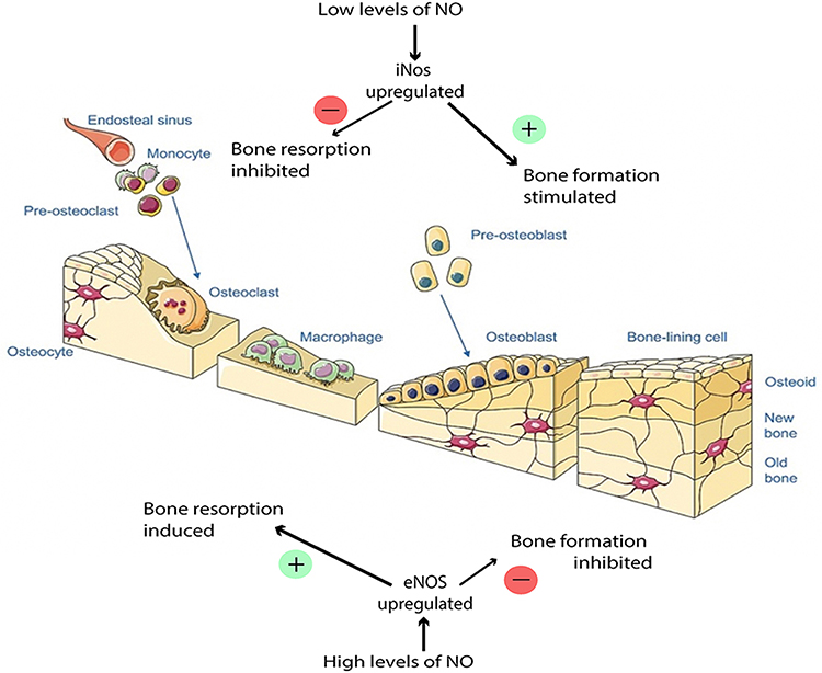 |
Figure 1 Nitric oxide as key regulatory molecule in bone remodeling by exerting dose-dependent effects on osteoblasts. At physiologically low concentrations, it promotes osteoblast proliferation and differentiation, thereby facilitating bone formation. Conversely at elevated NO levels trigger apoptosis in osteoblasts, contributing to the natural bone resorption process. Dual functionality underscores precise NO regulation in maintaining bone homeostasis and design of targeted NO-based therapeutic strategies for bone regeneration. Reproduced from Anastasio AT, Paniagua A, Diamond C, et al. Nanomaterial nitric oxide delivery in traumatic orthopedic regenerative medicine. Frontiers in Bioengineering and Biotechnology. 2021;8:592008.44 licensed under CC BY 4.0. The figure was created using Servier Medical Art templates, which are licensed under a Creative Commons Attribution 3.0 Unported License (https://creativecommons.org/licenses/by-sa/3.0/legalcode); https://smart.servier.com. |
NO plays a pivotal, concentration-dependent role in bone metabolism, affecting both osteoblasts and osteoclasts.14, At low concentrations, NO promotes osteogenesis by enhancing osteoblast proliferation, differentiation, and mineralization through the activation of key transcription factors such as runt-related transcription factor 2 (Runx2) and osterix.45–47 These effects are primarily mediated via the activation of soluble guanylate cyclase (sGC) and the subsequent increase in cyclic guanosine monophosphate (cGMP) levels, which trigger downstream signaling pathways involved in bone formation and repair.48 NO also facilitates the expression of alkaline phosphatase, a marker of osteoblastic activity, contributing to functional bone development.49,50 Additionally, NO plays a critical role in maintaining bone mass by modulating osteoclastogenesis. It inhibits the differentiation of osteoclast precursors through the receptor activator of nuclear factor kappa B ligand (RANKL) pathway, reducing osteoclast formation and activity.51–53 This dual function of NO stimulating bone formation via osteoblast activation while suppressing bone resorption by osteoclasts ensures the balance necessary for skeletal homeostasis. Beyond its effects on bone cells, NO is a potent mediator of angiogenesis.54–56 It stimulates endothelial cell proliferation, migration, and the formation of capillary-like structures, essential for the development of a functional vascular network within healing bone tissue.57 Proper vascularization is crucial for effective bone regeneration, ensuring the delivery of nutrients and oxygen to the site of injury, while also facilitating the recruitment of osteoprogenitor cells required for tissue repair.11,58–60 NO therefore functions as a molecular link between bone formation and neovascularization, both of which must occur in a coordinated manner for effective tissue regeneration Figure 2.
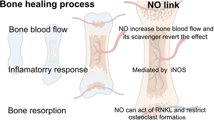 |
Figure 2 Nitric oxide’s role in coordinating both bone formation and neovascularization, processes essential for effective bone regeneration. As signaling molecule, it stimulates osteoblast activity, promoting new bone synthesis, simultaneously enhancing growth of new blood vessels. This dual function ensures regenerating bone tissue receives adequate nutrients and oxygen, facilitating efficient healing and restoration of bone integrity. Reproduced from Nascimento MH, T. Pelegrino M, C. Pieretti J, et al. How can nitric oxide help osteogenesis? AIMS Molecular Science. 2020;7(1):29–48.61 licensed under CC BY 4.0 (http://creativecommons.org/licenses/by/4.0). |
In addition to its osteogenic and angiogenic functions, NO plays a central role in regulating the inflammatory response during the early phases of bone healing.44,62 At physiologically relevant concentrations, it supports the resolution of inflammation by modulating cytokine production, inhibiting the adhesion and infiltration of leukocytes, and limiting the extent of tissue damage.63,64 This immunomodulatory effect contributes to the creation of a regenerative microenvironment that favors tissue repair rather than chronic inflammation.65 Taken together, the multifunctional biological activities of NO highlight its potential as a therapeutic agent in bone tissue engineering.44,66 However, due to its short biological half-life, chemical instability, and localized activity, effective delivery strategies are essential to achieve sustained and controlled NO signaling within bone defects.22,44,67 While polymeric nanoformulations hold significant promise for overcoming the challenges in bone regeneration, their successful application still faces numerous hurdles. These challenges, ranging from stability issues to controlled release, are crucial to address for clinical success.
Challenges in NO Delivery for Bone Regeneration
NO therapy for bone regeneration presents a unique pharmacological challenge due to the molecule’s paradoxical behaviour in biological systems. While NO demonstrates well-documented osteogenic and angiogenic properties at optimal concentrations, its therapeutic potential is constrained by several intrinsic limitations.13 The molecule’s gaseous nature and small size facilitate rapid diffusion across biological membranes, making containment and targeted delivery particularly difficult. This characteristic, combined with its reactive oxygen species (ROS) scavenging ability, creates a complex dosing paradigm where the molecule’s beneficial effects can quickly transition to cytotoxicity.68,69 The development of effective delivery systems account for NO’s concentration-dependent duality in bone metabolism. At physiological levels, NO promotes osteoblast differentiation through the upregulation of key transcription factors while simultaneously inhibiting osteoclast activity.70 However, these effects are reversed at higher concentrations, creating a narrow therapeutic window that demands precise spatiotemporal control.39 This challenge is compounded by the dynamic nature of bone healing, which progresses through distinct inflammatory, reparative, and remodelling phases each requiring different NO signaling patterns for optimal tissue regeneration.71
Current research focuses on overcoming these barriers through innovative biomaterial approaches. Smart delivery platforms incorporating stimuli-responsive polymers show promise in addressing the temporal aspects of NO delivery, with some formulations capable of detecting and responding to microenvironmental changes in pH, redox potential, or enzyme activity.72–74 Hybrid systems combining NO donors with structural biomaterials like hydroxyapatite or bioactive glasses attempt to solve the spatial control challenge by providing localized release at the defect site.33,66 Emerging technologies such as 3D-printed scaffold systems with gradient release profiles aim to mimic the natural healing cascade by delivering differential NO concentrations to distinct tissue zones Figure 3.
 |
Figure 3 Types of nanocarriers incorporated into 3D printed scaffold for nitric oxide delivery Reproduced from Suleman A, Kondiah PPD, Mabrouk M, et al. The application of 3D-printing and nanotechnology for the targeted treatment of osteosarcoma. Frontiers in Materials. 2021;8:668834.75 licensed under CC BY 4.0 license (https://creativecommons.org/licenses/by/4.0/). |
However, significant challenges remain in clinical translation. The field must develop standardized protocols for evaluating NO release kinetics and biological effects across different model systems.76 There is need for delivery platforms that can maintain NO stability during long-term storage while ensuring precise activation upon implantation.77 Furthermore, the potential for donor molecule degradation products to influence bone regeneration outcomes requires thorough investigation.78 By optimizing controlled release mechanisms, these therapies can fully leverage the pleiotropic effects of NO.
Polymer-Based Controlled Release Strategies
Polymers serve as the foundation of controlled drug delivery, with their selection dictated by biocompatibility, degradation behavior, and intended release kinetics.79 Natural polymers, such as chitosan, alginate, and gelatin, are derived from biological sources and offer inherent biocompatibility, making them suitable for applications like wound healing and mucosal delivery.80,81 However, they may exhibit batch-to-batch variability and occasional immunogenicity. In contrast, synthetic polymers like PLGA, PEG, and PCL provide precise control over mechanical properties and degradation rates, enabling reproducible and scalable drug delivery systems.82,83 Biodegradable polymers, including PLGA and polycaprolactone, are particularly advantageous as they break down into non-toxic byproducts, eliminating the need for surgical removal.84,85 Non-biodegradable polymers, such as silicone and ethylene-vinyl acetate, are reserved for long-term implants but carry risks of chronic inflammation and eventual retrieval.86,87 While PLGA remains most investigated polymer for NO delivery in bone repair, recent studies indicate promising developments using alternative materials. PEG-based nano-particles (mPEG-P) embedded within an injectable thermosensitive hydrogels to deliver NO through photothermal-responsive mechanism, demonstrating enhanced angiogenesis and osteogenesis in bone defect models.13 Hybrid scaffolds combining inorganic agents like zinc oxide combined with bioactive polymers and ECM components provide anti-inflammation, osteoinduction, and improved healing in osteoporotic bone models.88 Understanding the mechanisms of drug release from polymeric systems is crucial to improving the design of safer and more effective delivery platforms.
Mechanisms of Drug Release from Polymeric Systems
The release of therapeutics from polymeric carriers can be governed by various stimuli-responsive mechanisms (Table 1). Thermally responsive polymers, such as poly(N-isopropylacrylamide) (PNIPAM), undergo phase transitions near body temperature, enabling on-demand drug release in hyperthermia-based cancer therapy.89,90 Hydrolytic degradation, commonly observed in polyesters like PLGA, results in gradual drug release as the polymer erodes in aqueous environments.91,92 Enzymatically triggered systems leverage disease-specific enzymes, hyaluronidase in tumor microenvironments, to degrade polymers like hyaluronic acid and release encapsulated drugs.93 pH-sensitive polymers, including Eudragit and poly(β-amino esters), dissolve selectively in acidic or alkaline conditions, allowing targeted delivery to stomach, intestines, or cancerous tissues.94 The structural design of polymeric carriers plays a critical role in determining drug release profiles. Matrix systems, where the drug is uniformly dispersed within the polymer, typically exhibit an initial burst release followed by sustained diffusion-controlled release.95,96
These systems are widely used in PLGA microspheres for long-acting injectable formulations. In contrast, reservoir systems feature a drug core encapsulated by a rate-controlling polymer membrane, facilitating zero-order release kinetics ideal for hormonal therapies like contraceptive implants.97,98 While matrix systems are simpler to fabricate, reservoir systems offer more consistent drug release over extended periods.99 The localization of drugs within polymeric carriers significantly influences release dynamics.100 Surface loading techniques, such as adsorption or layer-by-layer assembly, enable rapid initial drug release, which is beneficial for applications requiring immediate therapeutic effects, such as antimicrobial coatings.101 Conversely, core loading strategies, where drugs are entrapped within the polymer matrix, provide prolonged release with minimal burst effects, making them suitable for sustained anticancer drug delivery.102
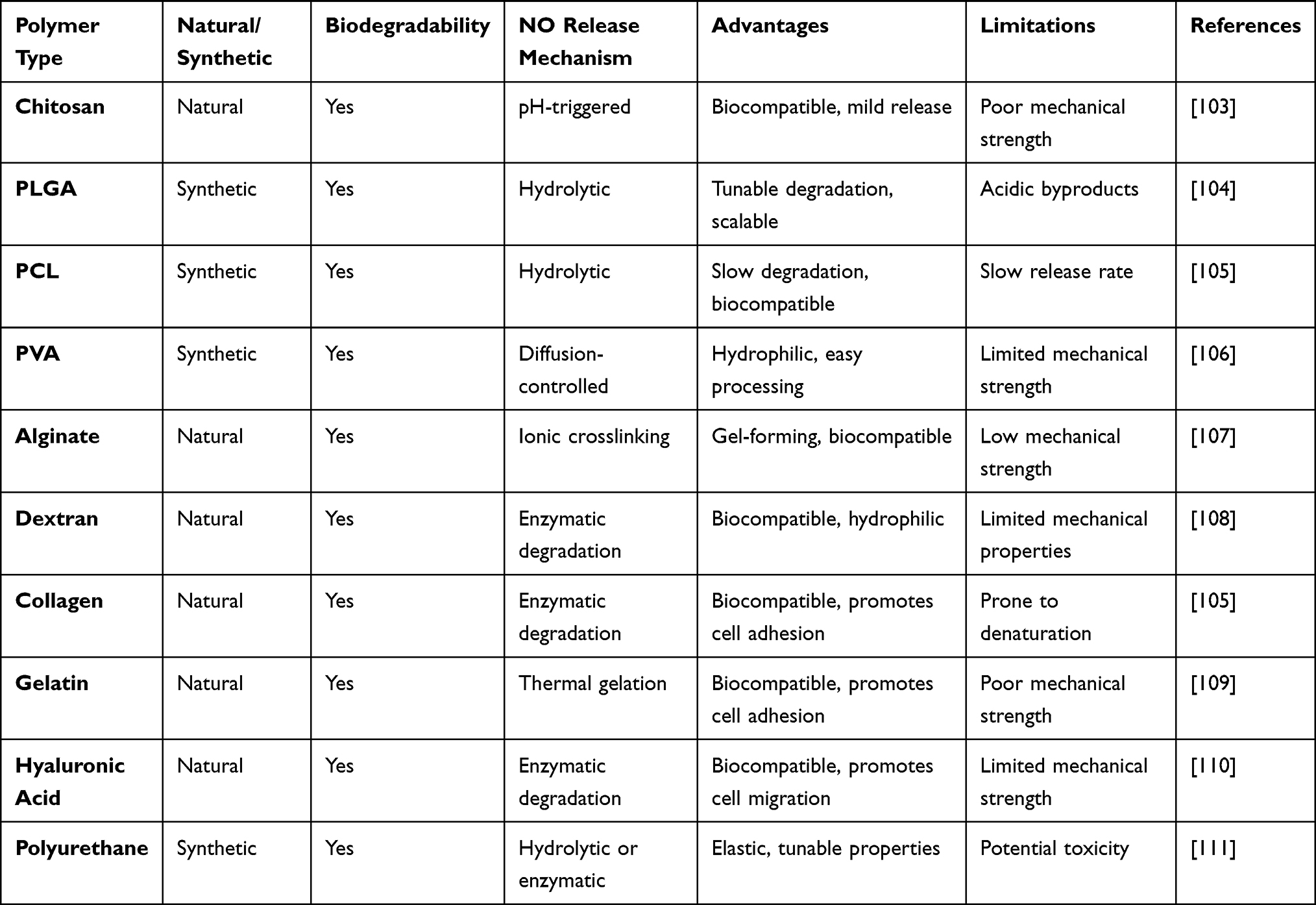 |
Table 1 Overview of Various Polymer Types Used in Nitric Oxide Delivery Systems Highlighting the Biodegradability, Release Mechanisms, Advantages, and Limitations of Both Natural and Synthetic Polymers Utilized for NO Release in Tissue Engineering Applications |
Polymeric nanocarriers intended for NO delivery are synthesized through various strategies including incorporation of NO donors during carrier formation or affix them afterward via surface modifications. Encapsulation during fabrication often employs emulsion-based techniques such as oil-in-water (O/W) or water-in-oil-in-water (W/O/W) methods where the NO donor is mixed with polymer solution prior to droplet formation, followed by solvent evaporation under stirring, sonication, or homogenization to yield NO-loaded nanoparticles. Alternatively, post loading includes conjugation of donors (S-nitrosothiols, diazeniumdiolates) onto functional groups of polymer scaffold, or blending of donor doped inorganic nanoparticles into polymer matrices. Polymer molecular weight, donor to polymer ratio, surfactant type, and polymer concentration influence entrapment efficiency, loading capacity, release rate, and stability of these nanoparticles.112,113
Due to highly reactive and transient nature of NO, confirming its successful loading within delivery systems requires highly sensitive and selective analytical techniques. Among widely adopted methods are chemiluminescence analysis, which provides quantitative assessment of NO release flux in real time, electrochemical sensors, capable of detecting NO via electrooxidation or electroreduction with modifications to enhance selectivity; and the Griess assay, which allows indirect quantification through nitrite measurement in oxygenated environments. In addition, fluorometric approaches employing diaminofluorescein (DAF) and diaminoanthraquinone (DAA)-based probes facilitate in vitro and in vivo imaging of NO release by exploiting photoinduced electron transfer (PET) quenching mechanisms. These methodologies are routinely utilized not only to verify NO encapsulation within nanocarriers but also to monitor its release kinetics, stability, and bioavailability, which are critical parameters in evaluating therapeutic performance of NO-delivery platforms.15
Recent advancements in polymer-based drug delivery emphasize hybrid systems that combine the biocompatibility of natural polymers with the tunability of synthetic ones.114,115 Stimuli-responsive platforms, including light-, redox-, and ultrasound-triggered systems, are gaining traction for precision medicine applications. Furthermore, 3D printing technology is enabling the fabrication of personalized drug-loaded implants with complex geometries, paving the way for patient-specific therapies.116
Nanotechnology Approaches for NO Delivery
NO is crucial in various physiological processes, including vasodilation, immune response, and wound healing.37 However, its gaseous nature and short half-life pose significant delivery challenges. Nanotechnology offers promising solutions by enabling controlled NO release through diverse nanocarriers and hybrid systems.117,118 Nanoscale delivery systems nanospheres, nanogels, dendrimers, and micelles have been extensively explored for NO storage and controlled release.119 Nanospheres, typically composed of polymers like PLGA or silica, encapsulate NO donors (S-nitrosothiols, N-diazeniumdiolates) within their core, providing sustained release kinetics.120,121 Nanogels, with their hydrophilic and highly tunable networks, allow for high NO payloads and stimuli-responsive release, particularly in response to pH or enzymatic triggers.119 Dendrimers, with their well-defined branched structures, enable precise NO donor conjugation at terminal functional groups, facilitating high loading efficiency and targeted delivery.122,123 Polymeric micelles, formed from amphiphilic block copolymers, can solubilize hydrophobic NO donors in their core while maintaining biocompatibility, making them suitable for systemic applications.124,125
Recent studies have demonstrated the efficacy of these nanocarriers in enhancing NO bioavailability while minimizing off-target effects. To further improve NO delivery efficiency, hybrid nanosystems combining polymers with inorganic materials have been developed. Polymer-ceramic composites, such as mesoporous silica nanoparticles coated with NO-releasing polymers, leverage the high surface area of ceramics for increased NO donor loading while utilizing polymers for controlled release.126 Polymer-metal hybrids, including gold or silver nanoparticles functionalized with NO-donating ligands, exploit the plasmonic properties of metals for light-triggered NO release, enabling spatiotemporal control.77 These hybrid systems not only enhance NO stability but also integrate additional functionalities of imaging capabilities or synergistic therapeutic effects.127 For instance, NO-releasing polymer-coated iron oxide nanoparticles have been explored for combined NO delivery and magnetic resonance imaging (MRI), highlighting their potential in theranostic applications.128 Recent studies have also highlighted the potential of NO-releasing nanocomposites in combination therapies, where NO synergizes with chemotherapy, photodynamic therapy, or immunotherapy to enhance treatment efficacy.128 These advancements underscore the versatility of nanotechnology in overcoming the challenges associated with NO delivery and expanding its therapeutic applications.129
Beyond NO-release systems, recent developments provide critical insights which reinforce the importance of material diversity in bone regeneration. Studies on PCL/bioactive glass or PCL/Fe3O4@ZIF-8 nanocomposites fabricated via 3D printing have demonstrated enhanced osteogenic size or differentiation of stem cells, antibacterial activity, and significant in vivo bone formation in infected defect models.130 GelMA, PCL nanofibers and bioactive glass have yielded higher bone volume/tissue volume (BV/TV) metrics in rat cranial defects relative to controls.131 Membranes of PCL/LAP nanosilicate blends exhibit favorable immunomodulation (M2 macrophage polarization), osteogenesis, and in vivo repair efficacy in periodontal and calvarial defect models.132
Polymer-NO Nanoformulations in Preclinical Bone Regeneration Models
The therapeutic potential of NO-releasing polymer nanoformulations for bone regeneration has been rigorously demonstrated across multiple preclinical models.77 Recent advances in controlled delivery systems have enabled precise modulation of osteogenic responses through tailored NO release kinetics and scaffold integration strategies.133
In vitro Findings: Cellular Responses and Osteogenic Potential
NO is a gaseous signaling molecule known for its multifaceted biological functions, including regulation of inflammation, vascular tone, and bone remodeling. Due to its ultra-short half-life and high reactivity, delivering NO in a controlled and sustained manner poses a significant challenge. In typical experimental studies, PLGA (poly(lactic-co-glycolic acid)) nanoparticles are used as carriers for NO donors like BPEI/NONOates.134 These are incorporated through nanoprecipitation or emulsion techniques, ensuring encapsulation efficiency and controlled release. In vitro cell culture experiments are conducted on pre-osteoblasts and macrophages to assess cytocompatibility, osteogenic differentiation (via alkaline phosphatase activity, calcium deposition), and polarization responses using qPCR and immunocytochemistry.134,135 In vitro studies have consistently shown that polymeric NO delivery systems do not exhibit significant cytotoxicity at therapeutic concentrations.15 On the contrary, NO-releasing PLGA nanoparticles promote proliferation and migration of osteoprogenitor cells. Moreover, these formulations influence macrophage polarization, favoring an M2-like, anti-inflammatory phenotype.12,14 This shift supports a regenerative microenvironment conducive to bone formation.
Enhanced proliferation and osteogenic differentiation in MC3T3-E1 pre-osteoblasts cultured with NO-releasing polymeric scaffolds.136 The scaffolds also induced VEGF expression and angiogenesis-related signaling, key factors in osteogenesis.69,137 Controlled release of NO has demonstrated a significant impact on osteoblast differentiation markers such as alkaline phosphatase (ALP), osteocalcin, and Runx2.88,138 Studies have shown elevated ALP activity and matrix mineralization following treatment with NO-loaded nanoparticles. A study using PLGA-based NO nanocarriers observed sustained NO release over several days, with a concomitant increase in calcium deposition and osteogenic gene expression.88,139,140 These findings underline the role of NO not only as an anti-inflammatory agent but also as a direct promoter of osteogenic pathways.141 Emerging in vitro studies on hybrid NO delivery systems integrating ZnO within polymeric scaffolds have demonstrated valuable outcomes in bone tissue engineering. These systems enhance endothelial cell migration and upregulate VEGF, promote osteogenic differentiation under co-culture conditions, and offer advancement in functionally integrated approaches for bone regeneration.88
In vivo Studies: Animal Models of Bone Repair
The translational potential of these systems has been validated in various animal models of bone regeneration (Table 2). A particularly compelling study in rat critical-sized calvarial defects, achieving greater bone volume compared to controls by micro-CT analysis.142 Histological evaluation revealed complete bridging of 5-mm defects by week 12, accompanied by a 3.2-fold increase in neovascularization as demonstrated through CD31 immunohistochemistry.143 For more complex regeneration scenarios requiring structural support, an advanced composite system combining poly(ε-caprolactone) nanofibers with NO-donating dendrimers.144 In a rabbit femoral condyle model, this approach achieved defect filling compared to in controls through histomorphometric analysis, with restored mechanical strength reaching of native bone.145 Molecular analysis revealed significant upregulation of both VEGF and BMP-2, suggesting the system’s ability to simultaneously promote angiogenesis and osteogenesis.146
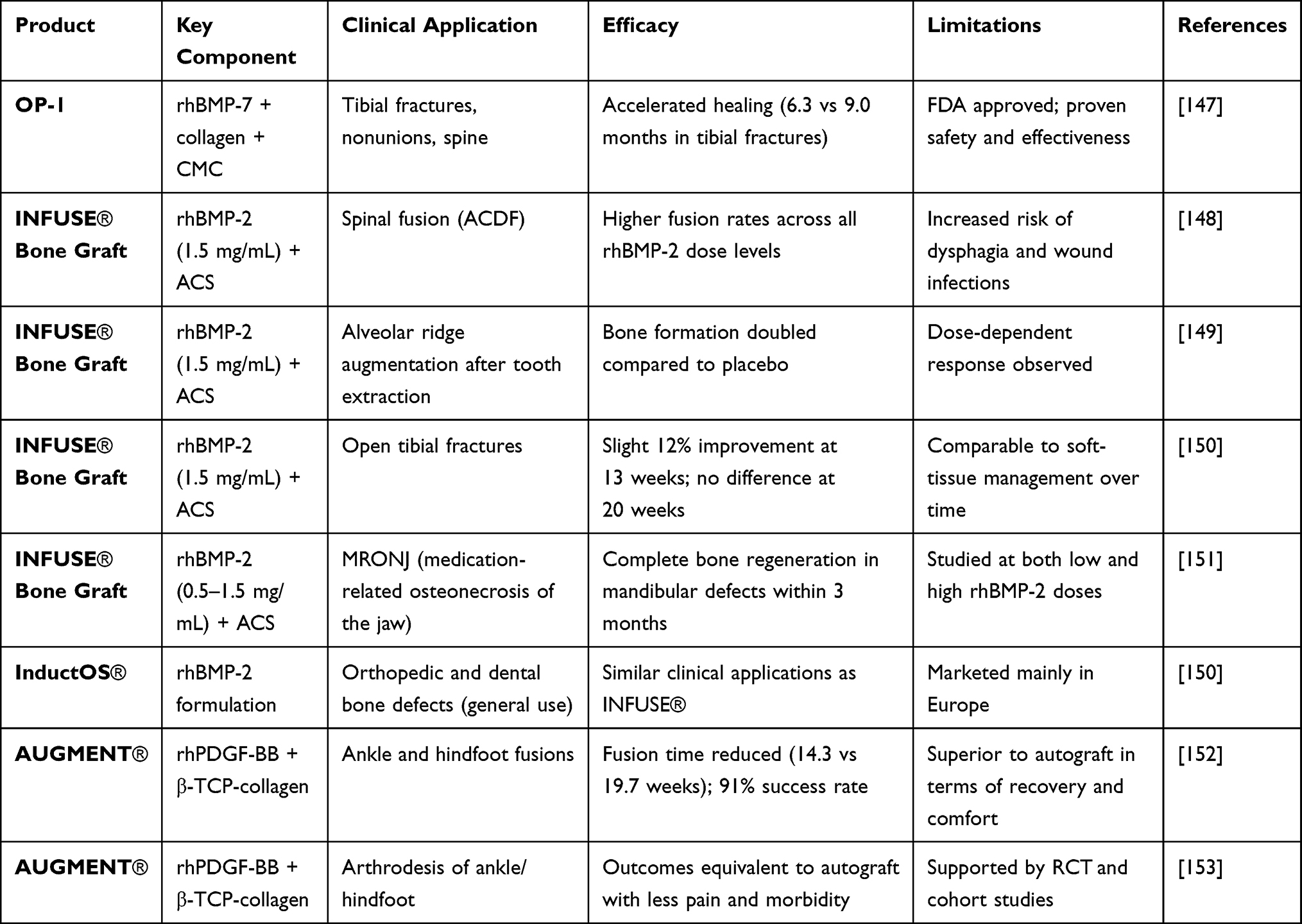 |
Table 2 Overview of Clinically Evaluated Polymer-Based Drug Delivery Systems for Bone Regeneration |
Bioinspired scaffold of PLGA/magnesium-modified hydroxyapatite/extracellular matrix composite, further functionalized with ZnO/ALN/BMP-2 nanoparticles exhibits robust repair in osteoporotic rat calvarial defects. NO release was confirmed by NO analyzer and DAF-FM fluorescence imaging. Compared to controls, treated group shown higher bone volume/tissue volume (BV/TV), bone mineral density (BMD), vascularization (VV/TV), and elevated expression of angiogenic and osteogenic genes (ALP, RUNX2, OCN). These in vivo findings illustrate integrating NO donors into multifunctional scaffold can restore bone (and vascular) architecture under osteoporotic conditions.88
Scaffold and Implant Integration
The successful integration of scaffolds and implants into host bone tissue is a critical determinant of the overall efficacy of bone regeneration therapies. NO-releasing polymer-based nanoformulations, when incorporated into scaffold systems (Figure 4), can significantly enhance the biological performance and integration of implants.121 Scaffold integration begins with the appropriate selection of biomaterials. Biocompatible materials such as titanium, hydroxyapatite, poly(lactic-co-glycolic acid) (PLGA), and bioactive ceramics have shown excellent results in facilitating osteointegration.154 The architecture of the scaffold—particularly its porosity and surface topology plays a key role in supporting cellular attachment, proliferation, and vascular infiltration.155 Ideal scaffolds mimic the extracellular matrix (ECM) and allow for nutrient diffusion and waste removal while providing mechanical support.156 Surface modification of scaffolds with bioactive molecules like growth factors (BMP-2), peptides (RGD sequences), or NO donors enhances their biofunctionality. NO release has been shown to stimulate osteoblast differentiation and angiogenesis while also modulating local immune responses. This bioactivity fosters a favorable microenvironment at the implant site, accelerating tissue regeneration and reducing the risk of fibrous tissue formation.156 Mechanical interlocking between the scaffold and host bone is essential for long-term implant stability. (Figure 4) This can be achieved by designing scaffolds with interconnected porosity and appropriate roughness, enabling deep bone ingrowth and tight integration. Studies have demonstrated that scaffolds with pore sizes between 100–400 μm are optimal for promoting osteoconduction and capillary infiltration.157,158
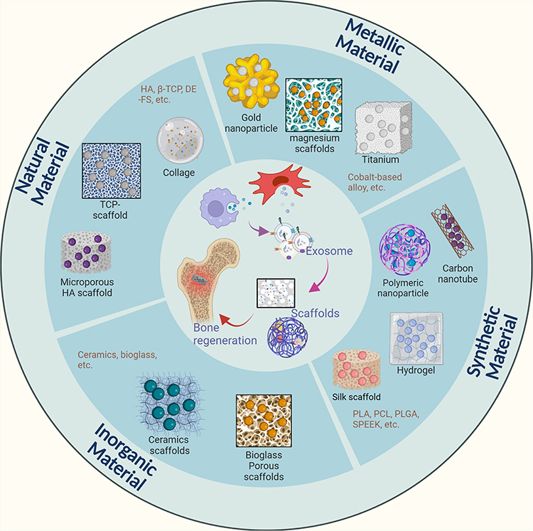 |
Figure 4 Exosome-based scaffolds combined with NO delivery offer bone regeneration without relying on transplanted cells. Exosomes are incorporated into scaffolds that release nitric oxide in a controlled manner to encourages new bone growth and the formation of blood vessels. This strategy not only promotes healing through enhanced cellular communication and angiogenesis but also helps regulate inflammation. Reproduced with permission from Deng L, Liu Y, Wu Q, et al. Exosomes to exosome-functionalized scaffolds: a novel approach to stimulate bone regeneration. Stem Cell Research & Therapy. 15(1):407. 2024, Springer Nature.159 |
Limitations, Toxicity & Regulatory Concerns
The clinical translation of polymer-based NO delivery systems is hindered by multiple limitations related to toxicity, material degradation, and regulatory inconsistency. Though PLGA is FDA-approved, its acidic degradation byproducts can significantly lower local pH, triggering pro-inflammatory responses and impairing osteoblast function.160 Research indicates that such acidic environments may promote M1 macrophage polarization and increased neutrophil infiltration.161 Similarly, chitosan degradation into glucosamine oligomers has been shown to activate TLR4-mediated inflammatory pathways when concentrations exceed physiological thresholds, raising concerns for chronic inflammation.162 In addition, NO itself has a narrow therapeutic window; sustained delivery beyond optimal levels can result in mitochondrial dysfunction, DNA damage, and apoptosis of osteogenic cells.163 High-output NO donors have been associated with the formation of peroxynitrite (ONOO−). These cytotoxic species aggravates oxidative stress and has been linked to increased bone resorption in diabetic models.164 Compounding these issues are manufacturing and regulatory barriers. Residual catalysts from synthesis, such as tin in polycaprolactone systems, have been observed to accumulate in hepatic tissue, necessitating stringent purification.165 Moreover, variability in release kinetics, poor reproducibility, and inconsistent preclinical testing protocols remain significant challenges. Lack of standardized protocols International Organization for Standardization (ISO) or American Society for Testing and Materials (ASTM) protocols and limited Good Laboratory Practice (GLP)-compliant toxicity evaluations further hinder progress.166
Future Directions and Emerging Trends
The next generation of NO-releasing polymer systems is moving towards advanced material designs, multimodal delivery strategies, and precision manufacturing to address current limitations in bone regeneration. Emerging smart nanoresponsive platforms are focusing on stimuli-triggered NO release, enabling spatiotemporal control (Table 3). For instance, poly(β-amino ester)-based nanogels exhibit a significant increase in NO release under acidic conditions (pH 6.0), which is ideal for targeting infected or inflammatory bone defects.167 Similarly, light-responsive systems incorporating upconversion nanoparticles (UCNPs) allow near-infrared (NIR) light-triggered NO liberation with rapid response times, facilitating non-invasive, on-demand therapy.72 Enzyme-cleavable linkers, such as matrix metalloproteinase (MMP)-sensitive peptides, further enhance specificity.168 Recent studies have shown that these systems can achieve higher NO release in osteoporotic bone compared to healthy tissue due to elevated protease activity.
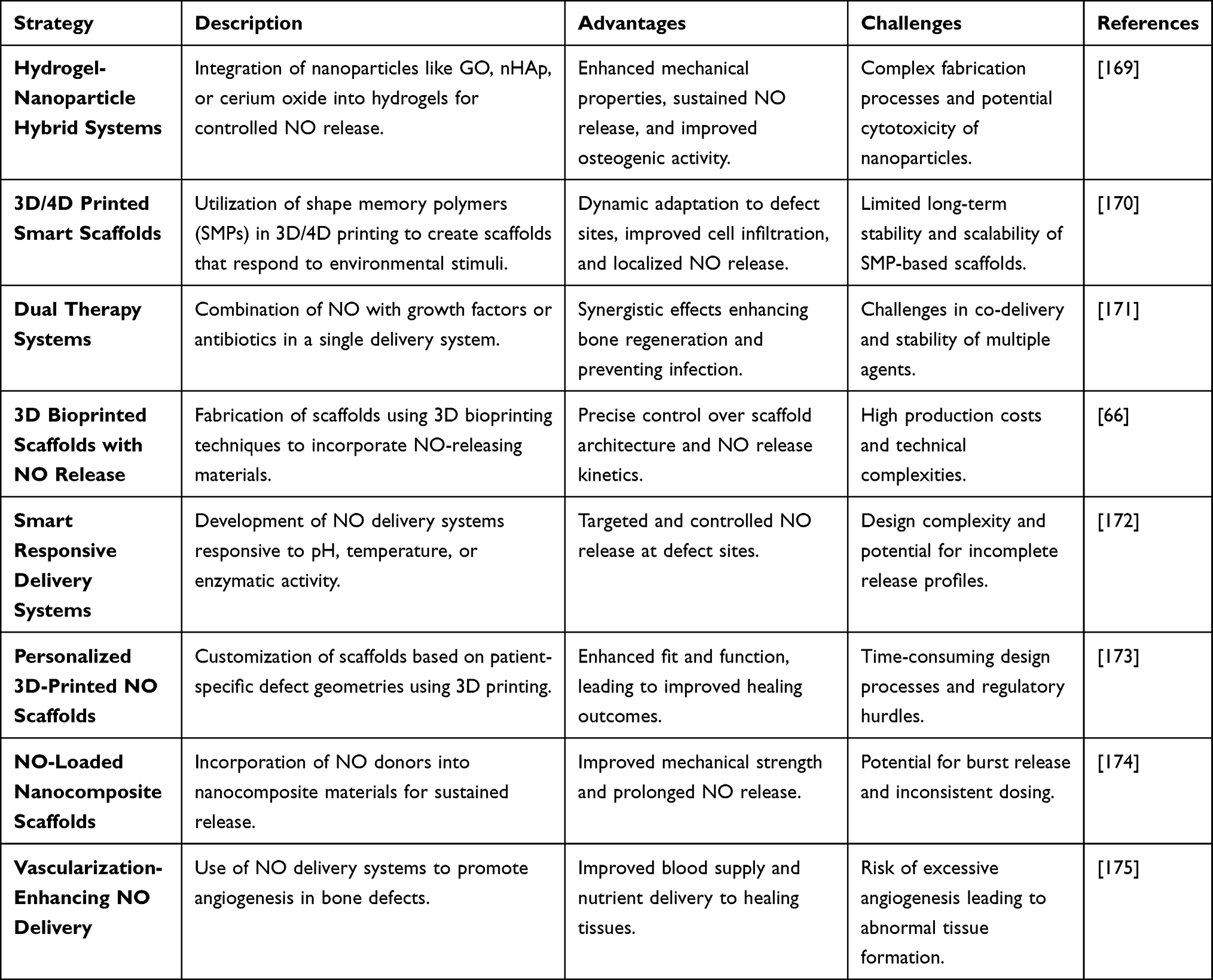 |
Table 3 Emerging Strategies for Nitric Oxide-Based Polymeric Delivery Systems in Bone Tissue Engineering. This Table Summarizes Recent Innovations Including Hybrid Platforms, Smart Scaffolds, and Personalized Systems, Highlighting Their Design Approach, Therapeutic Advantages, and Translational Challenges |
Conclusion
Polymer-based NO delivery systems represent a transformative frontier in bone regeneration, bridging the gap between advanced biomaterials engineering and targeted therapeutic intervention. These systems have demonstrated compelling preclinical efficacy by promoting osteogenesis, enhancing angiogenesis, and mitigating infection through localized, sustained NO release. By integrating NO with osteoinductive cues and responsive release mechanisms, these platforms offer precise spatiotemporal control aligned with the dynamic requirements of bone healing. However, their translation into clinical practice demands progress on multiple fronts. Critical challenges include manufacturing scalability, ensuring batch-to-batch consistency, and the development of standardized regulatory frameworks tailored to combination products. Furthermore, long-term safety, immunological compatibility, and efficacy must be rigorously validated in large-animal and disease-specific models. Recent advancements in additive manufacturing, AI-driven formulation design, and real-time biosensing are accelerating the evolution of these systems toward patient-specific, precision-guided therapies. With the convergence of material innovation, computational modeling, and personalized medicine, polymer-NO platforms are well-positioned to redefine the therapeutic landscape of bone repair. Their success will hinge not only on scientific innovation but also on regulatory clarity, interdisciplinary collaboration, and sustained clinical investment. If these align, the next decade could witness the transition of polymer-based NO delivery systems from bench to bedside, offering a sophisticated, responsive, and highly effective alternative to conventional bone grafting techniques.
Data Sharing Statement
This is a review article, and all relevant information is provided in the article.
Ethical Approval and Consent to Participate
This is a review paper and does not involve direct research on humans or animals.
Acknowledgment
This work was supported by Zhejiang Provincial Natural Science Foundation of China (QN25H040001 to LYX), Medical and Health Research Project of Zhejiang Province (2025KY1807 to CZ, 2025KY1804 to LYX), Traditional Chinese Medicine Science and Technology Projects of Zhejiang Province (2025ZR218 to LYX), Science and Technology Project of Zhoushan (2025C31060 to SQ, 2025C31079 to LC, 2024C31020 to HML), Research Fund Projects of The Affiliated Hospital of Zhejiang Chinese Medicine University (2024FSYYZZ14 to YJY, 2024FSYYZY34 to HML).
Author Contributions
All authors made a significant contribution to the work reported, whether that is in the conception, study design, execution, acquisition of data, analysis and interpretation, or in all these areas; took part in drafting, revising or critically reviewing the article; gave final approval of the version to be published; have agreed on the journal to which the article has been submitted; and agree to be accountable for all aspects of the work. All the authors listed meet the criteria for authorship as per the ICMJE guidelines, read the final manuscript and agree to publish this work.
Disclosure
The Authors declare that they have no competing interests financial or non-financial or any other interests that might be perceived to influence the results and/or discussion reported in this paper.
References
1. Salari N, Ghasemi H, Mohammadi L, et al. The global prevalence of osteoporosis in the world: a comprehensive systematic review and meta-analysis. J Orthopaedic Surg Res. 2021;16(1):609. doi:10.1186/s13018-021-02772-0
2. Fukuba S, Okada M, Nohara K, et al. Alloplastic bone substitutes for periodontal and bone regeneration in dentistry: current status and prospects. Materials. 2021;14(5):1096. doi:10.3390/ma14051096
3. Georgeanu VA, Gingu O, Antoniac IV, et al. Current options and future perspectives on bone graft and biomaterials substitutes for bone repair, from clinical needs to advanced biomaterials research. Appl Sci. 2023;13(14):8471. doi:10.3390/app13148471
4. Sadat‐Shojai M, Asadnia M, Shahsavani MB, et al. Bone regenerative medicine: an emerging field with opportunities and challenges. J Ame Ceramic Soc. 2025;108(11):e20508. doi:10.1111/jace.20508
5. Migliorini F, Cuozzo F, Torsiello E, et al. Autologous bone grafting in trauma and orthopaedic surgery: an evidence-based narrative review. J Clin Med. 2021;10(19):4347. doi:10.3390/jcm10194347
6. Kim Y-H, Kim K-W, Rhyu K-W, et al. Bone fusion materials: past, present, and future. Asian Spine J. 2025;19(3):490–18. doi:10.31616/asj.2024.0520
7. Nayak VV, Slavin B, Bergamo ETP, et al. Bone tissue engineering (BTE) of the craniofacial skeleton, Part I: evolution and optimization of 3D-printed scaffolds for repair of defects. J Craniofacial Surg. 2023;34(7):2016–2025. doi:10.1097/SCS.0000000000009593
8. Santoro A, Voto A, Fortino L, et al. Bone defect treatment in regenerative medicine: exploring natural and synthetic bone substitutes. Inter J Molecular Sci. 2025;26(7):3085. doi:10.3390/ijms26073085
9. Hung C-Y, Hsueh T-Y, Rethi L, et al. Advancements in regenerative medicine: a comprehensive review of stem cell and growth factor therapies for osteoarthritis. J Mater Chem B. 2025;13(15):4494–4526. doi:10.1039/D4TB01769B
10. Zhou Z, Liu J, Xiong T, et al. Engineering Innervated Musculoskeletal tissues for regenerative orthopedics and Disease modeling. Small. 2024;20(23):2310614. doi:10.1002/smll.202310614
11. Lee E-J, Jain M, Alimperti S. Bone microvasculature: stimulus for tissue function and regeneration. Tissue Engineering Part B. Reviews. 2021;27(4):313–329.
12. Jang H-J, Yoon J-K. The Role of Vasculature and Angiogenic Strategies in Bone Regeneration. Biomimetics. 2024;9(2):75. doi:10.3390/biomimetics9020075
13. Cheng Y, Huo Y, Yu Y, et al. A photothermal responsive system accelerating nitric oxide release to enhance bone repair by promoting osteogenesis and angiogenesis. Materials Today Bio. 2024;28:101180. doi:10.1016/j.mtbio.2024.101180
14. Won JE, Kim WJ, Shim JS, et al. Guided bone regeneration with a nitric‐oxide releasing polymer inducing angiogenesis and osteogenesis in critical‐sized bone Defects. Macromole Biosci. 2022;22(10):2200162. doi:10.1002/mabi.202200162
15. Paul S, Pan S, Mukherjee A, et al. Nitric oxide releasing delivery platforms: design, detection, biomedical applications, and future possibilities. Mole Pharmace. 2021;18(9):3181–3205. doi:10.1021/acs.molpharmaceut.1c00486
16. Bolívar-Monsalve EJ, Alvarez MM, Hosseini S, et al. Engineering bioactive synthetic polymers for biomedical applications: a review with emphasis on tissue engineering and controlled release. Mater Adv. 2021;2(14):4447–4478. doi:10.1039/D1MA00092F
17. Niculescu A-G, Grumezescu AM. Polymer-based nanosystems—A versatile delivery approach. Materials. 2021;14(22):6812. doi:10.3390/ma14226812
18. Elmowafy M, Shalaby K, Elkomy MH, et al. Polymeric nanoparticles for delivery of natural bioactive agents: recent advances and challenges. Polymers. 2023;15(5):1123. doi:10.3390/polym15051123
19. Beach MA, Nayanathara U, Gao Y, et al. Polymeric nanoparticles for drug delivery. Chem Rev. 2024;124(9):5505–5616. doi:10.1021/acs.chemrev.3c00705
20. Haider A, Khan S, Iqbal DN, et al. Advances in chitosan-based drug delivery systems: a comprehensive review for therapeutic applications. Euro Polymer J. 2024;210:112983. doi:10.1016/j.eurpolymj.2024.112983
21. Li M, Yu B, Wang S, et al. Microenvironment-responsive nanocarriers for targeted bone disease therapy. Nano Today. 2023;50:101838. doi:10.1016/j.nantod.2023.101838
22. Liu S, Li G, Ma D. Controllable nitric oxide‐delivering platforms for biomedical applications. Adv Therapeutics. 2022;5(3):2100227. doi:10.1002/adtp.202100227
23. Fattahi R, Mohebichamkhorami F, Taghipour N, et al. The effect of extracellular matrix remodeling on material-based strategies for bone regeneration. Tissue Cell. 2022;76:101748. doi:10.1016/j.tice.2022.101748
24. De R, Mahata MK, Kim KT. Structure‐based varieties of polymeric nanocarriers and influences of their physicochemical properties on drug delivery profiles. Adv Sci. 2022;9(10):2105373. doi:10.1002/advs.202105373
25. Mitchell MJ, Billingsley MM, Haley RM, et al. Engineering precision nanoparticles for drug delivery. Nat Rev Drug Discovery. 2021;20(2):101–124. doi:10.1038/s41573-020-0090-8
26. Visan AI, Popescu-Pelin G, Socol G. Degradation behavior of polymers used as coating materials for drug delivery—A basic review. Polymers. 2021;13(8):1272. doi:10.3390/polym13081272
27. Kim T, Nah Y, Kim J, et al. Nitric-oxide-modulatory materials for biomedical applications. Accf Chem Res. 2022;55(17):2384–2396. doi:10.1021/acs.accounts.2c00159
28. Coats JP, Cochereau R, Dinu IA, et al. Trends in the synthesis of polymer nano‐and microscale materials for bio‐related applications. Macromole Biosci. 2023;23(8):2200474. doi:10.1002/mabi.202200474
29. Machtakova M, Thérien-Aubin H, Landfester K. Polymer nano-systems for the encapsulation and delivery of active biomacromolecular therapeutic agents. Chem Soc Rev. 2022;51(1):128–152. doi:10.1039/D1CS00686J
30. Kumar R, Chhillar M. Nano-bio interface, bioadaptability of different nanoparticles, nanokicking and extracellular matrix mimicking: a biological and medicinal front to promote the concept of a cell, having a better defense system inbuilt by nature. Aditum J Clin Biomed Res. 2021;1:1–7.
31. Khalilov R. A comprehensive review of advanced nano-biomaterials in regenerative medicine and drug delivery. Advances in Biology & Earth Sciences. 2023;8(1):1.
32. Farjaminejad S, Farjaminejad R, Garcia-Godoy F. Nanoparticles in bone regeneration: a narrative review of current advances and future directions in tissue engineering. Journal of Functional Biomaterials. 2024;15(9):241. doi:10.3390/jfb15090241
33. Wang Y, Zhang H, Qiang H, et al. Innovative biomaterials for bone tumor treatment and regeneration: tackling postoperative challenges and charting the path forward. Advanced Healthcare Materials. 2024;13(16):2304060. doi:10.1002/adhm.202304060
34. Frączek W, Kotela A, Kotela I, et al. Nanostructures in Orthopedics: advancing Diagnostics, Targeted Therapies, and Tissue Regeneration. Materials. 2024;17(24):6162. doi:10.3390/ma17246162
35. Du Z, Cao G, Li K, et al. Nanocomposites for the delivery of bioactive molecules in tissue repair: vital structural features, application mechanisms, updated progress and future perspectives. Journal of Materials Chemistry B. 2020;8(45):10271–10289. doi:10.1039/D0TB01670E
36. Oyovwi MO, Atere AD. Exploring the medicinal significance of l-Arginine mediated nitric oxide in preventing health disorders. European Journal of Medicinal Chemistry Reports. 2024;12:100175. doi:10.1016/j.ejmcr.2024.100175
37. Lundberg JO, Weitzberg E. Nitric oxide signaling in health and disease. Cell. 2022;185(16):2853–2878. doi:10.1016/j.cell.2022.06.010
38. Shepherd M, Giordano D, Verde C, et al. The evolution of nitric oxide function: from reactivity in the prebiotic earth to examples of biological roles and therapeutic applications. Antioxidants. 2022;11(7):1222. doi:10.3390/antiox11071222
39. Cheng K, et al. The bone nonunion microenvironment: a place where osteogenesis struggles with osteoclastic capacity. Heliyon. 2024;10(10):1.
40. Hosseini N, Kourosh-Arami M, Nadjafi S, et al. Structure, distribution, regulation, and function of splice variant isoforms of nitric oxide synthase family in the nervous system. Current Protein and Peptide Science. 2022;23(8):510–534. doi:10.2174/1389203723666220823151326
41. Solanki K, Rajpoot S, Bezsonov EE, et al. The expanding roles of neuronal nitric oxide synthase (NOS1). PeerJ. 2022;10:e13651. doi:10.7717/peerj.13651
42. Villadangos L, Serrador JM. Subcellular Localization Guides eNOS Function. International Journal of Molecular Sciences. 2024;25(24):13402. doi:10.3390/ijms252413402
43. Roy R, Wilcox J, Webb AJ, et al. Dysfunctional and dysregulated nitric oxide synthases in cardiovascular disease: mechanisms and therapeutic potential. International Journal of Molecular Sciences. 2023;24(20):15200. doi:10.3390/ijms242015200
44. Anastasio AT, Paniagua A, Diamond C, et al. Nanomaterial nitric oxide delivery in traumatic orthopedic regenerative medicine. Frontiers in Bioengineering and Biotechnology. 2021;8:592008. doi:10.3389/fbioe.2020.592008
45. Chan WCW, Z Tan, MKT To, et al. Regulation and role of transcription factors in osteogenesis. International Journal of Molecular Sciences. 2021;22(11):5445. doi:10.3390/ijms22115445
46. Jing Z, Liang Z, Yang L, et al. Bone formation and bone repair: the roles and crosstalk of osteoinductive signaling pathways. Process Biochemistry. 2022;118:252–262. doi:10.1016/j.procbio.2022.04.033
47. Tang C-Y, M Wu, D Zhao, et al. Runx1 is a central regulator of osteogenesis for bone homeostasis by orchestrating BMP and WNT signaling pathways. PLoS Genetics. 2021;17(1):e1009233. doi:10.1371/journal.pgen.1009233
48. Arya PN, Saranya I, Selvamurugan N. RUNX2 regulation in osteoblast differentiation: a possible therapeutic function of the lncRNA and miRNA-mediated network. Differentiation. 2024;2024:100803.
49. Sekaran S, Vimalraj S, Thangavelu L. The physiological and pathological role of tissue nonspecific alkaline phosphatase beyond mineralization. Biomolecules. 2021;11(11):1564. doi:10.3390/biom11111564
50. Bartlett C-L, Cave EM, Crowther NJ, et al. A new perspective on the function of Tissue Non-Specific Alkaline Phosphatase: from bone mineralization to intra-cellular lipid accumulation. Molecular and Cellular Biochemistry. 2022;477(8):2093–2106. doi:10.1007/s11010-022-04429-w
51. Miriouni E, Trifonidi I, Chronopoulos E, et al. Alkaline Phosphatase-Biochemistry, Biological Functions Measurement and Clinical Relevance. Journal of Research and Practice on the Musculoskeletal System. 2025;9(1):33–45. doi:10.22540/JRPMS-09-033
52. Yao Z, Getting SJ, Locke IC. Regulation of TNF-induced osteoclast differentiation. Cells. 2021;11(1):132. doi:10.3390/cells11010132
53. Qu Z, Zhang B, Kong L, et al. Receptor activator of nuclear factor-κB ligand-mediated osteoclastogenesis signaling pathway and related therapeutic natural compounds. Frontiers in Pharmacology. 2022;13:1043975. doi:10.3389/fphar.2022.1043975
54. Singh S, Sarma DK, Verma V, et al. From cells to environment: exploring the interplay between factors shaping bone health and disease. Medicina. 2023;59(9):1546. doi:10.3390/medicina59091546
55. Li S, Cai X, Guo J, et al. Cell communication and relevant signaling pathways in osteogenesis–angiogenesis coupling. Bone Research. 2025;13(1):45. doi:10.1038/s41413-025-00417-0
56. Sharma A, Sharma L, Goyal R. Molecular signaling pathways and essential metabolic elements in bone remodeling: an implication of therapeutic targets for bone diseases. Current Drug Targets. 2021;22(1):77–104. doi:10.2174/18735592MTA5uODcAy
57. Banoriya GK, Singh VK, Maurya R, et al. Neuro-Immuno-Endocrine Regulation of Bone Homeostasis. Discovery Medicine. 2025;37(194):464–485. doi:10.24976/Discov.Med.202537194.39
58. Lv N, Zhou Z, Hou M, et al. Research progress of vascularization strategies of tissue-engineered bone. Frontiers in Bioengineering and Biotechnology. 2024;11:1291969. doi:10.3389/fbioe.2023.1291969
59. He Y, Liang L, Luo C, et al. Strategies for in situ tissue engineering of vascularized bone regeneration. Biomedical Reports. 2023;18(6):42. doi:10.3892/br.2023.1625
60. Huang J, Han Q, Cai M, et al. Effect of angiogenesis in bone tissue engineering. Annals of Biomedical Engineering. 2022;50(8):898–913. doi:10.1007/s10439-022-02970-9
61. Nascimento MH, T. Pelegrino M, C. Pieretti J, et al. How can nitric oxide help osteogenesis? AIMS Molecular Science. 2020;7(1):29–48. doi:10.3934/molsci.2020003
62. Jiang H, Ji P, Shang X, et al. Connection between osteoarthritis and nitric oxide: from pathophysiology to therapeutic target. Molecules. 2023;28(4):1683. doi:10.3390/molecules28041683
63. Kim ME, Lee JS. Advances in the Regulation of Inflammatory Mediators in Nitric Oxide Synthase: implications for Disease Modulation and Therapeutic Approaches. International Journal of Molecular Sciences. 2025;26(3):1204. doi:10.3390/ijms26031204
64. Tu Z, Zhong Y, Hu H, et al. Design of therapeutic biomaterials to control inflammation. Nature Reviews Materials. 2022;7(7):557–574. doi:10.1038/s41578-022-00426-z
65. Masoomikarimi M, Salehi M. Modulation of the immune system promotes tissue regeneration. Molecular Biotechnology. 2022;64(6):599–610. doi:10.1007/s12033-021-00430-8
66. de Almeida HV, Bomediano MP, Catori DM, et al. Integrating 3D printing of biomaterials with nitric oxide release. Biomaterials Science. 2025;13(4):858–874. doi:10.1039/D4BM01304B
67. Sun J, Wang W, Hu X, et al. Local delivery of gaseous signaling molecules for orthopedic disease therapy. Journal of Nanobiotechnology. 2023;21(1):58. doi:10.1186/s12951-023-01813-6
68. Xiong Y, Mi B, Liu G, et al. Microenvironment-sensitive nanozymes for tissue regeneration. Biomaterials. 2024;309:122585. doi:10.1016/j.biomaterials.2024.122585
69. Abdollahi F, Saghatchi M, Paryab A, et al. Angiogenesis in bone tissue engineering via ceramic scaffolds: a review of concepts and recent advancements. Biomaterials Advances. 2024;159:213828. doi:10.1016/j.bioadv.2024.213828
70. Zhu S, W Chen, A Masson, et al. Cell signaling and transcriptional regulation of osteoblast lineage commitment, differentiation, bone formation, and homeostasis. Cell Discovery. 2024;10(1):71.
71. Kim JH, Kim K, Kim I, et al. Nodal negatively regulates osteoclast differentiation by inducing stat1 phosphorylation. Journal of Cellular Physiology. 2024;239(6):e31268. doi:10.1002/jcp.31268
72. Hu S, Zhao R, Shen Y, et al. Revolutionizing drug delivery: the power of stimulus-responsive nanoscale systems. Chemical Engineering Journal. 2024;496:154265. doi:10.1016/j.cej.2024.154265
73. Shi Y, Zhang Y, Zhu L, et al. Tailored drug delivery platforms: stimulus‐responsive core–shell structured nanocarriers. Advanced Healthcare Materials. 2024;13(1):2301726. doi:10.1002/adhm.202301726
74. Shahi F, Afshar H, Dawi EA, et al. Smart Microneedles in Biomedical Engineering: harnessing Stimuli‐Responsive Polymers for Novel Applications. Polymers for Advanced Technologies. 2024;35(12):e70020. doi:10.1002/pat.70020
75. Suleman A, Kondiah PPD, Mabrouk M, et al. The application of 3D-printing and nanotechnology for the targeted treatment of osteosarcoma. Frontiers in Materials. 2021;8:668834. doi:10.3389/fmats.2021.668834
76. Neuber C, Niedenzu L, Schulze S, et al. Exploring a Nitric Oxide-Releasing Celecoxib Derivative as a Potential Modulator of Bone Healing: insights from Ex Vivo and In Vivo Imaging Experiments. International Journal of Molecular Sciences. 2025;26(6):2582. doi:10.3390/ijms26062582
77. Parisi C, Laneri F, Martins TJ, et al. Nitric Oxide-Photodelivering Materials with Multiple Functionalities: from Rational Design to Therapeutic Applications. ACS Applied Materials & Interfaces. 2024;16(44):59697–59720. doi:10.1021/acsami.4c13478
78. Łuczak JW, Palusińska M, Matak D, et al. The Future of Bone Repair: emerging Technologies and Biomaterials in Bone Regeneration. International Journal of Molecular Sciences. 2024;25(23):12766. doi:10.3390/ijms252312766
79. Gupta AK, A Choudhari, A Kumar, et al. Composites for drug-eluting devices: emerging biomedical applications. Applications of Biotribology in Biomedical Systems. 2024;251–311.
80. Khane Y, S Albukhaty, GM Sulaiman, et al. Fabrication, characterization and application of biocompatible nanocomposites: a review. Eur Polym J. 2024;113187.
81. Trucillo P. Biomaterials for drug delivery and human applications. Materials. 2024;17(2):456. doi:10.3390/ma17020456
82. Glisoni R. Encapsulation and Structural Modifications of Biotherapeutics. Springer; 2024.
83. Ding S, Alexander E, Liang H, et al. Synthetic and Biogenic Materials for Oral Delivery of Biologics: from Bench to Bedside. Chem Rev. 2025;1966–1978.
84. Garkal A, et al. Biodegradable Polymers in Tissue Engineering and Regenerative Medicine. In Handbook of Biodegradable Polymers. Jenny Stanford Publishing; 2025:449–524.
85. Ershad-Langroudi A, N Babazadeh. Polymers for implantable devices. Journal of Industrial and Engineering Chemistry. 2024.
86. Bedse A, Dhamane S, Raut S, et al. Polymeric Implants and Prosthetics. In: Polymers in Modern Medicine (Part 1). Bentham Science Publishers; 2024:140–185.
87. Saadat E. Pharmaceutical implants: from concept to commercialization. In: Advances in Drug Delivery Systems for Healthcare: From Concept to Clinic. Bristol, UK: IOP Publishing; 2024:1–42.
88. Lee JK, et al. Nitric oxide‐releasing bioinspired scaffold for exquisite regeneration of osteoporotic bone via regulation of homeostasis. Adv Sci. 2023;10(6):2205336. doi:10.1002/advs.202205336
89. Baghali M, Ziyadi H, Di Martino A. Engineering smart hydrogels for intelligent drug delivery. Polymer Bulletin. 2024; 2024:1–42.
90. Singh D. Dynamic Covalent Macromolecular Networks for Adaptive Drug Delivery Systems: an Informative Review. Journal of Macromolecular Science, Part B. 2024;2024:1–14.
91. Velu AJ, Thirunavukkarasu P, Rahman T, et al. Computational Analysis of Biodegradable Polyester Materials for Biomedical Applications: investigating Molecular Weight Change due to Hydrolysis. ACS omega. 2024;9(17):19108–19116. doi:10.1021/acsomega.3c10112
92. Nifant’ev IE, Tavtorkin AN, Shlyakhtin AV, et al. Chemical features of the synthesis, degradation, molding and performance of poly (lactic-co-glycolic) acid (PLGA) and PLGA-based articles. European Polymer Journal. 2024;215:113250. doi:10.1016/j.eurpolymj.2024.113250
93. El-Tanani M, Satyam SM, Rabbani SA, et al. Revolutionizing Drug Delivery: the Impact of Advanced Materials Science and Technology on Precision Medicine. Pharmaceutics. 2025;17(3):375. doi:10.3390/pharmaceutics17030375
94. Gomte SS, Agnihotri TG, Khopade S, et al. Exploring the potential of pH-sensitive polymers in targeted drug delivery. Journal of Biomaterials Science Polymer. 2024;35(2):228–268. doi:10.1080/09205063.2023.2279792
95. A Kumar, SK Jain, DK Mishra, et al. Influence of Drug Properties and Routes of Drug Administration on Design of Sustained and Controlled Release Systems. In: Novel Carrier Systems for Targeted and Controlled Drug Delivery. Springer; 2024:1–46.
96. Visan AI, Negut I. Development and applications of PLGA hydrogels for sustained delivery of therapeutic agents. Gels. 2024;10(8):497. doi:10.3390/gels10080497
97. Attia MS, NN Mohammad, M Ghonem, et al. Implantable drug delivery systems: design and applications. In: Novel Formulations and Future Trends. Elsevier; 2024:85–123.
98. Abdurashtov AS, Proshin PI, Sukhorukov GB. The pursuit of linear dosage in pharmacy: reservoir-based drug delivery systems from macro to micro scale. Expert Opinion on Drug Delivery. 2025;22(2):219–238. doi:10.1080/17425247.2024.2448026
99. Mehrotra S, Pathak K. Controlled release drug delivery systems: principles and design. In: Novel Formulations and Future Trends. Elsevier; 2024:3–30.
100. Jafarzadeh F, Peyman H, Roshanfekr H, et al. Fabrication of a nanomagnetic smart polymer carrier as a potential candidate for a drug delivery system. Arabian Journal for Science and Engineering. 2024;49(7):9381–9394. doi:10.1007/s13369-024-08724-0
101. Borges J, Zeng J, Liu XQ, et al. Recent Developments in Layer‐by‐Layer Assembly for Drug Delivery and Tissue Engineering Applications. Advanced Healthcare Materials. 2024;13(8):2302713. doi:10.1002/adhm.202302713
102. Ewii UE, AL Onugwu, VC Nwokpor, et al. Novel drug delivery systems: insight into self-powered and nano-enabled drug delivery systems. Nano TransMed. 2024;2024:100042.
103. Pérez-Pacheco Y, Tylkowski B, García-Valls R. Chitosan Micro/Nanocapsules in Action: linking Design, Production, and Therapeutic Application. Molecules. 2025;30(2):252. doi:10.3390/molecules30020252
104. Li Y, Liu J, He J, et al. Recent Advances in Stimuli-Responsive Self-Immolative Polymers for Drug Delivery and Molecular Imaging. Chemistry of Materials. 2024;36(9):4054–4077. doi:10.1021/acs.chemmater.4c00583
105. Ali M, Mohd Noor SNF, Mohamad H, et al. Advances in guided bone regeneration membranes: a comprehensive review of materials and techniques. Biomedical Physics & Engineering Express. 2024;10(3):032003. doi:10.1088/2057-1976/ad1e75
106. Wang Y. Biofabrication of 3D Functional Hybrid-Construct for Osteochondral Tissue Regeneration. The University of Manchester (United Kingdom); 2024.
107. Liying Q, Yining Y, Yongjian S, et al. Incorporation of carboxymethyl chitosan (CMCS) for the modulation of physio-chemical characteristics and cell proliferation environment of the composite hydrogel microspheres. Biomedical Materials. 2024;19(6):065003. doi:10.1088/1748-605X/ad7565
108. Cota Quintero JL, Ramos-Payán R, Romero-Quintana JG, et al. Hydrogel-Based Scaffolds: advancing Bone Regeneration Through Tissue Engineering. Gels. 2025;11(3):175. doi:10.3390/gels11030175
109. Wang Y, Lv H, Ren S, et al. Biological Functions of Macromolecular Protein Hydrogels in Constructing Osteogenic Microenvironment. ACS Biomaterials Science & Engineering. 2024;10(9):5513–5536. doi:10.1021/acsbiomaterials.4c00910
110. Srinivasan S, Vijayalekha A, Anandasadagopan S, et al. Hyaluronic Acid: a Comprehensive Review of Its Osteogenic Potential and Diverse Biomedical Applications. Current Pharmacology Reports. 2025;11(1):1–15. doi:10.1007/s40495-025-00408-z
111. Skleničková K, Abbrent S, Halecký M, et al. Biodegradability and ecotoxicity of polyurethane foams: a review. Critical Reviews in Environmental Science and Technology. 2022;52(2):157–202. doi:10.1080/10643389.2020.1818496
112. Gao M, Liu S, Fan A, et al. Nitric oxide-releasing graft polymer micelles with distinct pendant amphiphiles. RSC Advances. 2015;5(82):67041–67048. doi:10.1039/C5RA13341F
113. Yang L, Lu Y, Soto RJ, et al. S-Nitrosothiol-modified hyperbranched polyesters. Polymer Chemistry. 2016;7(46):7161–7169. doi:10.1039/C6PY01516F
114. Harun-Or-Rashid M, Aktar MN, Hossain MS, et al. Recent advances in micro-and nano-drug delivery systems based on natural and synthetic biomaterials. Polymers. 2023;15(23):4563. doi:10.3390/polym15234563
115. Quinn JF, Whittaker MR, Davis TP. Delivering nitric oxide with nanoparticles. Journal of Controlled Release. 2015;205:190–205. doi:10.1016/j.jconrel.2015.02.007
116. Gao Q, Lee J-S, Kim BS, et al. Three-dimensional printing of smart constructs using stimuli-responsive biomaterials: a future direction of precision medicine. International Journal of Bioprinting. 2022;9(1):638. doi:10.18063/ijb.v9i1.638
117. Tang Y, Li Q, Zhou Z, et al. Nitric oxide-based multi-synergistic nanomedicine: an emerging therapeutic for anticancer. Journal of Nanobiotechnology. 2024;22(1):674. doi:10.1186/s12951-024-02929-z
118. Kim J, Saravanakumar G, Choi HW, et al. A platform for nitric oxide delivery. Journal of Materials Chemistry B. 2014;2(4):341–356. doi:10.1039/C3TB21259A
119. Delgado-Pujol EJ, Martínez G, Casado-Jurado D, et al. Hydrogels and Nanogels: pioneering the Future of Advanced Drug Delivery Systems. Pharmaceutics. 2025;17(2):215. doi:10.3390/pharmaceutics17020215
120. Yang Y, Huang Z, Li -L-L. Advanced nitric oxide donors: chemical structure of NO drugs, NO nanomedicines and biomedical applications. Nanoscale. 2021;13(2):444–459. doi:10.1039/D0NR07484E
121. Qian Y, Kumar R, Chug MK, et al. Therapeutic delivery of nitric oxide utilizing saccharide-based materials. ACS Applied Materials & Interfaces. 2021;13(44):52250–52273. doi:10.1021/acsami.1c10964
122. Mittal P, Saharan A, Verma R, et al. Dendrimers: a new race of pharmaceutical nanocarriers. BioMed Research International. 2021;2021(1):8844030. doi:10.1155/2021/8844030
123. Pérez-Ferreiro M, M. Abelairas A, Criado A, et al. Dendrimers: exploring their wide structural variety and applications. Polymers. 2023;15(22):4369. doi:10.3390/polym15224369
124. Kaur J, Mishra V, Singh SK, et al. Harnessing amphiphilic polymeric micelles for diagnostic and therapeutic applications: breakthroughs and bottlenecks. Journal of Controlled Release. 2021;334:64–95. doi:10.1016/j.jconrel.2021.04.014
125. Figueiras A, Domingues C, Jarak I, et al. New advances in biomedical application of polymeric micelles. Pharmaceutics. 2022;14(8):1700. doi:10.3390/pharmaceutics14081700
126. Zhou X, Zhang J, Feng G, et al. Nitric oxide-releasing biomaterials for biomedical applications. Current Medicinal Chemistry. 2016;23(24):2579–2601. doi:10.2174/0929867323666160729104647
127. Pan Y, Zhao H, Huang W, et al. Metal‐Protein Hybrid Materials: unlocking New Frontiers in Biomedical Applications. Adv Healthc Mater. 2025;14(6):2404405. doi:10.1002/adhm.202404405
128. Jiang C, Zhou L, Hu W, et al. Recent advances in NO-triggered gas therapy by metal-organic frameworks. Materials Today Chemistry. 2024;36:101964. doi:10.1016/j.mtchem.2024.101964
129. Kaplish D, Vagha JD, Meshram RJ, et al. A comprehensive review of inhaled nitric oxide therapy: current trends, challenges, and future directions. Cureus. 2024;16(2):1.
130. Xiao Y, Ding Y, Qiu J, et al. Treatment effects of 3D-printed PCL/Fe3O4@ ZIF-8 magnetic nanocomposite on infected bone defect. International Journal of Bioprinting. 2024;10(4):2271. doi:10.36922/ijb.2271
131. Yu C, Chen J, Wang T, et al. GelMA hydrogels reinforced by PCL@ GelMA nanofibers and bioactive glass induce bone regeneration in critical size cranial defects. Journal of Nanobiotechnology. 2024;22(1):696. doi:10.1186/s12951-024-02980-w
132. Xu X, Chen Z, Xiao L, et al. Nanosilicate-functionalized nanofibrous membrane facilitated periodontal regeneration potential by harnessing periodontal ligament cell-mediated osteogenesis and immunomodulation. Journal of Nanobiotechnology. 2023;21(1):223. doi:10.1186/s12951-023-01982-4
133. Percival KM, Paul V, Husseini GA. Recent Advancements in Bone Tissue Engineering: integrating Smart Scaffold Technologies and Bio-Responsive Systems for Enhanced Regeneration. International Journal of Molecular Sciences. 2024;25(11):6012. doi:10.3390/ijms25116012
134. Oh Y, Park K, Jung S, et al. Inhalable nitric oxide delivery systems for pulmonary arterial hypertension treatment. Small. 2024;20(20):2308936. doi:10.1002/smll.202308936
135. Yang S, Guo L, Su Y, et al. Nitric oxide balances osteoblast and adipocyte lineage differentiation via the JNK/MAPK signaling pathway in periodontal ligament stem cells. Stem Cell Research & Therapy. 2018;9(1). doi:10.1186/s13287-018-0869-2
136. Sabzehmeidani MM, Kazemzad M. Recent advances in surface-mounted metal–organic framework thin film coatings for biomaterials and medical applications: a review. Biomaterials Research. 2023;27(1):115. doi:10.1186/s40824-023-00454-y
137. Zhang Z-Y, An Y-L, Wang X-S, et al. In vitro degradation, photo-dynamic and thermal antibacterial activities of Cu-bearing chlorophyllin-induced Ca–P coating on magnesium alloy AZ31. Bioactive Materials. 2022;18:284–299. doi:10.1016/j.bioactmat.2022.01.050
138. Abnos MH, Sargolzaei J, Maleklou M. Exogenous nitric oxide up-regulates the runx2 via bmp7 overexpression to increase the osteoblast matrix production in vitro. Avicenna Journal of Medical Biochemistry. 2022;10(1):58–64. doi:10.34172/ajmb.2022.08
139. Bernardo MP, da Silva BCR, Hamouda AEI, et al. PLA/Hydroxyapatite scaffolds exhibit in vitro immunological inertness and promote robust osteogenic differentiation of human mesenchymal stem cells without osteogenic stimuli. Scientific Reports. 2022;12(1):2333. doi:10.1038/s41598-022-05207-w
140. Yan B, Hua Y, Wang J, et al. Surface modification progress for PLGA-Based cell scaffolds. Polymers. 2024;16(1):165. doi:10.3390/polym16010165
141. Amroodi MN, et al. Unraveling the molecular and immunological landscape: exploring signaling pathways in osteoporosis. Biomedicine Pharmacotherapy. 2024;177:116954.
142. Bei H-P, et al. ECM-based Ca2+/l-arginine/NO periosteum nourishes bone defect microenvironment, directs macrophage polarity, and accelerates osteogenesis and angiogenesis. Composites Part B Engineering. 2024;278:111410.
143. Zhang X, Lan Z, Jiang S, et al. A highly versatile bioenergetic-active scaffold system for customized orthopedic applications. Materials Today. 2025;82:109–122. doi:10.1016/j.mattod.2024.12.008
144. Tabish TA, Crabtree MJ, Townley HE, et al. Nitric oxide releasing nanomaterials for cardiovascular applications. Basic to Translational Science. 2024;9(5):691–709. doi:10.1016/j.jacbts.2023.07.017
145. Sapoznikov L, Humphrey M. Progress in Dentin-Derived Bone Graft Materials: a New Xenogeneic Dentin-Derived Material with Retained Organic Component Allows for Broader and Easier Application. Cells. 2024;13(21):1806. doi:10.3390/cells13211806
146. Liu X, Zhou J, Chen M, et al. 3D-printed biomimetic bone scaffold loaded with lyophilized concentrated growth factors promotes bone defect repair by regulation the VEGFR2/PI3K/AKT signaling pathway. International Journal of Biological Macromolecules. 2024;282:136938. doi:10.1016/j.ijbiomac.2024.136938
147. White AP, Vaccaro AR, Hall JA, et al. Clinical applications of BMP-7/OP-1 in fractures, nonunions and spinal fusion. International Orthopaedics. 2007;31(6):735–741. doi:10.1007/s00264-007-0422-x
148. Malham GM, Louie PK, Brazenor GA, et al. Recombinant human bone morphogenetic protein-2 in spine surgery: recommendations for use and alternative bone substitutes—a narrative review. Journal of Spine Surgery. 2022;8(4):477. doi:10.21037/jss-22-23
149. Ogay V, Mun EA, Kudaibergen G, et al. Progress and prospects of polymer-based drug delivery systems for bone tissue regeneration. Polymers. 2020;12(12):2881. doi:10.3390/polym12122881
150. Peckham S, Zanella JM, McKay WF. Infuse® Bone Graft. Drug‐Device Combinations for Chronic Diseases. 2015;2015:241–260.
151. Min S-H, Kang N-E, Song S-I, et al. Regenerative effect of recombinant human bone morphogenetic protein-2/absorbable collagen sponge (rhBMP-2/ACS) after sequestrectomy of medication-related osteonecrosis of the jaw (MRONJ). Journal of the Korean Association of Oral and Maxillofacial Surgeons. 2020;46(3):191–196. doi:10.5125/jkaoms.2020.46.3.191
152. Daniels TR, Younger ASE, Penner MJ, et al. Prospective randomized controlled trial of hindfoot and ankle fusions treated with rhPDGF-BB in combination with a β-TCP-collagen matrix. Foot & Ankle International. 2015;36(7):739–748. doi:10.1177/1071100715576370
153. DiGiovanni CW, Baumhauer J, Lin SS, et al. Prospective, randomized, multi-center feasibility trial of rhPDGF-BB versus autologous bone graft in a foot and ankle fusion model. Foot & Ankle International. 2011;32(4):344–354. doi:10.3113/FAI.2011.0344
154. Maadani AM, Salahinejad E. Performance comparison of PLA-and PLGA-coated porous bioceramic scaffolds: mechanical, biodegradability, bioactivity, delivery and biocompatibility assessments. Journal of Controlled Release. 2022;351:1–7. doi:10.1016/j.jconrel.2022.09.022
155. Zhou J, Xiong S, Liu M, et al. Study on the influence of scaffold morphology and structure on osteogenic performance. Frontiers in bioengineering and Biotechnology. 2023;11:1127162. doi:10.3389/fbioe.2023.1127162
156. Gu R, Liu H, Zhu Y, et al. Is extracellular matrix (ECM) a promising scaffold biomaterial for bone repair?. Histol Histopathol. 2021;36(12):1219–1234. doi:10.14670/HH-18-370
157. Jin Y, J Li, H Fan, et al. Biomechanics and Mechanobiology of Additively Manufactured Porous Load‐Bearing Bone Implants. Small. 2025;21(20):2409955.
158. Gui X, Zhang B, Song P, et al. 3D printing of biomimetic hierarchical porous architecture scaffold with dual osteoinduction and osteoconduction biofunctions for large size bone defect repair. Applied Materials Today. 2024;37:102085. doi:10.1016/j.apmt.2024.102085
159. Deng L, Liu Y, Wu Q, et al. Exosomes to exosome-functionalized scaffolds: a novel approach to stimulate bone regeneration. Stem Cell Research & Therapy. 2024;15(1):407. doi:10.1186/s13287-024-04024-4
160. Md yusop AH, Ulum MF, Al Sakkaf A, et al. Insight into the bioabsorption of Fe‐based materials and their current developments in bone applications. Biotechnology Journal. 2021;16(12):2100255. doi:10.1002/biot.202100255
161. Lu X, et al. Interaction of macrophages with bone healing microenvironment: mechanism and biomaterials. Tissue Eng. B Rev. 2024;30(3):285–298.
162. Austin JW. Evaluation of the Use of a Bioengineered Hydrogel Containing Hyaluronan to Reduce Inflammation and Scarring Following Spinal Cord Injury Associated with Arachnoiditis. University of Toronto (Canada); 2012.
163. Andrabi SM, Sharma NS, Karan A, et al. Nitric oxide: physiological functions, delivery, and biomedical applications. Advanced Science. 2023;10(30):2303259. doi:10.1002/advs.202303259
164. Sharma P, Sri Swetha Victoria V, Praneeth Kumar P, et al. Cross-talk between insulin resistance and nitrogen species in hypoxia leads to deterioration of tissue and homeostasis. International Immunopharmacology. 2023;122:110472. doi:10.1016/j.intimp.2023.110472
165. Esmaeili J, Jalise SZ, Pisani S, et al. Development and characterization of Polycaprolactone/chitosan-based scaffolds for tissue engineering of various organs: a review. International Journal of Biological Macromolecules. 2024;272:132941. doi:10.1016/j.ijbiomac.2024.132941
166. Funk KA, Hampshire VA, Schuh JC. Nonclinical safety evaluation of medical devices. In: Toxicologic Pathology. CRC Press; 2018:95–152.
167. Latanza S. Fabrication and Efficacy of Poly (Beta-amino Esters)-Based Nanoparticles to Enhance Antimicrobial Agents Delivery to Orthopaedic Acquired Infections Sites. Cardiff University; 2020.
168. Chandrawati R. Enzyme-responsive polymer hydrogels for therapeutic delivery. Experimental Biology and Medicine. 2016;241(9):972–979. doi:10.1177/1535370216647186
169. Wang Y, Liu J, Zhang C, et al. Recent development of chitosan-based biomaterials for treatment of osteomyelitis. Journal of Polymer Engineering. 2024;44(8):542–558. doi:10.1515/polyeng-2023-0294
170. Yuan X, Zhu W, Yang Z, et al. Recent advances in 3D printing of smart scaffolds for bone tissue engineering and regeneration. Advanced Materials. 2024;36(34):2403641. doi:10.1002/adma.202403641
171. Afewerki S, et al. Advances in dual functional antimicrobial and osteoinductive biomaterials for orthopaedic applications. Nanomedicine: nanotechnology. Biology and Medicine. 2020;24:102143.
172. Karimi M, Ghasemi A, Sahandi Zangabad P, et al. Smart micro/nanoparticles in stimulus-responsive drug/gene delivery systems. Chemical Society Reviews. 2016;45(5):1457–1501. doi:10.1039/c5cs00798d
173. Borthakur PP, A Das, JJ Sahariah, et al. Revolutionizing Patient Care: 3D Printing for Customized Medical Devices and Therapeutics. Biomedical Materials & Devices. 2025;20251–28.
174. Poh WH, Rice SA. Recent developments in nitric oxide donors and delivery for antimicrobial and anti-biofilm applications. Molecules. 2022;27(3):674. doi:10.3390/molecules27030674
175. Huang X, Wang H, Wang C, et al. The applications and potentials of extracellular vesicles from different cell sources in periodontal regeneration. International Journal of Molecular Sciences. 2023;24(6):5790. doi:10.3390/ijms24065790
 © 2026 The Author(s). This work is published and licensed by Dove Medical Press Limited. The
full terms of this license are available at https://www.dovepress.com/terms
and incorporate the Creative Commons Attribution
- Non Commercial (unported, 4.0) License.
By accessing the work you hereby accept the Terms. Non-commercial uses of the work are permitted
without any further permission from Dove Medical Press Limited, provided the work is properly
attributed. For permission for commercial use of this work, please see paragraphs 4.2 and 5 of our Terms.
© 2026 The Author(s). This work is published and licensed by Dove Medical Press Limited. The
full terms of this license are available at https://www.dovepress.com/terms
and incorporate the Creative Commons Attribution
- Non Commercial (unported, 4.0) License.
By accessing the work you hereby accept the Terms. Non-commercial uses of the work are permitted
without any further permission from Dove Medical Press Limited, provided the work is properly
attributed. For permission for commercial use of this work, please see paragraphs 4.2 and 5 of our Terms.