
Back to Journals » Medical Devices: Evidence and Research » Volume 18
A Recently Patented Sleeve-Type Endotracheal Tube: Innovative Design and Clinical Prospects for Improving One-Lung Ventilation
Authors Zhang S, Li N, Su N, Yun L, Li L, Zhi J, Lin Q, Zhu M, Li K, Zhao Z
Received 6 March 2025
Accepted for publication 29 April 2025
Published 6 May 2025 Volume 2025:18 Pages 281—290
DOI https://doi.org/10.2147/MDER.S526736
Checked for plagiarism Yes
Review by Single anonymous peer review
Peer reviewer comments 2
Editor who approved publication: Dr Scott Fraser
Shengmao Zhang,1,* Na Li,2,* Nan Su,1 Letian Yun,1 Lele Li,1 Jing Zhi,1 Qihang Lin,1 Mengxin Zhu,1 Keer Li,1 Zhihui Zhao1
1Inner Mongolia People’s Hospital, Hohhot, Inner Mongolia Autonomous Region, People’s Republic of China; 2Pain Department, Hohhot First Hospital, Hohhot, Inner Mongolia Autonomous Region, People’s Republic of China
*These authors contributed equally to this work
Correspondence: Zhihui Zhao, Inner Mongolia People’s Hospital, Hohhot, Inner Mongolia Autonomous Region, People’s Republic of China, Email [email protected]
Abstract: The Sleeve-Type Endotracheal Tube (STET) is an innovative airway management device designed to address the limitations associated with traditional double-lumen tubes (DLTs) and bronchial blockers (BBs). Utilizing a modular structure comprising an outer sheath tube, inner sheath tube, and blocking tube, the STET offers increased flexibility in one-lung ventilation (OLV) and airway clearance, thereby reducing airway trauma and improving ventilation efficiency. Its dual-cuff system ensures stable positioning, while its versatile design accommodates diverse patient populations, including pediatric patients and those with difficult airway anatomy. This review explores the structural characteristics, manufacturing processes, clinical advantages, potential limitations, and practical applications of the STET. After multiple rounds of structural optimization, the STET design has successfully advanced into pilot production. Preliminary validation results from small-scale manufacturing and functional testing demonstrate promising structural stability, operational flexibility, and safety performance. The STET represents a significant advancement in airway management, holding substantial potential to improve patient outcomes and transform clinical practice in thoracic surgery and critical care anesthesiology.
Keywords: sleeve-type endotracheal tube, one-lung ventilation, airway management, modular design, thoracic surgery
The successful implementation of thoracic surgery relies on anesthesiologists’ precise management of lung isolation and one-lung ventilation (OLV).1 OLV is a critical technique that allows ventilation of one lung while collapsing the other, providing a static surgical field and protecting healthy lung tissue from cross-contamination, all while maintaining adequate oxygenation.2 This technique is widely used in pulmonary resections, minimally invasive cardiac surgery, esophagectomy, and certain spinal surgeries. Currently, double-lumen tubes (DLTs) and bronchial blockers (BBs) are the primary airway devices employed to achieve OLV in clinical practice.3
Both DLTs and BBs have their advantages and limitations. DLTs are favored for their rapid and stable positioning. Their dual-lumen design facilitates easy switching between bilateral lung ventilation and OLV, making them particularly suitable for surgeries requiring alternating operation on both lungs.4 Additionally, DLTs support continuous positive airway pressure (CPAP), aiding in the management of hypoxemia during OLV. DLTs also allow for efficient suctioning of secretions and blood, which is especially beneficial for patients with significant distal airway secretions or bleeding.5 However, due to their larger external diameter, DLTs pose a risk of laryngeal and tracheal injury, with postoperative complications such as sore throat, hoarseness, and airway edema commonly reported.6 Moreover, DLT use is limited in pediatric patients and those with difficult airways or abnormal tracheobronchial anatomy, and the limited range of available sizes can increase the difficulty of tube selection and placement.7
In contrast, BBs, which are inserted through single-lumen tubes (SLTs), are often the preferred choice for pediatric patients and those with complex airway anatomy.8 The smaller external diameter of BBs reduces the risk of laryngeal and tracheal injuries, while their larger internal diameter improves ventilation efficiency. Furthermore, BBs can be easily removed after OLV, minimizing the need for tube exchange in patients requiring prolonged postoperative mechanical ventilation.9 However, BBs have significant drawbacks, including more complex placement procedures, longer positioning times compared to DLTs, and a higher likelihood of intraoperative displacement requiring frequent readjustments.10 When patients’ positions change during surgery, BBs are more prone to dislocation,11 which may interfere with surgical operations. BBs also have limited capacity for clearing secretions from the non-ventilated lung, making them less ideal for surgeries requiring alternating lung isolation.12
In summary, both DLTs and BBs are effective tools for achieving OLV, yet each has its inherent limitations. Optimizing airway management tools to enable safer, more stable, and efficient OLV remains a critical scientific challenge in the field of anesthesiology for thoracic surgery.
To address these challenges, we have developed the Sleeve-Type Endotracheal Tube (STET), a recently patented airway device that integrates several innovative design elements. Unlike existing double-sleeve ET or traditional devices, the STET features a modular dual-lumen architecture, detachable inner components, and a dual-cuff structure. These patented innovations enhance operational flexibility, improve sealing performance, and increase procedural safety. The STET aims to combine the advantages of both DLT and BB while minimizing their respective shortcomings, such as airway trauma, complex positioning, and secretion management difficulties. By optimizing both structural design and functional performance, the STET represents a promising advancement in airway management for one-lung ventilation.
Design of the Sleeve-Type Endotracheal Tube (STET)
STET is a recently patented, novel airway device specifically designed to overcome the limitations of existing endotracheal tools. Unlike traditional devices, the STET integrates the advantages of the DLT, such as ease of positioning and reduced displacement, with the benefits of the BB, including a smaller external diameter, larger ventilation lumen, and minimized airway trauma. Furthermore, through its modular dual-lumen architecture, detachable inner components, and dual-cuff structure, the STET provides enhanced flexibility and operational efficiency, effectively addressing the shortcomings of both DLT and BB.
Structural Features
The STET design consists of an outer sheath tube (1) and an inner sheath tube (7), where the inner tube is inserted into the lumen of the outer sheath tube and forms a detachable, sealed connection with the outer tube. The length of the inner sheath tube is fixed and does not exceed the length of the outer sheath tube (Figure 1).
 |
Figure 1 The outer sheath tube. 1. Outer sheath tube; 2. First cuff; 3. Second cuff; 4. First inflation port; 5. Second inflation port; 6. Branch tube; 7. Inner sheath tube; 8. Connector sealing cap; 9. Breathing hole. |
The front end of the outer sheath tube is equipped with two cuffs: the first cuff (2) and the second cuff (3). A breathing hole (9) is located between the two cuffs. The rear end of the outer sheath tube is fixed with a first inflation port (4) and a second inflation port (5), which are connected to the first and second cuffs through inflation tubes embedded within the wall of the outer sheath tube. The rear end of the outer sheath tube is inclined and equipped with a branch tube (6) that passes through it, with the end of the branch tube fixed to a ventilator interface. This branch tube not only meets ventilation needs but also provides a passage for bronchoscopy and airway clearance (Figure 2).
 |
Figure 2 The inner sheath tube. 7. Inner sheath tube; 10. Ventilator interface; 11. Sealing cuff; 12. Inflation tube; 13. Third inflation port. |
The inner sheath tube is a hollow, fixed-length tube structure with openings at both ends. The front end of the inner tube is equipped with a sealing cuff (11), and the rear end is fixed with a third inflation port (13), which connects through tubes embedded within the inner tube to control cuff inflation.
Operation and Flexibility
The STET supports a straightforward and adaptable operation. First, the outer sheath tube is inserted into one side of the bronchus, ensuring that the first cuff (2) enters the bronchus, with the breathing hole (9) positioned in the trachea. Both cuffs (2 and 3) are inflated to seal them. Next, the inner sheath tube is inserted into the outer sheath tube, and the sealing cuff (11) at the front of the inner tube is positioned across the breathing hole (9) of the outer sheath tube. The sealing cuff (11) is inflated to block the inner lumen of the outer sheath tube.
Based on surgical needs, the appropriate inner sheath tube can be selected and swapped to alternate between OLV and airway clearance. Due to significant anatomical variations in the right upper lobe bronchus, left-sided insertion of the outer sheath tube is preferred in clinical practice, except when a right-sided OLV is required. If left-sided OLV is needed, a larger inner sheath tube is chosen, and the ventilator interface (10) at the tail end of the inner tube is connected to the ventilator. Ventilation is performed through the outer sheath tube branch (6), collapsing the right lung. Conversely, if right-sided OLV is required, a smaller inner sheath tube is used to seal the outer sheath tube and collapse the left lung, with the ventilator connected to the outer sheath tube branch (6) for ventilation (Figure 3).
 |
Figure 3 Schematic diagram of operation method. 2. First cuff; 6. Branch tube; 7. Inner sheath tube; 8. Connector sealing cap; 9. Breathing hole; 10. Ventilator interface; 11. Sealing cuff; 14. Blocking tube; 15. Inflated cuffs. |
For bilateral lung ventilation, the inner sheath tube (2) is removed, and a sealing cap (8) is placed on either the outer sheath tube’s ventilator port (7) or the branch tube port (6) to block it. The remaining port is then connected to the ventilator for ventilation.
Pilot Production and Preliminary Validation
After multiple rounds of structural and technical refinement, the STET design was finalized and advanced to a small-scale pilot production phase. The objective of this pilot study was twofold: first, to evaluate the structural durability and material endurance of the device under simulated operational conditions; and second, to assess its functional performance in airway management procedures using simulated airway models. This preliminary evaluation focused on validating the design and operational feasibility of the STET, establishing a foundation for subsequent development and future clinical application.
In selecting materials and manufacturing processes, we employed a range of polymers, including polyvinyl chloride (PVC), polyurethane (TPU), polypropylene (PP), and nylon (PA), tailored specifically for the outer sheath tube, inner sheath tube, and blocking tube. The materials and manufacturing processes were shown in Table 1. The outer sheath tube was extruded from PVC and featured a thermally molded tip, which ensured sufficient flexibility and compatibility with airway anatomy (Figure 4A). The inner sheath tube, designed to fit precisely within the outer sheath, enabled accurate lung-lobe isolation (Figure 4B). The blocking tube, fabricated by extruding PA with an integrated TPU balloon, facilitated localized bronchial occlusion or airway clearance (Figure 4C).
 |
Table 1 Materials and Manufacturing Processes of the Main STET Components |
 |
Figure 4 STET and its main components. (A) Outer sheath tube with dual. cuff structure for secure placement in the airway; (B) Inner sheath tube designed for precise lung. lobe isolation; inserted within the outer sheath tube; (C) Blocking tube (blocker) used for localized bronchial occlusion or airway clearance; (D) Complete STET assembly showing the integrated arrangement of the outer sheath tube; inner sheath tube; and blocking tube. |
Manufacturing methods such as extrusion, injection molding, blow molding, and thermal forming were carefully chosen to meet specific requirements concerning hardness, flexibility, transparency, and biocompatibility. Both the outer and inner sheath tubes utilized PVC extrusion combined with thermal forming techniques to maintain optimal flexibility and structural stability within the airway. Balloon components, fabricated through TPU or PVC blow molding based on their functional placement, effectively balanced expansion characteristics with durability.
Following individual component production and rigorous quality inspections, semi-automated assembly was conducted under clean-room conditions to produce the final STET device. As illustrated in Figure 4D, the completed STET integrates the outer sheath tube, inner sheath tube, and blocking tube into a cohesive unit. Each balloon underwent independent leak and pressure testing to verify safety and reliability standards essential for clinical application. Concurrently, repeated adjustments were made to material formulas and manufacturing processes to optimize transparency and visualization, thus enabling clinicians to achieve a consistently clear view during direct laryngoscopy or fiber-optic bronchoscopy.
During the preliminary functional validation, we focused on several key aspects: we first assessed the ease of intubation and operational flexibility by testing STET insertion, inner sheath replacement, and blocker placement in simulated airways and anatomical models, thereby confirming its navigability and positioning capability under both routine and difficult airway conditions. We then evaluated sealing integrity and lung isolation performance by inflating the two cuffs of the outer sheath tube and the balloon on the inner sheath tube (or blocker) separately, verifying their capacity to effectively block airflow and achieve targeted lung isolation while accommodating a collapsed non-ventilated lung or maintaining CPAP. In terms of safety and durability, we repeatedly inflated and deflated the balloons and monitored the catheter walls in a 37°C environment over several hours, observing no significant material degradation, deformation, or balloon rupture. Finally, regarding cleaning and visualization, the branch interface on the outer sheath tube and the removable design of the inner sheath tube facilitated suctioning, secretion clearance, and management of intraoperative bleeding, while fiber-optic bronchoscopy ensured prompt aspiration of secretions and preservation of a clear view.
Overall, the results from the small-scale pilot production and preliminary tests demonstrate that the STET has achieved the expected goals in terms of structural stability, operational flexibility, and lung isolation efficiency. Larger-scale pilot production and clinical trials will be conducted in the future to collect multicenter data and further refine the STET’s manufacturing parameters and material formulations.
Advantages of the Sleeve-Type Endotracheal Tube (STET)
The STET overcomes many limitations of traditional airway devices, offering a range of distinct advantages that improve safety, efficiency, and versatility in clinical practice. By combining the strengths of DLTs and BBs while mitigating their respective drawbacks, the STET represents a significant advancement in airway management for thoracic surgery and other complex procedures.13 To provide an overall comparison of the STET with existing airway devices, Table 2 summarizes the functional features of the STET in relation to the DLT and BB.
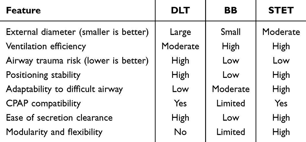 |
Table 2 Comparative Analysis Highlighting the Advantages of STET Over DLT and BB |
Enhanced Safety and Reduced Airway Trauma
One of the primary advantages of the STET is its smaller external diameter, which significantly reduces the risk of laryngeal and bronchial trauma during intubation. This is particularly important for patients with difficult airways, airway compression, or anatomical abnormalities where traditional DLTs may pose challenges.14 By minimizing trauma to the airway structures, the STET helps reduce the incidence of postoperative complications such as sore throat, hoarseness, and airway edema.15 This advantage also extends to pediatric patients, for whom appropriately sized DLTs are often unavailable, making the STET a safer and more feasible option.16
Superior Ventilation Efficiency
The STET features a larger ventilation lumen compared to traditional DLTs, enhancing ventilation efficiency during OLV.17 This improvement ensures adequate oxygenation and ventilation, particularly in patients with compromised respiratory function. For instance, an outer sheath tube with a 7.5 mm internal diameter, paired with an inner sheath tube of 2.3 mm internal diameter, provides ventilation performance equivalent to a SLT with a 7.14 mm internal diameter. This configuration is especially beneficial for patients requiring high ventilation demands or prolonged surgical procedures.18 Alternatively, using an inner sheath tube with a 5 mm internal diameter achieves ventilation performance comparable to a 39Fr DLT, offering flexibility for varying clinical scenarios.
Modular Design and Surgical Adaptability
The modular design of the STET, featuring interchangeable inner sheath tubes, offers exceptional flexibility to accommodate various surgical needs. This design enables rapid transitions between OLV and airway clearance, minimizing procedural interruptions and allowing anesthesiologists to efficiently adapt to dynamic surgical requirements.19 For example, the inner sheath tube can be quickly replaced to switch between ventilating different lungs or to facilitate airway clearance without necessitating a complete tube replacement.
Additionally, the modularity allows the STET to handle diverse surgical scenarios. When the inner sheath tube is used for ventilation, the outer sheath tube can serve to collapse the non-ventilated lung or clear the airway. Conversely, when ventilating through the outer sheath tube, the inner sheath tube can be employed for lung isolation. This adaptability is particularly beneficial in complex surgeries requiring frequent transitions, such as bilateral thoracic procedures.20 By reducing the risks associated with tube exchange—such as airway trauma or loss of ventilation—the STET ensures safer and more efficient intraoperative management.21
Stable Positioning and Reduced Adjustments
The dual-cuff structure of the outer sheath tube ensures stable positioning, significantly reducing the likelihood of displacement during surgery.22 This stability is particularly valuable in thoracic procedures, where changes in patient positioning, such as transitioning from supine to lateral decubitus, are common. The reduced need for intraoperative adjustments enhances surgical workflow efficiency and minimizes disruptions. Additionally, the outer sheath tube’s secure fit decreases the reliance on fiberoptic bronchoscopy for positioning verification, further simplifying the procedure.
Versatility for Diverse Clinical Scenarios
The STET’s versatility makes it suitable for a wide range of patients and clinical scenarios. It is especially advantageous for patients with difficult airways, including those with airway compression due to tumors or tracheobronchial anomalies. Its smaller external diameter and modular inner sheath tube design provide a practical solution for pediatric patients, addressing a significant limitation of traditional DLTs. Moreover, the STET supports nasal and awake intubation, offering flexibility in cases where oral intubation may not be feasible.
The STET is also compatible with CPAP for the non-ventilated lung, an essential feature for managing hypoxemia during OLV. This capability enhances its utility in complex surgeries and critical care settings, ensuring better patient outcomes.
Application Prospects of the Sleeve-Type Endotracheal Tube (STET)
The STET holds significant promise for advancing airway management, particularly in thoracic surgical anesthesia. Its unique design, combining the strengths of DLTs and BBs, positions it as an innovative solution to meet diverse clinical demands.22 The STET’s versatility, safety profile, and operational efficiency make it a valuable tool in a variety of clinical scenarios, including difficult airway management, pediatric anesthesia, and surgeries requiring precise lung isolation.
Enhanced Safety and Efficiency in Thoracic Surgery
Thoracic surgery often requires meticulous airway management to achieve effective OLV and ensure optimal surgical exposure.23 The STET’s dual-cuff design and modular inner sheath tube allow for stable positioning, reduced risk of airway trauma, and seamless transitions between OLV and bilateral ventilation. These features significantly enhance patient safety and minimize complications such as laryngeal and bronchial injuries. Furthermore, the STET’s compatibility with CPAP supports the management of hypoxemia during OLV, a critical challenge in thoracic surgery.24
Broad Clinical Applicability
The STET’s adaptability extends its application beyond thoracic surgery. It is particularly advantageous in patients with difficult airways, airway compression due to tumors, or tracheobronchial anomalies, where traditional DLTs may be difficult or impossible to use.25 Its smaller external diameter and larger ventilation lumen make it suitable for pediatric patients, addressing a significant gap in airway management for younger populations. The STET also supports nasal and awake intubation, offering anesthesiologists greater flexibility in managing complex cases.26
Practical Benefits in Resource-Limited Settings
The STET’s simple and efficient design makes it ideal for use in primary care hospitals and resource-limited settings. Unlike DLTs, which require precise sizing and carry higher risks of complications, the STET’s modular structure and ease of insertion reduce the technical demands on anesthesiologists.27 Its compatibility with both fiberoptic bronchoscopy and traditional auscultation methods simplifies the learning curve, enabling broader adoption across healthcare settings. Additionally, the reduced need for intraoperative repositioning enhances surgical workflows and reduces operating room time.
Potential for Broader Adoption
As healthcare systems increasingly emphasize patient safety, efficiency, and cost-effectiveness, the STET aligns well with these priorities. Its ability to combine the advantages of existing airway devices while mitigating their limitations highlights its potential for widespread adoption. Future clinical studies comparing the STET to DLTs and BBs in terms of safety, efficacy, and cost-benefit will be critical to establishing its role as a standard airway management tool.28
Driving Innovation in Airway Management
The STET not only addresses current challenges in thoracic anesthesia but also sets the stage for further innovations in airway management. Its modular design could inspire the development of next-generation devices tailored to specific surgical needs, such as robotic-assisted thoracic surgery or minimally invasive procedures. Additionally, the integration of advanced materials and technologies, such as real-time pressure sensors or biofeedback systems, could further enhance the STET’s functionality and safety.
Potential Limitations of the Sleeve-Type Endotracheal Tube (STET)
While the STET presents significant advantages in airway management, it is essential to acknowledge its potential limitations to provide a balanced perspective and identify areas for improvement.
Increased Complexity in Design and Manufacturing
The STET’s modular design and dual-component structure, while innovative, may increase manufacturing complexity and costs compared to conventional airway devices like DLTs and BBs. The integration of multiple cuffs, inflation ports, and the detachable inner sheath tube requires precise engineering and quality control, which could pose challenges in large-scale production. This may affect its affordability and accessibility, particularly in resource-limited settings.
Learning Curve for Clinical Implementation
The STET introduces a new approach to airway management, which may require additional training for anesthesiologists unfamiliar with its operation. Proper insertion and management, especially the precise coordination between the inner and outer sheath tubes, could initially lengthen procedural times in clinical settings. The reliance on specific insertion techniques, such as fiberoptic bronchoscopy (FOB) or auscultation, may further complicate its adoption in hospitals with limited expertise or equipment.
Risk of Airway Leakage and Misalignment
Although the STET is designed for secure and stable positioning, the detachable connection between the inner and outer sheath tubes introduces a potential risk of airway leakage or misalignment. During complex or prolonged surgeries, shifts in patient positioning or repeated adjustments could disrupt the seal, potentially compromising ventilation or lung isolation. Additional clinical studies are needed to evaluate the robustness of the STET in these scenarios.
Limited Evidence From Clinical Trials
As a novel device, the STET currently lacks extensive clinical trial data to fully validate its efficacy, safety, and long-term outcomes compared to established devices. While its design addresses known limitations of DLTs and BBs, empirical evidence is essential to confirm its superiority in diverse patient populations and surgical contexts. The lack of robust data may initially limit its acceptance and adoption among anesthesiologists.
Compatibility With Existing Ventilators and Equipment
The STET’s dual-component structure and modular design may raise compatibility concerns with standard ventilators and other airway management equipment. For example, ensuring a secure and efficient connection between the STET and various ventilator models could require additional adapters or modifications, potentially complicating its use in different clinical settings.
Prospects and Outlook
The STET holds great promise for transforming airway management in thoracic surgery and beyond. Its innovative modular design, combining versatility and efficiency, makes it a strong candidate for addressing diverse clinical needs. Beyond thoracic anesthesia, the STET’s adaptability offers advantages for managing difficult airways, pediatric cases, and airway compression, positioning it as a versatile tool in a wide range of surgical and critical care scenarios. As healthcare systems emphasize safety and efficiency, the STET is well-aligned to meet these demands and improve patient outcomes.
Future advancements could further enhance the STET’s potential by integrating smart technologies like real-time pressure monitoring and automated cuff adjustments. Comprehensive clinical trials and comparative studies are crucial to validating its safety, efficacy, and cost-effectiveness. Efforts to address current limitations, such as manufacturing complexity and compatibility with existing equipment, will enable broader adoption. With its simplicity and accessibility, the STET could also benefit resource-limited settings, making advanced airway management more widely available. As these developments unfold, the STET is poised to become a cornerstone of modern airway management, advancing the field of anesthesiology and improving surgical care.
Conclusion
The STET offers an innovative approach to airway management, combining the strengths of traditional devices like DLTs and BBs while addressing their limitations. Its modular design ensures adaptability, safety, and efficiency, making it particularly valuable for OLV and airway clearance in thoracic surgery. With ongoing refinement and clinical validation, the STET has the potential to set a new standard in airway management, improving patient outcomes and advancing the field of anesthesiology.
Funding
This study was supported by the Inner Mongolia Autonomous Region “14th Five-Year Plan” Key R&D and Achievement Transformation Project in the Social Welfare Field (Project No. 2022YFSH0016), and The Inner Mongolia Autonomous Region Health and Wellness Science and Technology Program (Project No. 202201007).
Disclosure
The authors declare no competing financial interests.
References
1. Mitzman B, Beller JP, Edgar L. Thoracic surgery milestones 2.0: rationale and revision. J Thorac Cardiov Sur. 2020;160(5):1399–1404. doi:10.1016/j.jtcvs.2019.12.132
2. Bernasconi F, Piccioni F. One-lung ventilation for thoracic surgery: current perspectives. Tumori J. 2017;103(6):495–503. doi:10.5301/tj.5000638
3. Szamos K, Balla B, Pálóczi B, et al. One-lung ventilation with fixed and variable tidal volumes on oxygenation and pulmonary outcomes: a randomized trial. J Clin Anesth. 2024;95:111465. doi:10.1016/j.jclinane.2024.111465
4. Zhang C, Qin X, Zhou W, et al. Prediction of Left Double-Lumen Tube Size by Measurement of Cricoid Cartilage Transverse Diameter by Ultrasound and CT Multi-Planar Reconstruction. Front Med Lausanne. 2021;8:657612. doi:10.3389/fmed.2021.657612
5. Yu W, Wang Z, Gao D, et al. A method for addressing right upper lobe obstruction with right-sided double-lumen endobronchial tubes during surgery: a randomized controlled trial. BMC Anesthesiol. 2018;18(1):130. doi:10.1186/s12871-018-0596-3
6. Mihatsch LL, Weiland S, Helmberger T, et al. Common double-lumen tube selection methods overestimate adequate tube sizes in individual patients - a 3D reconstruction study. BMC Anesthesiol. 2024;24(1):215. doi:10.1186/s12871-024-02605-7
7. Zango BA, Morales SP, Raimundo MG, et al. Correct placement of left-sided double lumen endotracheal tubes: a simple verification technique. Rep Esp Anast Ren. 2008;55(5):277–281. doi:10.1016/s0034-9356(08)70570-x
8. Kim TK, Hong DM, Lee SH, et al. Effect-site concentration of remifentanil required to blunt haemodynamic responses during tracheal intubation: a randomized comparison between single- and double-lumen tubes. J Int Med Res. 2017;46(1):430–439. doi:10.1177/0300060517721072
9. Mirzabeigi E, Johnson C, Ternian A. One-lung anesthesia update. Semin Cardiothorac. 2005;9(3):213–226. doi:10.1177/108925320500900304
10. Luther DGP, Robertson HF, Suchett-Kaye I, et al. Double-lumen tracheal tubes and bougies: a bench study to investigate factors that influence the risk of shearing. Anaesthesia. 2019;74(7):891–895. doi:10.1111/anae.14672
11. Zhang R, Liu S, Sun H, et al. The application of single-lumen endotracheal tube anaesthesia with artificial pneumothorax in thoracolaparoscopic oesophagectomy. Interact Cardiov Th. 2014;19(2):308–310. doi:10.1093/icvts/ivu100
12. Hoshijima H, Maruyama K, Mihara T, et al. Airtraq® reduces the hemodynamic response to tracheal intubation using single-lumen tubes in adults compared with the Macintosh laryngoscope: a systematic review and meta-analysis of randomized control trials. J Clin Anesth. 2018;47:86–94. doi:10.1016/j.jclinane.2018.03.022
13. Heidegger T, Longo DL. Management of the Difficult Airway. New Engl J Med. 2021;384(19):1836–1847. doi:10.1056/NEJMra1916801
14. Weingart SD, Levitan RM. Preoxygenation and prevention of desaturation during emergency airway management. Ann Emerg Med. 2011;59(3):165–75.e1. doi:10.1016/j.annemergmed.2011.10.002
15. Mushambi MC, Athanassoglou V, Kinsella SM. Anticipated difficult airway during obstetric general anaesthesia: narrative literature review and management recommendations. Anaesthesia. 2020;75(7):945–961. doi:10.1111/anae.15007
16. Scott JA, Heard SO, Zayaruzny M, et al. Airway Management in Critical Illness: an Update. Chest. 2019;157(4):877–887. doi:10.1016/j.chest.2019.10.026
17. Gigengack RK, Cleffken BI, Loer SA. Advances in airway management and mechanical ventilation in inhalation injury. Curr Opin Anesthesio. 2020;33(6):774–780. doi:10.1097/ACO.0000000000000929
18. Xu Z, Ma W, Hester DL, et al. Anticipated and unanticipated difficult airway management. Curr Opin Anesthesio. 2018;31(1):96–103. doi:10.1097/ACO.0000000000000540
19. Müller SH, Defosse JM, Gerbershagen MU, et al. Difficult Airway Management in Thoracic Anaesthesia. Anasth Intensiv Notf. 2018;53(3):187–197. doi:10.1055/s-0043-114679
20. Mushambi MC, Kinsella SM, Popat M, et al. Obstetric Anaesthetists’ Association and Difficult Airway Society guidelines for the management of difficult and failed tracheal intubation in obstetrics. Anaesthesia. 2015;70(11):1286–1306. doi:10.1111/anae.13260
21. Gibbins M, Kelly FE, Cook TM. Airway management equipment and practice: time to optimise institutional, team, and personal preparedness. Brit J Anaesth. 2020;125(3):221–224. doi:10.1016/j.bja.2020.06.012
22. Cumberworth A, Lewith H, Sud A, et al. Major complications of airway management: a prospective multicentre observational study. Anaesthesia. 2022;77(6):640–648. doi:10.1111/anae.15668
23. Schieren M, Böhmer A, Dusse F, et al. New Approaches to Airway Management in Tracheal Resections-A Systematic Review and Meta-analysis. J Cardiothor Vasc An. 2017;31(4):1351–1358. doi:10.1053/j.jvca.2017.03.020
24. Jagannathan N, Sohn L, Fiadjoe JE. Paediatric difficult airway management: what every anaesthetist should know! Brit J Anaesth. 2016;117 Suppl 1:i3–i5. doi:10.1093/bja/aew054
25. Asai T. Airway management inside and outside operating rooms-circumstances are quite different. Brit J Anaesth. 2017;120(2):207–209. doi:10.1016/j.bja.2017.10.010
26. Liu Y, Liang L, Yang H. Airway management in “tubeless” spontaneous-ventilation video-assisted thoracoscopic tracheal surgery: a retrospective observational case series study. J Cardiothorac Surg. 2023;18(1):59. doi:10.1186/s13019-023-02157-w
27. Murselović T, Makovšek A. AIRWAY MANAGEMENT IN NEUROANESTHESIA. Acta Clin Croat. 2023;62(Suppl1):119–124. doi:10.20471/acc.2023.62.s1.15
28. Huitema AN, Koers L, Preckel B. Data Interpretation on the Use of Double-Lumen Tube (DLT) Versus Bronchial Blocker (BB) for One-Lung Ventilation. J Cardiothor Vasc An. 2015;31(1):e2. doi:10.1053/j.jvca.2015.12.018
 © 2025 The Author(s). This work is published and licensed by Dove Medical Press Limited. The
full terms of this license are available at https://www.dovepress.com/terms
and incorporate the Creative Commons Attribution
- Non Commercial (unported, 4.0) License.
By accessing the work you hereby accept the Terms. Non-commercial uses of the work are permitted
without any further permission from Dove Medical Press Limited, provided the work is properly
attributed. For permission for commercial use of this work, please see paragraphs 4.2 and 5 of our Terms.
© 2025 The Author(s). This work is published and licensed by Dove Medical Press Limited. The
full terms of this license are available at https://www.dovepress.com/terms
and incorporate the Creative Commons Attribution
- Non Commercial (unported, 4.0) License.
By accessing the work you hereby accept the Terms. Non-commercial uses of the work are permitted
without any further permission from Dove Medical Press Limited, provided the work is properly
attributed. For permission for commercial use of this work, please see paragraphs 4.2 and 5 of our Terms.