Back to Journals » Clinical Ophthalmology » Volume 14
A Novel Method of Video Recording Ophthalmic Surgical Procedures
Authors Kruger AD, Hendler K, Kruger JM
Received 16 June 2020
Accepted for publication 4 August 2020
Published 24 August 2020 Volume 2020:14 Pages 2481—2483
DOI https://doi.org/10.2147/OPTH.S267951
Checked for plagiarism Yes
Review by Single anonymous peer review
Peer reviewer comments 2
Editor who approved publication: Dr Scott Fraser
Video abstract presented by Joshua M. Kruger.
Views: 510
Avishai D Kruger, 1 Karen Hendler, 2 Joshua M Kruger 2
1Independent Researcher, Modiin, Israel; 2Division of Ophthalmology, Hadassah-Hebrew University Medical Center, Jerusalem 91120, Israel
Correspondence: Joshua M Kruger Email [email protected]
Background: Modern surgical microscopes are equipped with video recording and broadcasting capability. We present a simple method for utilizing these systems even in circumstances where the surgeon is operating with surgical loupes.
Methods: A divergent lens is suspended immediately below the objective of the microscope, thereby increasing the microscope’s working distance. The microscope can be suspended high above the patient, out of the surgeon’s field of view, yet still provide excellent video recording of the surgical procedure.
Results: The technique has been used successfully in over 30 surgical cases.
Conclusion: This method offers a simple solution for recording surgical procedures that do not use the operating microscope. The implications are relevant to surgeons who operate with surgical loupes.
Keywords: teaching, operating microscope, recording, video, strabismus, oculoplastics
Introduction
The surgical microscope has become an integral component of the ophthalmology operating theater. Modern day surgical microscopes are equipped to record surgeries. They also allow for live broadcast of the surgery on display monitors for the benefit of the auxiliary staff and students. For some surgical procedures, such as strabismus and orbital surgery, surgical loupes are often used in place of the surgical microscope. In such circumstances, recording and broadcasting the surgery becomes a challenge.1
Dedicated commercial video recording systems for the operating theater are available, but costly. Head-mounted options, such as a GoPro camera or Google Glass, have been advocated by others.2–4 We have developed an alternative method that is much less expensive yet highly effective. The technique allows utilization of the surgical microscope with a simple modification.
Technique
Our goal was to devise a method to record surgeries through the operating microscope without impeding the surgeon’s ability to operate with surgical loupes. The operating microscope has a relatively limited working distance. Without modifications, the microscope would need to be located so close to the patient that it would interfere with the surgeon’s view through the surgical loupes. If elevated out of the surgical viewing field, the location would be unobtrusive, but beyond the working distance, and the recorded images would be completely out of focus. Our solution was to place a diverging lens immediately below the microscope objective. Optically, this increases the working distance of the microscope. This allows the surgeon to elevate the microscope to a comfortable height, while still maintaining a clear view through the microscope.
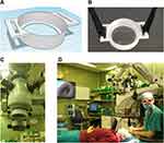
The optimal spherical power can be determined by simple “trial and error” with different lenses from an optometry trial lens set. We found that a lens power of −3.50 sphere was most optimal for our working conditions (Zeiss Lumera 700). The distance from the microscope to the eye was approximately 60 cm (with a −4.00 lens the distance increased to approximately 85 cm). We then ordered a corresponding 62 mm diameter lens from a local supplier. The lens can simply be firmly taped in place, but due to concern of slippage and contamination of the surgical field, we custom-designed a simple adapter (Figure 1). We 3D printed a plastic piece in which the lens can be securely seated. The digital design file can be accessed from the following link: https://www.thingiverse.com/thing:4290576. The dimensions are such that the piece can slide over the lower part of the surgical microscope. The plastic piece has handles to which we attached fabric straps with complementary Velcroed ends. The straps are wrapped around the top of the microscope and secured to each other to hold the adapter firmly in place (Video 1). The cost for all the supplies was approximately 25 USD. The focus and magnification/minification are accomplished in the exact same way that is typically used for the microscope – by adjusting the height of the microscope with the handles or the foot pedal.
Development of the technique did not involve experimentation on human subjects, and it was exempted by the local institutional review board.
Outcomes
We successfully implemented the method to broadcast and record over 30 surgical procedures, mainly strabismus and orbital. The recording was broadcast on the operating room’s surgical monitor display, allowing medical students and nurses to view the surgery with ease and comfort. Video 2 demonstrates a recording of an inferior oblique recession. The patient provided written consent for the surgery to be recorded and published.
We tested both biconcave and meniscus designs for diverging lenses. The image clarity of the meniscus lens was greatest when it was oriented with the concave surfaces directed downward (toward the patient). The image clarity with a biconcave lens was judged to be slightly better than a meniscus lens.
Discussion
The ability to record and broadcast surgeries allows for surgeon self-assessment and teaching.1 We furthermore believe that a live broadcast in the operating room increases the auxiliary staff’s awareness of the surgical details and improves teamwork. A number of options have been previously described for surgeons that use surgical loupes.1–4 We have developed a simple method to achieve these goals by simply placing a diverging lens under the surgical microscope. Because our method allows recording from the surgical microscope, it confers a number of benefits relative to a head-mounted camera: 1) A head-mounted camera will cause a degree of shaking in the recording, due to head motion; 2) there is less risk of neck strain compared to using a head-mounted camera; and 3) the video is captured in the typical “surgeon’s view” that is familiar to ophthalmologists.
In addition, the method that we present is far less expensive and still extremely effective.
Acknowledgment
The authors would like to thank Ms. Hila Miriam for preparing the velcro straps of the microscope adapter.
Disclosure
The authors report no conflicts of interest in this work.
References
1. Thia BC, Wong NJ, Sheth SJ. Video recording in ophthalmic surgery. Surv Ophthalmol. 2019;64(4):570–578. doi:10.1016/j.survophthal.2019.01.005
2. Lin LK. Surgical video recording with a modified GoPro Hero 4 camera. Clin Ophthalmol Auckl NZ. 2016;10:117–119. doi:10.2147/OPTH.S95666
3. Rahimy E, Garg SJ. Google glass for recording scleral buckling surgery. JAMA Ophthalmol. 2015;133(6):710–711. doi:10.1001/jamaophthalmol.2015.0465
4. Birnbaum FA, Wang A, Brady CJ. Stereoscopic surgical recording using GoPro cameras: a low-cost means for capturing external eye surgery. JAMA Ophthalmol. 2015;133(12):1483–1484. doi:10.1001/jamaophthalmol.2015.3865
 © 2020 The Author(s). This work is published and licensed by Dove Medical Press Limited. The full terms of this license are available at https://www.dovepress.com/terms.php and incorporate the Creative Commons Attribution - Non Commercial (unported, v3.0) License.
By accessing the work you hereby accept the Terms. Non-commercial uses of the work are permitted without any further permission from Dove Medical Press Limited, provided the work is properly attributed. For permission for commercial use of this work, please see paragraphs 4.2 and 5 of our Terms.

