Back to Journals » International Journal of Nanomedicine » Volume 20
A Minireview on Nanosized Hypericin-Based Inducer of Immune Cell Death Under ROS-Based Therapies
Received 9 September 2025
Accepted for publication 29 November 2025
Published 9 December 2025 Volume 2025:20 Pages 14695—14705
DOI https://doi.org/10.2147/IJN.S566489
Checked for plagiarism Yes
Review by Single anonymous peer review
Peer reviewer comments 2
Editor who approved publication: Dr RDK Misra
Chuanshan Xu,1,* Xiaowen Cai,2,* Lingran Du1
1Key Laboratory of Molecular Target and Clinical Pharmacology, State Key Laboratory of Respiratory Disease, School of Pharmaceutical Sciences, Guangzhou Medical University, Guangzhou, Guangdong, 511436, People’s Republic of China; 2School of Applied Biology, Shenzhen City Polytechnic, Shenzhen, People’s Republic of China
*These authors contributed equally to this work
Correspondence: Lingran Du, Key Laboratory of Molecular Target and Clinical Pharmacology, State Key Laboratory of Respiratory Disease, School of Pharmaceutical Sciences, Guangzhou Medical University, Guangzhou, Guangdong, 511436, People’s Republic of China, Tel/Fax +8620-37106238, Email [email protected]
Abstract: Immunotherapy is emerging as a powerful strategy against cancer; however, its efficacy is often blunted by the immunosuppressive tumor microenvironment (TME). Immunogenic cell death (ICD) can tilt this balance by releasing tumor-associated antigens and damage-associated molecular patterns that enhance TME immunogenicity, promote antigen-presenting cell maturation, and activate effector T cells. Ionizing radiation and doxorubicin (Dox) are two types of the common ICD inducers. However, they have severe off-target toxicities and limited therapeutic indices. To overcome these challenges, safe and natural products are now drawing widespread attention. Hypericin, a naturally occurring photosensitizer derived from the traditional Chinese herb Hypericum perforatum (St. John’s wort), has been used medicinally for centuries, and is now recognized for its potent antimicrobial, antiviral, anti-inflammatory, and anticancer properties. Recent studies have revealed that hypericin can modulate tumor immunity, and when employed in photodynamic therapy (PDT) or sonodynamic therapy (SDT) it generates reactive oxygen species that trigger endoplasmic reticulum stress-mediated ICD. Nanocarrier-mediated delivery further amplified these effects by enhancing hypericin solubility, tumor accumulation, and ROS yield upon light irradiation. This minireview synthesizes the current knowledge on the immunomodulatory actions of hypericin within the tumor microenvironment, evaluates its performance as a PDT/SDT-based ICD inducer, and highlights that nanosized formulations of hypericin may accelerate the development of novel ICD inducers and immunomodulators.
Keywords: hypericin, photodynamic therapy, sonodynmamic therapy, nanosizing technology, immunogenic cell death, tumor immune microenvironment, cancer immunotherapy
Introduction
Cancer remains a serious health challenge for humans. The International Agency for Research on Cancer (IARC) reported in 2022 that nearly one in five men or women developed cancer in a lifetime.1,2 In the United States, there are an estimated 2.04 million new cases and 618,120 cancer-related deaths by 2025,3 underscoring the escalating societal burden. Surgery, chemotherapy, and radiotherapy, as conventional modalities, achieve only transient responses with substantial side-effects.4 Immunotherapy offers an alternative treatment that leverages the immune system to recognize and eliminate malignant cells with unprecedented specificity.5–7 However, cancer cells frequently escape immune control by creating an immunosuppressive microenvironment and by presenting inadequate immunogenic cues.5–9 Immunogenic cell death (ICD) can overcome these barriers by triggering the rapid release of tumor-associated antigens (TAAs) and damage-associated molecular patterns (DAMPs). This cascade enhances the maturation of antigen-presenting cells, activates cytotoxic T lymphocytes, and ultimately reprograms the tumor microenvironment toward an antitumor state.10–13 Consequently, the research and development of safe and potential ICD inducers has emerged as a promising strategy to augment immunotherapeutic efficacy.
ICD inducers, such as therapeutic modalities or drugs, can convert malignant cells into potent vaccines, eliciting robust tumor-specific adaptive immunity. This process is governed by three hallmark molecular events: (1) lysosome-mediated ATP secretion, which acts as ann “find-me” signal for dendritic-cell (DC) precursors; (2) pre-apoptotic translocation of calreticulin (CRT) to the plasma membrane, functioning as an “eat-me” signal; and (3) secondary necrosis–driven release of high-mobility group box 1 (HMGB1), which engages TLR4 on DCs to amplify antigen processing and presentation.10–13 Together, these danger signals mature DCs, stimulate tumor-antigen cross-presentation to CD4⁺ and CD8⁺ T cells (Figure 1), and transform immunologically “cold” tumors into “hot” ones, thereby synergizing with immune-checkpoint inhibitors (eg, anti-PD-1/PD-L1) and adoptive T-cell therapies.10–22 Ionizing radiation and doxorubicin (Dox) are commonly used ICD inducers (Table 1); however, their serious toxicity to normal tissues limits their therapeutic efficacy in clinical settings. Ionizing radiation typically induces myelosuppression. Dox is one of the most important ICD inducers and shows significant cardiotoxicity side effects, which seriously affect its clinical outcomes, leading to discontinuous Dox therapy in several cancer patients.23–25 Recently, naturally derived agents are drawing widespread attention as an alternative to develop safer ICD inducers. Hypericin (C30H16O8), a naturally occurring photosensitizer from traditional Chinese herb Hypericum perforatum (St. John’s Wort), exemplifies this opportunity. It has been used for centuries as a herbal remedy for depression and infection and licensed by Commission E (Germany) for anxiety, depression, and insomnia. Pharmacological studies have revealed that hypericin exhibits broad pharmacological activities, including antimicrobial, anti-inflammatory, antidepressant, and anticancer effects.26–28 Growing evidence shows hypericin-based photodynamic therapy (PDT) / sonodynamic therapy (SDT) generates cytotoxic reactive oxygen species (ROS) to provoke ICD and remodel the tumor immune microenvironment.11,12,29–32
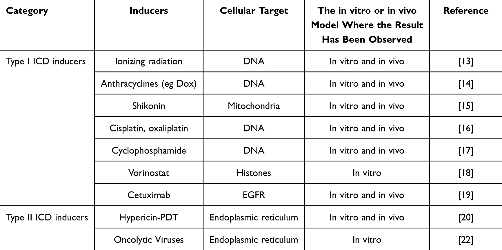 |
Table 1 Representative ICD Inducers |
 |
Figure 1 A schematic representation of ICD-induced antitumor immunity. |
In this review, we present the current knowledge on the immunomodulatory actions of hypericin within the tumor microenvironment, evaluate its performance as a PDT/SDT-based ICD inducer, and highlight that nanosized formulations of hypericin may accelerate the development of next-generation ICD inducers and immunomodulators. Our goal was to guide the further development of plant-derived agents and traditional Chinese medicines in photodynamic and sonodynamic immunotherapy.
Role of Hypericin in Tumor Immune Microenvironment
The tumor immune microenvironment dictates whether the cancer progresses or regresses. The TME mainly consists of malignant cells, stromal elements (fibroblasts and endothelial cells), and a heterogeneous immune infiltrate, including CD8⁺ cytotoxic T lymphocytes (CTLs), regulatory T cells, dendritic cells, tumor-associated macrophages (TAMs), and myeloid-derived suppressor cells. In TME, CTLs are often sequestered at the tumor margin and blocked from the core by M2-polarized TAMs, resulting in an immunosuppressive microenvironment. Therefore, reducing M2 macrophages is an attractive strategy for “treating” cold tumors and prevent metastasis.33,34
Emerging data have shown that hypericin can re-educate the TME. In the dark, hypericin downregulates the M2 marker CD206 and other M2-associated signatures via PI3K/AKT inhibition, thereby impeding macrophage-driven proliferation, invasion, and migration of cancer cells.35 In murine models, hypericin alone decreased intratumoral M2 macrophages, slowed tumor growth, and enhanced CTL infiltration into the tumor core.35 In addition to myeloid cells, hypericin in the dark suppresses endothelial proliferation and angiogenesis, destabilizes hypoxia-inducible factors (HIF-1/2α), and diminishes the side population of chemoresistant cancer stem cells.36
Upon light exposure, hypericin generates ROS to trigger robust cytokine cascades including IL-1α/β, IL-6, TNF-α, IFN-γ, and multiple chemokines in HepG2 hepatocellular carcinoma cells.37 A similar light-dependent upregulation of IL-6 mRNA occurs in CNE-2 nasopharyngeal carcinoma cells and xenografts,38 whereas human dermal fibroblasts and keratinocytes exhibit light-dependent decreases in IL-8, MMP-1, IL-19, and IL-22, along with increases in IL-11.39 These findings suggest that hypericin is a versatile, dual-mode immunomodulator, both in the dark and under illumination, that can reprogram the TME and augment anticancer immunity. PDT with hypericin could inhibit MMP-9 expression and the activity of nuclear factor kappa-B (NF-κB), a transcription factor that regulates the expression of pro-inflammatory cytokines and other genes involved in immune responses.40 This inhibition of NF-κB downregulates tumor-promoting cytokines, thereby reducing the immunosuppressive environment within the tumor and enhancing the efficacy of the immune response. Moreover, PDT with hypericin can also modulate the tumor microenvironment by affecting the integrity of the tumor vasculature and promoting the infiltration of neutrophils and other immune cells into the tumor tissues.41 This modulation is essential for the establishment of a pro-inflammatory environment that supports the activation of the adaptive immune system. The release of pro-inflammatory signals into the tumor-draining lymph nodes further enhances the systemic anti-tumor immune response.
Photodynamic Therapy with Hypericin: An Effective ICD Inducer
Cell death is an essential physiological process that maintains tissue homeostasis by balancing the cell proliferation and metabolic turnover. The 2018 guidelines of the Nomenclature Committee on Cell Death (NCCD) recognized 12 distinct modalities: apoptosis, parthanatos, necroptosis, ferroptosis, pyroptosis, mitochondrial-permeability-transition-driven necrosis, entosis, NETosis, lysosome-dependent death, autophagy-dependent death, mitotic catastrophe, cellular senescence, and ICD.42,43 ICD is unique because it can convert dying cancer cells into de facto vaccines by simultaneously providing antigenicity (TAAs), adjuvanticity (DAMPs), and a microenvironment permissive to antigen presentation. Effective type II ICD typically requires ROS and endoplasmic-reticulum stress, which together drive the surface exposure or extracellular release of TAAs and DAMPs, thereby potentiating adaptive immunity and improving the efficacy of cancer immunotherapy.20,30,31
PDT is a clinically validated, minimally invasive strategy that exploits the photochemical interaction of three harmless components–light, photosensitizer, and tissue oxygen–to generate cytotoxic ROS.44–47 Because both visible light and most photosensitizers are innocuous in isolation, PDT provides an exceptional therapeutic window for eradicating tumors while sparing normal tissues. The exact activated cell death modality depends on the subcellular localization of the photosensitizer, local oxygen tension, and light dose.31 Upon light irradiation, photosensitizers localized in the endoplasmic reticulum (ER) generate large amounts of ROS and trigger ER stress to induce type II ICD. Hypericin is a natural photosensitizer that is predominantly localized in the ER of cancer cells. When hypericin accumulates in cancer cells, light activation produces a burst of ROS within the ER lumen, unleashing intense ER stress that drives bona fide ICD.20,30,31 The underlying mechanisms of PDT with hyericin involve the generation of ROS predominantly in the ER, where hypericin localizes, leading to focused ER stress,48,49 subsequently activating the PERK signaling pathway, which is crucial for the release of DAMPs such as CRT, HMGB1, and ATP. CRT, in particular, acts as an “eat me” signal, facilitating the phagocytosis of dying tumor cells by DCs. This process is essential for the maturation of DCs, which then present tumor antigens to CTLs, initiating a robust anti-tumor immune response. The release of DAMPs following PDT with hypericin not only promotes the maturation of DCs but also recruits T immune cells to the TEM. ATP acts as a “find me” signal, attracting T immune cells to the site of tumor cell death.49–52 This recruitment is crucial for the establishment of an inflammatory milieu that supports the activation of the adaptive immune system. Additionally, the release of HMGB1 enhances the antigen-presenting capability of DCs, further amplifying the immune response against the tumor53 (Figure 2). Consequently, PDT with hypericin not only destroys the primary tumor but also releases TAAs and danger signals that reprogram the TME and potentiate systemic antitumor immunity. Therefore, hypericin-based PDT is a safe, minimally invasive, and highly effective inducer of ICD.
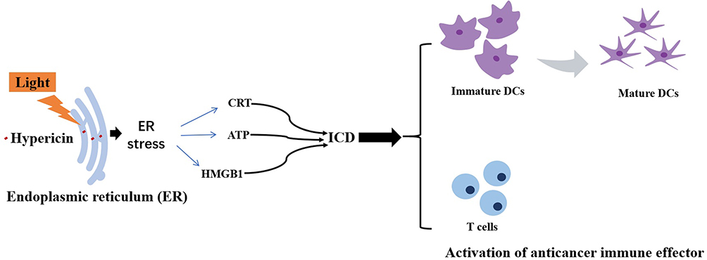 |
Figure 2 A schematic representation of photodynamic therapy with hypericin-induced ICD and antitumor immunity. |
Sonodynamic Therapy with Hypericin: An Innovative ICD Inducer
Similar to PDT, sonodynamic therapy (SDT) is a promising approach for inducing ICD. SDT employs a triad of sensitizers, oxygen, and ultrasound to generate cytotoxic ROS that triggers ER stress and mitochondrial dysfunction, ultimately releasing DAMPs that promote DC maturation and T cell infiltration. In addition, the cavitation effect of ultrasound can directly disrupt cell membranes and liberate immunogenic DAMPs, further enhancing ICD induction.31,54
SDT-induced ICD has been validated in various cancer models including glioblastoma and colon cancer. For example, temozolomide (TMZ), a first-line chemotherapeutic agent for glioblastoma (GBM), produces large amounts of ROS under ultrasonication. Zhou et al reported that SDT with TMZ elicited an ER stress response (ERSR), nuclear DNA damage, and mitochondrial permeability transition pore opening, releasing danger signals that trigger ICD and activate bone marrow-derived dendritic cells (BMDCs).54 However, TMZ is burdened by substantial systemic toxicity and multi-drug resistance (MDR). More recently, SDT with hypericin has been investigated in two- and three-dimensional HT-29 colon cancer models. Hypericin-based SDT generated robust ROS-mediated anticancer activity and hallmark-specific DAMPs including CRT exposure and HMGB1 release. Notably, P-gp overexpression in HT-29/MDR cells did not compromise cancer cell responsiveness to hypericin-based SDT.32 These data establish that hypericin is a potent sonosensitizer, and its SDT regimen effectively induces ICD and circumvents MDR. Unlike TMZ, hypericin is a naturally derived compound with tumor-selective cytotoxicity and minimal side effects, positioning hypericin-based SDT as a safe, non-invasive, and innovative ICD inducer.
Nanosized Hypericin-Mediated PDT/SDT: Novel and Promising ICD Inducers
Hypericin exerts potent immunomodulatory effects in cancer therapy by suppressing M2 macrophage polarization, reshaping the cytokine/chemokine milieus, and eliciting ICD. However, its clinical translation is hindered by severe hydrophobicity. In aqueous media, hypericin rapidly self-aggregates through hydrophobic interactions, diminishing water solubility, systemic bioavailability, and ultimately ROS yield upon light activation or ultrasound sonication, which significantly reduces its immunotherapeutic impact.55 Nanotechnology offers a versatile solution for this problem. The use of inorganic or organic materials encapsulating or conjugating hypericin within engineered nanocarriers,47 dispersibility, and tumor-directed delivery has markedly improved. These nanoformulations can be fabricated through straightforward physical encapsulation or covalent attachment, thereby providing a scalable platform to unlock the full immunomodulatory potential of hypericin-based PDT/SDT.
Among inorganic nanomaterials, mesoporous silica (eg, SBA-15) stands out for its large, tunable pores that accommodate both hydrophilic and hydrophobic cargos. Guo et al and Pevná et al exploited this feature to construct SBA-15-hypericin nanoparticles that remained biologically inert during circulation, yet released hypericin in a sustained manner inside cancer cells, providing a safe, targeted depot.56–58 Metal-organic frameworks, such as MIL-101(Al)-NH2 have also been loaded with hypericin, yielding MIL-101(Al)-NH2-hypericin composites with simultaneous anticancer, antibacterial, and antiviral activities.59 Graphene oxide functionalized with hypericin has been engineered to potentiate anticancer activity of Dox.60,61 Upconversion nanoparticles (UCNPs) further expand the utility of hypericin; CS-UCNP@P(DMA-AEA)-Ale-hypericin can induce substantially more apoptosis upon light irradiation than free hypericin, while enabling deep-tissue imaging and NIR-triggered PDT.62,63 Gold nanoparticles (AuNPs), prized for their biocompatibility and facile surface chemistry, have been covalently conjugated with hypericin to create AuNP-hypericin hybrids, which display enhanced solubility, prolonged circulation, and superior photodynamic activity.64–66
Recently, organic nanocarriers have been developed to overcome the poor aqueous solubility of hypericin and sharpen their phototherapeutic index. The oil-in-water nanoemulsions prepared by Salawi et al remained stable for months and retained their antidepressant activity in vivo.26 The amphiphilic block copolymer Pluronic F127 self-assembles into nanomicelles that encapsulate hypericin (F127/HYP), increasing its circulation time and driving preferential accumulation in the endoplasmic reticulum (ER) and mitochondria. Upon light irradiation, the F127/HYP micelles generated localized oxidative bursts that caused necrotic cell death in a dose-dependent fashion.67 Similarly, hypericin-loaded nanoparticles assembled from biodegradable amphiphilic copolymers exhibit markedly elevated ROS production and superior photodynamic potency compared with free hypericin.68 Gradient copoly(2-oxazoline)s engineered by Huntošová et al delivered hypericin for simultaneous tumor imaging and PDT, thus validating the versatility of self-assembled polymeric systems.69
Lipid-based vectors offer an additional clinical translatability. Liposomes and solid-lipid nanoparticles have been widely adopted to package hypericin, improving its solubility, tumor targeting, and safety by minimizing off-target photosensitization.70–74 More recently, the FDA-approved maize protein zein was used to formulate zein-hypericin nanoparticles, which demonstrated potent photodynamic eradication of hepatocellular carcinoma cells.75 These organic platforms provide tunable, biocompatible, and scalable solutions for next-generation hypericin-based cancer immunotherapy.
Nanotechnology endows hydrophobic hypericin with “stealth” blood-circulation properties. Amphiphilic block copolymers self-assembled into nanomicelles that cloaked hypericin from reticuloendothelial clearance, sheltered it from premature degradation, and exploited the enhanced permeability and retention (EPR) effect to preferentially accumulate in tumors. To confer active targeting of hypericin, lactose-decorated magnetic iron-oxide nanoparticles (Lac-PHM) have been engineered to deliver hypericin to asialoglycoprotein-receptor-positive HepG2 cells.76–78 Hypericin-loaded transferrin nanoformulations (HTfNPs) were prepared to deliver hypericin to transferrin-receptor–overexpressing colorectal cancer cells. In the presence of light irradiation, HTfNPs induced significant BMI1 degradation and robust tumor retardation.79 Folate-receptor-targeted P123 nanomicelles delivered hypericin to melanoma cells with high selectivity and photoinactivation efficiency.80,81 More recently, plant-derived exosome-like nanovesicles from Hypericum perforatum were shown to naturally ferry hypericin, achieving superior photodynamic efficacy against multiple malignancies.82
Lipoprotein shuttles further refine the targeted delivery of hypericin. LDL-hypericin nanocomplexes are internalized 2-fold more rapidly by glioma and breast cancer cells than HDL-hypericin, resulting in stronger photodynamic killing action on malignant cells.83–85 Smart stimuli-responsive nanoparticles now add temporal and spatial precisions. pH-, redox-, and enzyme-cleavable nanophotosensitizers can amplify ROS generation by photosensitizers within the acidic, glutathione-rich, or protease-abundant tumor milieu.86–89 A pH-responsive metal–drug nanocomplex integrating hypericin, apigenin, PVP, and Fe3⁺ has recently demonstrated markedly enhanced PDT potency and metastasis suppression.90 These nano-enabled hypericin systems not only improve solubility and enhance targeting ability but also induce widespread ICD, positioning nanosized hypericin as a next-generation ICD inducer and immunomodulator for photodynamic immunotherapy.
Recently, nanosized sonosensitizer-mediated SDT using phase-transformation nanoparticles or folate-targeted systems has been shown to induce ICD and stimulate DC maturation, resulting in increased CD8+ T-cell activation and tumor growth inhibition.91,92 Yuan et al used a hematoporphyrin monomethyl ether (HMME)-based liposomal nanosystem as a sonosensitizer for colon cancer treatment. They found that SDT extensively induced ICD, causing enhanced T-cell recruitment and infiltration to improve the immunosuppressive TME and promote antitumor immunity. Both in vitro and in vivo results demonstrated that SDT with an HMME-based liposomal nanosystem induced ferroptosis, apoptosis, and ICD via ROS generation during SDT and reprogrammed the TME.93 Tian et al also reported that the aggregation-induced emission (AIE) nanosonosensitizers under sonication produced abundant ROS-mediated mitochondrial oxidative stress to trigger significant ICD.94 Huang et al used the thin-film hydration method to prepare lipid nanobubbles (NBs) loading chlorin e6 (Ce6) and anti-PD-L1 Ab (Ce6@aPD-L1 NBs). They found that Ce6@aPD-L1 NBs in the presence of ultrasound sonication effectively induced ROS generation and ICD, thereby activating DC maturation.95 Moreover, red blood cell membrane-camouflaged nanoparticles (SB-IR-PLGA@RM) encapsulating the sonosensitizer IR780 and ER-targeting iridium(III) nanosonosensitizers were shown to effectively induce ICD in cancer cells, remodel the immunosuppressive TME, and amplify antitumor immunity.96,97 Although there are no reports on the direct study of nanosized hypericin-based SDT on ICD, the inspiring findings on nanosonosensitizers-mediated SDT inducing ICD induction and immunomodulation indicate that nanosized hypericin has great potential for developing an innovative ICD inducer and immunomodulator for sonodynamic immunotherapy.
Summary and Future Directions
Hypericin is the only FDA-approved natural photosensitizer with anticancer, antiviral, and immunomodulatory activity. Recent studies have confirmed that it elicits robust ICD upon light activation and reshapes the TME. However, hypericin’s hydrophobicity, which causes aggregation in aqueous environments, reducing bioavailability and ROS production. Nanoformulations could improve its solubility and targeting, systemic half-life, and ROS yield, positioning nanosized hypericin as a highly promising ICD inducer. For example, superparamagnetic iron oxide nanoparticles (SPIONs) as an inorganic nanoformulation enable magnetic-guided targeting of hypericin. Hypericin-loaded SPIONs enhanced the amount of hypericin in the tumor tissues under an external magnetic field, which resulted intratumoral hypericin accumulation, boosted ROS production, and amplified ICD via DNA damage and DAMP release.77 Mokoena et al found that Gold nanoparticles improved hypericin’s hydrophobicity and colloidal stability, allowing deeper tumor penetration and prolonged retention in tumor interstitial spaces.64 Organic nanoformulations, such as glyconanoparticles or polymeric systems, also enhance hypericin solubility and targeting. For instance, polymeric carriers also facilitate prolonged blood circulation and controlled release. Hypericin-entrapped glyconanoparticles improve aqueous solubility and tumor-specific delivery by leveraging surface ligands like folic acid, which binds to tumor-overexpressed receptors, enhancing cellular uptake and photodynamic efficacy.76 Lipid-based nanoformulations, particularly liposomes, effectively solubilize hypericin within their lipid bilayers, preventing aggregation and ROS loss. Olszowy et al found tetraether liposome-encapsulated hypericin exhibits superior tumor vasculature targeting due to passive tumor targeting via the enhanced permeability and retention (EPR) effect, thereby improving biodistribution and hypericin delivery.98 Surface modifications of nanoformulations using antibody or ligand can endow the active targeting of hypericin. Sardoiwala MNet al prepared a hypericin-loaded transferrin nanoparticles (HTfNPs) and found the improvement in half-time (t1/2) of hypericin from 2.16 to 4.29 h using transferrin nanocarrier, indicating the prolonged circulation time of hypericin for better distribution to its target site. And HTfNPs also show a significant photodynamic efficacy against colorectal cancer by inducing PP2A-mediated BMI1 ubiquitination/degradation.79 These encouraging findings indicate a great potential in developing nanosized hypericin-based PDT as an innovative ICD inducer. Nevertheless, the current proof-of-concept is largely derived from conventional in vitro and small-animal models; rigorous demonstration that nano-hypericin triggers bona fide ICD and remodels the TME in clinically relevant settings remains limited. Moreover, the multistep synthesis and batch-to-batch variability of existing nanocarriers hinders scalable translation, highlighting the need for a sustainable, eco-friendly preparation method and large-scale clinical trials to establish safety and efficacy.
A second hurdle of hypericin-based PDT in the clinic application is the limited tissue penetration of the 595 nm light required for classic PDT. SDT replaces light with ultrasound waves and offers an elegant solution to overcome some of the limitations of PDT. Similar to PDT, sonosensitizers can generate cytotoxic ROS to induce ICD upon ultrasound sonication; however, ultrasound penetrates centimeters deep with negligible systemic toxicity. Emerging evidence has demonstrated that hypericin retains its sonosensitizing capacity,31,32,99–107 producing ER-localized ROS under ultrasound sonication that provoke ER stress-mediated ICD. Therefore, nanosized hypericin engineered for SDT could overcome the depth restriction of light, showing a promising opportunity to induce ICD for the treatment of deep-seated malignancies. However, rigorously engineered quality-controlled nanohypericin-SDT systems require intensive investigation to develop nanohypericin-based SDT as a novel next-generation ICD inducer and immunomodulator.
Beyond delivery and activation issues, the clinical translation of ICD inducers like hypericin faces validated biomarkers and standardization challenges. There is a critical need for validated biomarkers to consistently measure ICD induction in patients, and the current lack of uniform evaluation metrics, such as standardized assays for DAMP release, hinders the clinical assessment of therapeutic efficacy.108 To bridge these gaps, future research must prioritize the development of innovative nanoformulations designed to improve hypericin’s solubility, stability, and tumor-specific targeting, thereby enhancing its pharmacokinetic profile.109,110 Developing ultrasound as an activating sources will potentially overcome the depth limitation of traditional PDT. Ultimately, a multidisciplinary effort combining advanced drug formulation, refined ultrasound delivery, and rigorous biomarker-driven clinical trial design is imperative to fully realize the potential of hypericin as a clinically viable ICD inducer.
Funding
This work was supported by the Research Ability Improvement Project of Guangzhou Medical University (01-410-250208) and the Graduate Education Innovation Plan Project of Guangdong Province, exemplary graduate course (2024SFKC_074).
Disclosure
The authors report no conflicts of interest in this work.
References
1. Bray F, Laversanne M, Sung H, et al. Global cancer statistics 2022: GLOBOCAN estimates of incidence and mortality worldwide for 36 cancers in 185 countries. CA Cancer J Clin. 2024;74(3):229–263. doi:10.3322/caac.21834
2. Filho AM, Laversanne M, Ferlay J, et al. GLOBOCAN 2022 cancer estimates: data sources, methods, and a snapshot of the cancer burden worldwide. Int J Cancer. 2025;156(7):1336–1346. doi:10.1002/ijc.35278
3. Siegel RL, Kratzer TB, Giaquinto AN, Sung H, Jemal A. Cancer statistics, 2025. CA Cancer J Clin. 2025;75(1):10–45. doi:10.3322/caac.21871
4. Liu H, Yang F, Chen W, et al. Enzyme-responsive materials as carriers for improving photodynamic therapy. Front Chem. 2021;9:763057. doi:10.3389/fchem.2021.763057
5. Hanahan D, Michielin O, Pittet MJ. Convergent inducers and effectors of T-cell paralysis in the tumour microenvironment. Nat Rev Cancer. 2025;25(1):41–58. doi:10.1038/s41568-024-00761-z
6. Arimoto KI, Miyauchi S, Liu M, Zhang DE. Emerging role of immunogenic cell death in cancer immunotherapy. Front Immunol. 2024;15:1390263. doi:10.3389/fimmu.2024.1390263
7. Linderman SW, DeRidder L, Sanjurjo L, et al. Enhancing immunotherapy with tumour-responsive nanomaterials. Nat Rev Clin Oncol. 2025;22(4):262–282. doi:10.1038/s41571-025-01000-6
8. Kimmel BR, Arora K, Chada NC, et al. Potentiating cancer immunotherapies with modular albumin-hitchhiking nanobody-STING agonist conjugates. Nat Biomed Eng. 2025;9:1719–1739. doi:10.1038/s41551-025-01400-0
9. Zeng Q, Liu M, Wang Z, Zhou R, Ai K. Enhancing radiotherapy-induced antitumor immunity via nanoparticle-edited STING agonist synergy. Mol Cancer. 2025;24(1):176. doi:10.1186/s12943-025-02366-y
10. Sukkurwala AQ, Adjemian S, Senovilla L, et al. Screening of novel immunogenic cell death inducers within the NCI Mechanistic Diversity Set. Oncoimmunology. 2014;3:e28473. doi:10.4161/onci.28473
11. Garg AD, Vandenberk L, Koks C, et al. Dendritic cell vaccines based on immunogenic cell death elicit danger signals and T cell-driven rejection of high-grade glioma. Sci Transl Med. 2016;8(328):328ra27. doi:10.1126/scitranslmed.aae0105
12. Diederich M. Natural compound inducers of immunogenic cell death. Arch Pharm Res. 2019;42(7):629–645. doi:10.1007/s12272-019-01150-z
13. Frey B, Rubner Y, Wunderlich R, et al. The induction of abscopal antitumor immunity and immunogenic tumor cell death by ionizing irradiation has implications for cancer therapies. Curr Med Chem. 2012;19(12):1751–1764. doi:10.2174/092986712800099811
14. Casares N, Pequignot MO, Tesniere A, et al. Caspase-dependent immunogenicity of doxorubicin-induced tumor cell death. J Exp Med. 2005;202(12):1691–1701. doi:10.1084/jem.20050915
15. Chen HM, Wang PH, Chen SS, et al. Shikonin induces immunogenic cell death in tumor cells and enhances dendritic cell-based cancer vaccines. Cancer Immunol Immunother. 2012;61(11):1989–2002. doi:10.1007/s00262-012-1258-9
16. Park SJ, Ye W, Xiao R, et al. Cisplatin and oxaliplatin induce similar immunogenic changes in preclinical models of head and neck cancer. Oral Oncol. 2019;95:127–135. doi:10.1016/j.oraloncology.2019.06.016
17. Du B, Waxman DJ. Medium dose intermittent cyclophosphamide induces immunogenic cell death and cancer cell autonomous type I interferon production in glioma models. Cancer Lett. 2020;470:170–180. doi:10.1016/j.canlet.2019.11.025
18. Laengle J, Kabiljo J, Hunter L, et al. Histone deacetylase inhibitors valproic acid and vorinostat enhance trastuzumab-mediated antibody-dependent cell-mediated phagocytosis. J Immunother Cancer. 2020;8(1):e000195. doi:10.1136/jitc-2019-000195
19. Pozzi C, Cuomo A, Spadoni I, et al. The EGFR-specific antibody cetuximab combined with chemotherapy triggers immunogenic cell death. Nat Med. 2016;22:624–631. doi:10.1038/nm.4078
20. Garg AD, Dudek AM, Ferreira GB, et al. ROS-induced autophagy in cancer cells assists in evasion from determinants of immunogenic cell death. Autophagy. 2013;9(9):1292–1307. doi:10.4161/auto.25399
21. Fang K, Yuan S, Zhang X, Zhang J, Sun SL, Li X. Regulation of immunogenic cell death and potential applications in cancer therapy. Front Immunol. 2025;16:1571212. doi:10.3389/fimmu.2025.1571212
22. Heinrich B, Klein J, Delic M, et al. Immunogenicity of oncolytic vaccinia viruses JX-GFP and TG6002 in a human melanoma in vitro model: studying immunogenic cell death, dendritic cell maturation and interaction with cytotoxic T lymphocytes. Onco Targets Ther. 2017;10:2389–2401. doi:10.2147/OTT.S126320
23. Belger C, Abrahams C, Imamdin A, Lecour S. Doxorubicin-induced cardiotoxicity and risk factors. Int J Cardiol Heart Vasc. 2023;50:101332. doi:10.1016/j.ijcha.2023.101332
24. Goje ID, Goje GI, Ordodi VL, et al. Doxorubicin-induced cardiotoxicity and the emerging role of SGLT2 inhibitors: from glycemic control to cardio-oncology. Pharmaceuticals. 2025;18(5):681. doi:10.3390/ph18050681
25. Bhutani V, Varzideh F, Wilson S, Kansakar U, Jankauskas SS, Santulli G. Doxorubicin-induced cardiotoxicity: a comprehensive update. J Cardiovasc Dev Dis. 2025;12(6):207. doi:10.3390/jcdd12060207
26. Salawi A, Almoshari Y, Sultan MH, et al. Production, characterization, and in vitro and in vivo studies of nanoemulsions containing St. John’s wort plant constituents and their potential for the treatment of depression. Pharmaceuticals. 2023;16(4):490. doi:10.3390/ph16040490
27. Choudhary N, Collignon TE, Tewari D, Bishayee A. Hypericin and its anticancer effects: from mechanism of action to potential therapeutic application. Phytomedicine. 2022;105:154356. doi:10.1016/j.phymed.2022.154356
28. Pan XG, Yin WF, Wang MN, et al. NOD-like receptor protein 3 inhibitors in St. John’s wort and their potential antidepressant effects. Int J Biol Macromol. 2025;306(Pt 4):141721. doi:10.1016/j.ijbiomac.2025.141721
29. Qiao C, Yang Z, Liu X, et al. Post-remedial oxygen supply: a new perspective on photodynamic therapy to suppress tumor metastasis. Nano Lett. 2022;22(20):8250–8257. doi:10.1021/acs.nanolett.2c02983
30. Verfaillie T, van Vliet A, Garg AD, et al. Pro-apoptotic signaling induced by photo-oxidative ER stress is amplified by Noxa, not Bim. Biochem Biophys Res Commun. 2013;438(3):500–506. doi:10.1016/j.bbrc.2013.07.107
31. Adkins I, Fucikova J, Garg AD, Agostinis P, Špíšek R. Physical modalities inducing immunogenic tumor cell death for cancer immunotherapy. Oncoimmunology. 2015;3(12):e968434. doi:10.4161/21624011.2014.968434
32. Foglietta F, Canaparo R, Cossari S, Panzanelli P, Dosio F, Serpe L. Ultrasound triggers hypericin activation leading to multifaceted anticancer activity. Pharmaceutics. 2022;14(5):1102. doi:10.3390/pharmaceutics14051102
33. Lin HJ, Liu Y, Caroland K, Lin J. Polarization of cancer-associated macrophages maneuvers neoplastic attributes of pancreatic ductal adenocarcinoma. Cancer. 2023;15(13):3507. doi:10.3390/cancers15133507
34. Zhou S, Hong M, Zhao D, et al. Reprogramming the tumor immune microenvironment with ICAM-1-targeted antibody-drug conjugates and B7-H3-CD3 bispecific antibodies. Adv Sci. 2025;12(16):e2415577. doi:10.1002/advs.202415577
35. Wen X, Wang X, Yao Q, et al. Hypericin impedes M2 macrophage polarization and protects against Hepatocellular carcinoma. Mol Immunol. 2025;181:160–168. doi:10.1016/j.molimm.2025.03.012
36. Buľková V, Vargová J, Babinčák M, et al. New findings on the action of hypericin in hypoxic cancer cells with a focus on the modulation of side population cells. Biomed Pharmacother. 2023;163:114829. doi:10.1016/j.biopha.2023.114829
37. Barathan M, Mariappan V, Shankar EM, Abdullah BJ, Goh KL, Vadivelu J. Hypericin-photodynamic therapy leads to interleukin-6 secretion by HepG2 cells and their apoptosis via recruitment of BH3 interacting-domain death agonist and caspases. Cell Death Dis. 2013;4(6):e697. doi:10.1038/cddis.2013.219
38. Du H, Bay BH, Mahendran R, Olivo M. Hypericin-mediated photodynamic therapy elicits differential interleukin-6 response in nasopharyngeal cancer. Cancer Lett. 2006;235(2):202–208. doi:10.1016/j.canlet.2005.04.013
39. Krupka-Olek M, Bożek A, Czuba ZP, Kłósek M, Cieślar G, Kawczyk-Krupka A. Cytotoxic and immunomodulatory effects of hypericin as a photosensitizer in photodynamic therapy used on skin cell cultures. Pharmaceutics. 2024;16(6):696. doi:10.3390/pharmaceutics16060696
40. Du HY, Olivo M, Mahendran R, et al. Hypericin photoactivation triggers down-regulation of matrix metalloproteinase-9 expression in well-differentiated human nasopharyngeal cancer cells. Cell Mol Life Sci. 2007;64(7–8):979–988. doi:10.1007/s00018-007-7030-1
41. Chen B, Roskams T, de Witte PA. Antivascular tumor eradication by hypericin-mediated photodynamic therapy. Photochem Photobiol. 2002;76(5):509–513. doi:10.1562/0031-8655(2002)076<0509:ATEBHM>2.0.CO;2
42. He R, Liu Y, Fu W, et al. Mechanisms and crosstalk of regulated cell death and their epigenetic modifications in tumor progression. Mol Cancer. 2024;23(1):267. doi:10.1186/s12943-024-02172-y
43. Galluzzi L, Vitale I, Aaronson SA, et al. Molecular mechanisms of cell death: recommendations of the nomenclature committee on cell death 2018. Cell Death Differ. 2018;25(3):486–541. doi:10.1038/s41418-017-0012-4
44. Zhou Y, Deng W, Mo M, et al. Stimuli-responsive nanoplatform-assisted photodynamic therapy against bacterial infections. Front Med. 2021;8:729300.
45. Liu H, Yao J, Guo H, et al. Tumor microenvironment-responsive nanomaterials as targeted delivery carriers for photodynamic anticancer therapy. Front Chem. 2020;8:758. doi:10.3389/fchem.2020.00758
46. Ain QT. Recent development of nanomaterials-based PDT to improve immunogenic cell death. Photochem Photobiol Sci. 2024;23(10):1983–1998. doi:10.1007/s43630-024-00638-y
47. Calixto GM, Bernegossi J, de Freitas LM, Fontana CR, Chorilli M. Nanotechnology-based drug delivery systems for photodynamic therapy of cancer: a review. Molecules. 2016;21(3):342. doi:10.3390/molecules21030342
48. Garg AD, Agostinis P. ER stress, autophagy and immunogenic cell death in photodynamic therapy-induced anti-cancer immune responses. Photochem Photobiol Sci. 2014;13(3):474–487. doi:10.1039/c3pp50333j
49. Ahmed A, Tait SWG. Targeting immunogenic cell death in cancer. Mol Oncol. 2020;14(12):2994–3006. doi:10.1002/1878-0261.12851
50. Alzeibak R, Mishchenko TA, Shilyagina NY, Balalaeva IV, Vedunova MV, Krysko DV. Targeting immunogenic cancer cell death by photodynamic therapy: past, present and future. J Immunother Cancer. 2021;9(1):e001926. doi:10.1136/jitc-2020-001926
51. Chou W, Sun T, Peng N, et al. Photodynamic therapy-induced anti-tumor immunity: influence factors and synergistic enhancement strategies. Pharmaceutics. 2023;15(11):2617. doi:10.3390/pharmaceutics15112617
52. Biteghe FAN, Chalomie NET, Mungra N, et al. Antibody-based immunotherapy: alternative approaches for the treatment of metastatic melanoma. Biomedicines. 2020;8(9):327. doi:10.3390/biomedicines8090327
53. Hirschberg H, Berg K, Peng Q. Photodynamic therapy mediated immune therapy of brain tumors. Neuroimmunol Neuroinflamm. 2018;5:27. doi:10.20517/2347-8659.2018.31
54. Zhou Y, Jiao J, Yang R, et al. Temozolomide-based sonodynamic therapy induces immunogenic cell death in glioma. Clin Immunol. 2023;256:109772. doi:10.1016/j.clim.2023.109772
55. Nazari MR, Abdossi V, Hargalani FZ, Larijani K. Antioxidant potential and essential oil properties of Hypericum perforatum L. assessed by application of selenite and nano-selenium. Sci Rep. 2022;12(1):6156. doi:10.1038/s41598-022-10109-y
56. Guo S, Shi Y, Liang Y, Liu L, Sun K, Li Y. Relationship and improvement strategies between drug nanocarrier characteristics and hemocompatibility: what can we learn from the literature? Asian J Pharm Sci. 2021;16(5):551–576. doi:10.1016/j.ajps.2020.12.002
57. Pevná V, Zauška Ľ, Almáši M, et al. Redistribution of hydrophobic hypericin from nanoporous particles of SBA-15 silica in vitro, in cells and in vivo. Int J Pharm. 2023;643:123288. doi:10.1016/j.ijpharm.2023.123288
58. Pevná V, Zauška Ľ, Benziane A, et al. Effective transport of aggregated hypericin encapsulated in SBA-15 nanoporous silica particles for photodynamic therapy of cancer cells. J Photochem Photobiol B. 2023;247:112785. doi:10.1016/j.jphotobiol.2023.112785
59. Huntošová V, Benziane A, Zauška L, et al. The potential of metal-organic framework MIL-101(Al)-NH2 in the forefront of antiviral protection of cells via interaction with SARS-CoV-2 spike RBD protein and their antibacterial action mediated with hypericin and photodynamic treatment. J Colloid Interface Sci. 2025;691:137454. doi:10.1016/j.jcis.2025.137454
60. Du X, Xiao R, Fu H, et al. Hypericin-loaded graphene oxide protects ducks against a novel duck reovirus. Mater Sci Eng C Mater Biol Appl. 2019;105:110052. doi:10.1016/j.msec.2019.110052
61. Han C, Zhang C, Ma T, et al. Hypericin-functionalized graphene oxide for enhanced mitochondria-targeting and synergistic anticancer effect. Acta Biomater. 2018;77:268–281. doi:10.1016/j.actbio.2018.07.018
62. Vasylyshyn T, Huntošová V, Patsula V, et al. Surface-engineered core-shell upconversion nanoparticles for effective hypericin delivery and multimodal imaging. Nanoscale. 2025;17(10):5838–5857. doi:10.1039/D4NR05348F
63. Yang X, Xiao Q, Niu C, et al. Multifunctional core-shell upconversion nanoparticles for targeted tumor cells induced by near-infrared light. J Mater Chem B. 2013;1(21):2757–2763. doi:10.1039/c3tb00575e
64. Mokoena D, George BP, Abrahamse H. Conjugation of hypericin to gold nanoparticles for enhancement of photodynamic therapy in MCF-7 breast cancer cells. Pharmaceutics. 2022;14(10):2212. doi:10.3390/pharmaceutics14102212
65. Kaundal B, Karmakar S, Roy Choudhury S. Mitochondria-targeting nano therapy altering IDH2-mediated EZH2/EZH1 interaction as precise epigenetic regulation in glioblastoma. Nat Biomater Sci. 2022;10(18):5301–5317. doi:10.1039/D1BM02006D
66. Mokoena DR, George BP, Abrahamse H. Enhancing breast cancer treatment using a combination of cannabidiol and gold nanoparticles for photodynamic therapy. Int J Mol Sci. 2019;20(19):4771. doi:10.3390/ijms20194771
67. De Souza MVF, Shinobu-Mesquita CS, Meirelles LEF, et al. Effects of hypericin encapsulated on Pluronic F127 photodynamic therapy against triple negative breast cancer. Asian Pacific J Cancer Prev. 2022;23(5):1741–1751. doi:10.31557/APJCP.2022.23.5.1741
68. Nafee N, Youssef A, Asem H, Kandil S. Antibiotic-free nanotherapeutics: hypericin nanoparticles thereof for improved in vitro and in vivo antimicrobial photodynamic therapy and wound healing. Int J Pharm. 2013;454(1):249–258. doi:10.1016/j.ijpharm.2013.06.067
69. Huntošová V, Datta S, Lenkavská L, et al. Alkyl chain length in Poly(2-oxazoline)-based amphiphilic gradient copolymers regulates the delivery of hydrophobic molecules: a case of the biodistribution and the photodynamic activity of the photosensitizer hypericin. Biomacromolecules. 2021;22(10):4199–4216. doi:10.1021/acs.biomac.1c00768
70. Loscertales E, Mateo J, España S. A comparative study of sensitizers and liposome composition in radiation-induced controlled drug release for cancer therapy. J Liposome Res. 2025;35(1):64–75. doi:10.1080/08982104.2024.2401800
71. de Morais FAP, Balbinot RB, Bakoshi ABK, et al. Hypericin-loaded modified theranostic liposome nanoplatform: a preliminary in vivo study of targeting and diagnosis. Naunyn Schmiedebergs Arch Pharmacol. 2025;398(1):1013–1021. doi:10.1007/s00210-024-03379-y
72. de Morais FAP, Gonçalves RS, Vilsinski BH, et al. Hypericin photodynamic activity in DPPC liposomes - part II: stability and application in melanoma B16-F10 cancer cells. Photochem Photobiol Sci. 2020;19(5):620–630. doi:10.1039/c9pp00284g
73. Lima AM, Pizzol CD, Monteiro FB, et al. Hypericin encapsulated in solid lipid nanoparticles: phototoxicity and photodynamic efficiency. J Photochem Photobiol. 2013;125:146–154. doi:10.1016/j.jphotobiol.2013.05.010
74. Youssef T, Fadel M, Fahmy R, Kassab K. Evaluation of hypericin-loaded solid lipid nanoparticles: physicochemical properties, photostability, and phototoxicity. Pharm Dev Technol. 2012;17(2):177–186. doi:10.3109/10837450.2010.529148
75. Abdelsalam AM, Somaida A, Ambreen G, et al. Surface tailored zein as a novel delivery system for hypericin: application in photodynamic therapy. Mater Sci Eng C Mater Biol Appl. 2021;129:112420. doi:10.1016/j.msec.2021.112420
76. Shao C, Shang K, Xu H, Zhang Y, Pei Z, Pei Y. Facile fabrication of hypericin-entrapped glyconanoparticles for targeted photodynamic therapy. Int J Nanomed. 2018;13:4319–4331. doi:10.2147/IJN.S161262
77. Mühleisen L, Alev M, Unterweger H, et al. Analysis of hypericin-mediated effects and implications for targeted photodynamic therapy. Int J Mol Sci. 2017;18(7):1388. doi:10.3390/ijms18071388
78. Unterweger H, Subatzus D, Tietze R, et al. Hypericin-bearing magnetic iron oxide nanoparticles for selective drug delivery in photodynamic therapy. Int J Nanomed. 2015;10:6985–6996. doi:10.2147/IJN.S92336
79. Sardoiwala MN, Kushwaha AC, Dev A, et al. Hypericin-loaded transferrin nanoparticles induce PP2A-regulated BMI1 degradation in colorectal cancer-specific chemo-photodynamic therapy. ACS Biomater Sci Eng. 2020;6(5):3139–3153. doi:10.1021/acsbiomaterials.9b01844
80. Benziane A, Huntošová V, Pevná V, et al. Synergistic effect of folic acid and hypericin administration to improve the efficacy of photodynamic therapy via folate receptors. J Photochem Photobiol B. 2024;261:113046. doi:10.1016/j.jphotobiol.2024.113046
81. de Oliveira ACV, de Morais FAP, Campanholi KDSS, et al. Melanoma-targeted photodynamic therapy based on hypericin-loaded multifunctional P123-spermine/folate micelles. Photodiagnosis Photodyn Ther. 2022;40:103103. doi:10.1016/j.pdpdt.2022.103103
82. Ma X, Chen N, Zeng P, et al. Hypericum Perforatum-derived exosomes-like nanovesicles: a novel natural photosensitizer for effective tumor photodynamic therapy. Int J Nanomed. 2025;20:1529–1541. doi:10.2147/IJN.S510339
83. Jutkova A, Chorvat D, Miskovsky P, Jancura D, Datta S. Encapsulation of anticancer drug curcumin and co-loading with photosensitizer hypericin into lipoproteins investigated by fluorescence resonance energy transfer. Int J Pharm. 2019;564:369–378. doi:10.1016/j.ijpharm.2019.04.062
84. Lenkavska L, Blascakova L, Jurasekova Z, et al. Benefits of hypericin transport and delivery by low-and high-density lipoproteins to cancer cells: from in vitro to ex ovo. Photodiagnosis Photodyn Ther. 2019;25:214–224. doi:10.1016/j.pdpdt.2018.12.013
85. Huntosova V, Buzova D, Petrovajova D, et al. Development of a new LDL-based transport system for hydrophobic/amphiphilic drug delivery to cancer cells. Int J Pharm. 2012;436(1–2):463–471. doi:10.1016/j.ijpharm.2012.07.005
86. Xie L, Jiang N, Liu Y, et al. Thermo-responsive hydrogel loading hypericin induces pro-inflammatory response against Trichinella spiralis infection via toll-like receptor 3 activation. Phytomedicine. 2025;136:156284. doi:10.1016/j.phymed.2024.156284
87. Zhou W, Fan T, Yan Y, et al. A manganese-oxide nano-rambutan as the intrinsic modifier for hypericin delivery and triple-negative breast cancer treatment. Int J Pharm. 2024;666:124824. doi:10.1016/j.ijpharm.2024.124824
88. Liang R, Wong KH, Yang Y, Duan Y, Chen M. ROS-responsive dexamethasone micelles normalize the tumor microenvironment, enhancing hypericin in cancer photodynamic therapy. Biomater Sci. 2022;10(4):1018–1025. doi:10.1039/D1BM01802G
89. Abu Dayyih A, Alawak M, Ayoub AM, et al. Thermosensitive liposomes encapsulating hypericin: characterization and photodynamic efficiency. Int J Pharm. 2021;609:121195. doi:10.1016/j.ijpharm.2021.121195
90. Yin Y, Wong KH, Wen L, Chen M. Active iron-drug nanocomplexes improve photodynamic and photothermal cancer therapy by mitigating tumor hypoxia and counteracting tumor heat resistance. Adv Healthc Mater. 2025;14(9):e2404485. doi:10.1002/adhm.202404485
91. Si Y, Yue J, Liu Z, et al. Phase-transformation nanoparticle-mediated sonodynamic therapy: an effective modality to enhance anti-tumor immune response by inducing immunogenic cell death in breast cancer. Int J Nanomed. 2021;16:1913–1926. doi:10.2147/IJN.S297933
92. Zheng J, Sun Y, Long T, et al. Sonosensitizer nanoplatform-mediated sonodynamic therapy induced immunogenic cell death and tumor immune microenvironment variation. Drug Deliv. 2022;29(1):1164–1175. doi:10.1080/10717544.2022.2058653
93. Yuan H, Ma J, Huang W, et al. Antitumor effects of a distinct sonodynamic nanosystem through enhanced induction of immunogenic cell death and ferroptosis with modulation of tumor microenvironment. JACS Au. 2023;3(5):1507–1520. doi:10.1021/jacsau.3c00156
94. Tian M, Li Y, Li Y, et al. Sonodynamic therapy-driven immunotherapy: constructing AIE organic sonosensitizers using an advanced receptor-regulated strategy. Small. 2024;20(37):e2400654. doi:10.1002/smll.202400654
95. Huang X, Chen Y, Zhong F, et al. Targeted ultrasound nanobubble therapy for prostate cancer via immuno-sonodynamic effect. Int J Nanomed. 2024;19:2793–2806. doi:10.2147/IJN.S451179
96. Liao Z, Liao H, Luo Y, et al. Red blood cell membrane-camouflaged nanoparticles for synergistic sonodynamic therapy and TGF-β inhibition to reprogram immunosuppressive tumor microenvironment. ACS Appl Mater Interfaces. 2025;17(29):41680–41695. doi:10.1021/acsami.5c07008
97. Li C, Gao Y, Wang Y, et al. Bifunctional nano-assembly of Iridium(III) phthalocyanine complex encapsulated with BSA: hypoxia-relieving/sonosensitizing effects and immunogenic sonodynamic therapy. Adv Funct Mater. 2022;33:2210348. doi:10.1002/adfm.202210348
98. Olszowy M, Nowak-Perlak M, Woźniak M. Current strategies in Photodynamic Therapy (PDT) and Photodynamic Diagnostics (PDD) and the future potential of nanotechnology in cancer treatment. Pharmaceutics. 2023;15(6):1712. doi:10.3390/pharmaceutics15061712
99. Cai X, Liu Y, Luo G, Yu Z, Jiang C, Xu C. Ultrasound-assisted immunotherapy for malignant tumour. Front Immunol. 2025;16:1547594. doi:10.3389/fimmu.2025.1547594
100. Pang X, Xiao Q, Cheng Y, et al. Bacteria-responsive nanoliposomes as smart sonotheranostics for multidrug resistant bacterial infections. ACS Nano. 2019;13(2):2427–2438. doi:10.1021/acsnano.8b09336
101. Pang X, Xu C, Jiang Y, Xiao Q, Leung AW. Natural products in the discovery of novel sonosensitizers. Pharmacol Ther. 2016;162:144–151. doi:10.1016/j.pharmthera.2015.12.004
102. Pourhajibagher M, Parker S, Pourakbari B, Valian NK, Raoofian R, Bahador A. Enhancement of hypericin nanoparticle-mediated sonoinduced disruption of biofilm and persister cells of Streptococcus mutans by dermcidin-derived peptide DCD-1L. Photodiagnosis Photodyn Ther. 2023;41:103308. doi:10.1016/j.pdpdt.2023.103308
103. Pourhajibagher M, Hosseini N, Bahador A. Antimicrobial activity of D-amino acid in combination with photo-sonoactivated hypericin nanoparticles against Acinetobacter baumannii. BMC Microbiol. 2023;23(1):23. doi:10.1186/s12866-023-02758-4
104. Geng C, Zhang Y, Hidru TH, et al. Sonodynamic therapy: a potential treatment for atherosclerosis. Life Sci. 2018;207:304–313. doi:10.1016/j.lfs.2018.06.018
105. Li X, Zhang X, Zheng L, et al. Hypericin-mediated sonodynamic therapy induces autophagy and decreases lipids in THP-1 macrophage by promoting ROS-dependent nuclear translocation of TFEB. Cell Death Dis. 2016;7(12):e2527. doi:10.1038/cddis.2016.433
106. Zheng X, Wu J, Shao Q, et al. Apoptosis of THP-1 macrophages induced by pseudohypericin-mediated sonodynamic therapy through the mitochondria-caspase pathway. Cell Physiol Biochem. 2016;38(2):545–557. doi:10.1159/000438649
107. Li X, Gao L, Zheng L, et al. The efficacy and mechanism of apoptosis induction by hypericin-mediated sonodynamic therapy in THP-1 macrophages. Int J Nanomed. 2015;10:821–838. doi:10.2147/IJN.S75398
108. Wang J, Ma J, Tai Z, et al. Nanocarrier-mediated immunogenic cell death for melanoma treatment. Int J Nanomed. 2023;18:7149–7172. doi:10.2147/IJN.S434582
109. Plenagl N, Duse L, Seitz BS, et al. Photodynamic therapy - hypericin tetraether liposome conjugates and their antitumor and antiangiogenic activity. Drug Deliv. 2019;26(1):23–33. doi:10.1080/10717544.2018.1531954
110. Peng J, Li S, Ti H. Sensitize tumor immunotherapy: immunogenic cell death inducing nanosystems. Int J Nanomed. 2024;19:5895–5930. doi:10.2147/IJN.S457782
 © 2025 The Author(s). This work is published and licensed by Dove Medical Press Limited. The
full terms of this license are available at https://www.dovepress.com/terms
and incorporate the Creative Commons Attribution
- Non Commercial (unported, 4.0) License.
By accessing the work you hereby accept the Terms. Non-commercial uses of the work are permitted
without any further permission from Dove Medical Press Limited, provided the work is properly
attributed. For permission for commercial use of this work, please see paragraphs 4.2 and 5 of our Terms.
© 2025 The Author(s). This work is published and licensed by Dove Medical Press Limited. The
full terms of this license are available at https://www.dovepress.com/terms
and incorporate the Creative Commons Attribution
- Non Commercial (unported, 4.0) License.
By accessing the work you hereby accept the Terms. Non-commercial uses of the work are permitted
without any further permission from Dove Medical Press Limited, provided the work is properly
attributed. For permission for commercial use of this work, please see paragraphs 4.2 and 5 of our Terms.
Recommended articles
Smart Nanosystem-Mediated Inhibition of Mitochondrial Respiration for Enhanced Phototherapy-Induced Antitumor Immunity
Zhao Y, Lv B, Xue G, Sun Y, Cao J
International Journal of Nanomedicine 2023, 18:3443-3457
Published Date: 26 June 2023
IL-12-Overexpressed Nanoparticles Suppress the Proliferation of Melanoma Through Inducing ICD and Activating DC, CD8+ T, and CD4+ T Cells
Shen HH, Peng JF, Wang RR, Wang PY, Zhang JX, Sun HF, Liang Y, Li YM, Xue JN, Li YJ, Sun GB, Xie SY
International Journal of Nanomedicine 2024, 19:2755-2772
Published Date: 18 March 2024
Advancements in Nano-Delivery Systems for Photodynamic and Photothermal Therapy to Induce Immunogenic Cell Death in Tumor Immunotherapy
Zhao R, Li S, Zhao J, Yao C
International Journal of Nanomedicine 2025, 20:8221-8248
Published Date: 26 June 2025