
Back to Journals » Clinical, Cosmetic and Investigational Dermatology » Volume 19
A Bidirectional Mendelian Randomization Study of Androgenetic Alopecia and Obesity

Received 27 January 2026
Accepted for publication 11 July 2026
Published 21 July 2026 Volume 2026:19 590767
DOI https://doi.org/10.2147/CCID.S590767
Checked for plagiarism Yes
Review by Single anonymous peer review
Peer reviewer comments 2
Editor who approved publication: Dr Jeffrey Weinberg
Hong Lin, Yuanjun Liu
Department of Dermatovenereology, Tianjin Medical University General Hospital/Tianjin Institute of Sexually Transmitted Disease, Tianjin, 300052, People’s Republic of China
Correspondence: Yuanjun Liu, Email [email protected]
Background: Androgenetic alopecia (AGA) is a prevalent hereditary hair follicle disorder. Multiple observational epidemiological researches have reported an observational correlation between obesity and AGA onset, whereas reliable evidence supporting their intrinsic causal association is lacking.
Methods: Bidirectional two-sample Mendelian randomization (MR) design was performed to explore causal links between obesity and AGA. The inverse variance weighted (IVW) random-effects model was set as the primary causal estimation method, supplemented with weighted median, MR-Egger, simple mode and weighted mode approaches. Multiple sensitivity analyses, including heterogeneity evaluation, horizontal pleiotropy detection and leave-one-out analysis, were conducted to verify result robustness.
Results: Forward MR (obesity→AGA) identified no statistically significant causal effect of obesity on AGA risk (IVW OR=1.01, 95% CI 0.98– 1.04, P=0.58). Consistently, reverse MR (AGA→obesity) failed to verify a causal influence of AGA on obesity (IVW OR=1.00, 95% CI 0.99– 1.01, P=0.83). Subsequent pleiotropy testing and leave-one-out sensitivity analyses yielded concordant outcomes without detectable horizontal pleiotropic bias.
Conclusion: This MR analysis excludes bidirectional causal associations between obesity and AGA. Previously observed epidemiological correlations between the two phenotypes are plausibly driven by confounding variables rather than direct causation, providing novel genetic epidemiological evidence for AGA pathogenic exploration.
Keywords: androgenetic alopecia, obesity, Mendelian randomization, Genome-wide association study
Introduction
Androgenetic alopecia (AGA) is one of the most common patterned alopecia diseases worldwide, with population-based epidemiological data demonstrating approximately 50% lifetime prevalence in adult males and roughly 20–30% prevalence in adult females across European cohorts based on published population statistics.1 Cumulative epidemiological evidence indicates an obvious racial discrepancy in AGA morbidity: AGA incidence is markedly lower in East Asian and African ancestral populations relative to Caucasian ethnic groups, supported by multiple cross-sectional and cohort-based epidemiological surveys.1,2 AGA presents distinct clinical phenotypic differences by gender: male patients commonly develop frontal M-type hairline recession alongside progressive vertex scalp thinning; female AGA features preserved frontal hairline and diffuse crown hair rarefaction accompanied by gradual follicular miniaturization.2 AGA initiation and progression are jointly modulated by genetic susceptibility, androgen metabolism and environmental exposure factors.
Accumulating contemporary clinical researches have observed epidemiological associations between AGA and multiple metabolic disorders, including hypertension, dyslipidaemia and metabolic syndrome.3–5
Obesity constitutes a critical global public health crisis with persistently climbing worldwide prevalence over recent decades.6,7 Mounting clinical evidence verifies obesity as an independent risk contributor to cardiovascular diseases, type 2 diabetes mellitus, multiple malignant tumours and osteoarthropathy.8,9 Within the field of dermatology, existing studies have validated positive associations between obesity and psoriasis, hidradenitis suppurativa as well as atopic dermatitis.10–12 Several observational cohort researches further proposed potential connection between obesity and AGA development,5,13 yet existing observational findings are susceptible to confounders and reverse‑causation bias, unable to establish a definite causal relationship.
Given unresolved causal inference between obesity and AGA, this study adopted Mendelian randomization (MR) methodology to eliminate the limitations of traditional observational studies. MR leverages genome-wide significant genetic variants as instrumental variables to deduce causal effects independent of confounder interference.14 Therefore, we implemented bidirectional two-sample MR to systematically assess potential bidirectional causal links between obesity and AGA.
Materials and Methods
Data Sources Etonogestrel Implant
AGA GWAS summary statistics were retrieved from GWAS Catalog (accession ID: GCST90043616, Nature Genetics, 2021), consisting of 66,172 European ancestry AGA cases and 140,864 European ancestry control participants with a total of 11,831,104 genotyped and imputed single-nucleotide polymorphisms (SNPs). Obesity-related GWAS summary data originated from Finnish population database (phenotype E4_OBESITY), containing 31,499 European obese cases and 468,693 non-obese controls, encompassing 21,327,062 qualified SNPs. All summary-level data were publicly available open-access resources; according to domestic ethical management specifications for biomedical research using published aggregated GWAS data, formal ethical approval was exempted for this analytical study.
Selection of Instrumental Variables (IVs)
All selected instrumental variables strictly complied with three core fundamental MR statistical assumptions:
Relevance assumption: Selected instrumental SNPs achieve genome-wide significant association with the corresponding exposure phenotype, satisfying robust genetic instrument correlation;
Independence assumption: Genetic variants are not correlated with any known or unknown confounding factors that jointly affect exposure and outcome traits;
Exclusion restriction assumption: Candidate SNPs affect disease outcome exclusively via modulating exposure phenotype, without any independent direct effect on outcome beyond exposure pathway (Figure 1).
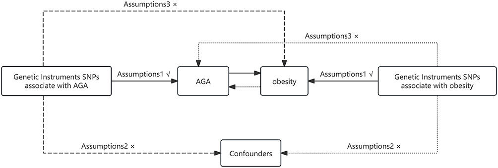 |
Figure 1 Overview of the study design. |
Concrete IV screening criteria: SNPs reaching genome-wide significance threshold P<5×10⁻⁸ for exposure trait; linkage disequilibrium clumping set as R2<0.001 within 10,000kb genomic window to exclude redundant correlated variants; F-statistic>10 was applied to guarantee strong instrument validity and avoid weak instrument bias.
Two-Sample Mendelian Randomization Analysis
Five MR analytical algorithms were applied in the current research: inverse-variance weighted (IVW), weighted median, MR-Egger regression, simple mode and weighted mode.15,16
IVW random-effects model: primary analytical approach, suitable under slight between-SNP heterogeneity without obvious directional horizontal pleiotropy;
MR-Egger regression: capable of detecting and adjusting unbalanced directional pleiotropy, relaxes exclusion restriction assumption;
Weighted median: robust against partial outlier instrumental SNPs, consistent causal estimation when over half IVs are valid genetic instruments;
Simple and Weighted mode: stable inference under extensive invalid instrumental variants.
Random-effects IVW was prioritized for primary causal estimation owing to detected between-variant heterogeneity in subsequent statistical testing.
Sensitivity Analysis
Three categories of sensitivity tests were comprehensively arranged: horizontal pleiotropy test, between-instrument heterogeneity assessment and leave-one-out iterative exclusion analysis, with detailed testing principles expanded as requested by peer reviewer:
MR-Egger intercept test (mr_pleiotropy_test): intercept P>0.05 indicates absence of statistically significant directional horizontal pleiotropy;
Cochran’s Q-based heterogeneity test (mr_heterogeneity): Q-statistic P<0.05 confirms remarkable heterogeneity across instrumental SNPs, necessitating random-effects IVW instead of fixed-effect model;
Leave-one-out analysis: sequentially eliminate individual single SNP from the IV set and repeat MR estimation to identify outlier variants dominating overall causal results.
All statistical calculations were finished via TwoSampleMR package (R software v4.4.2).
Results
After standardized IV screening, total 53 qualified SNPs were enrolled as obesity instrumental variables for forward MR analysis (Figure 2). Cochran Q heterogeneity test demonstrated significant inter-SNP heterogeneity (P< 0.05), so random-effects IVW was implemented as the primary outcome evaluation. Random-effects IVW showed no causal effect of obesity on AGA risk (OR=1.01,95% CI 0.98–1.04,P=0.58). Estimation outcomes from the weighted median, MR-Egger, simple mode and weighted mode all exhibited P>0.05, consistently ruling out causal association (Figure 3A).
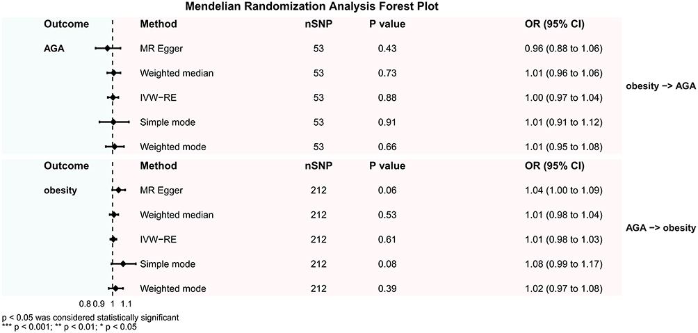 |
Figure 2 Forest plot of MR and reverse MR analyses for AGA and obesity. |
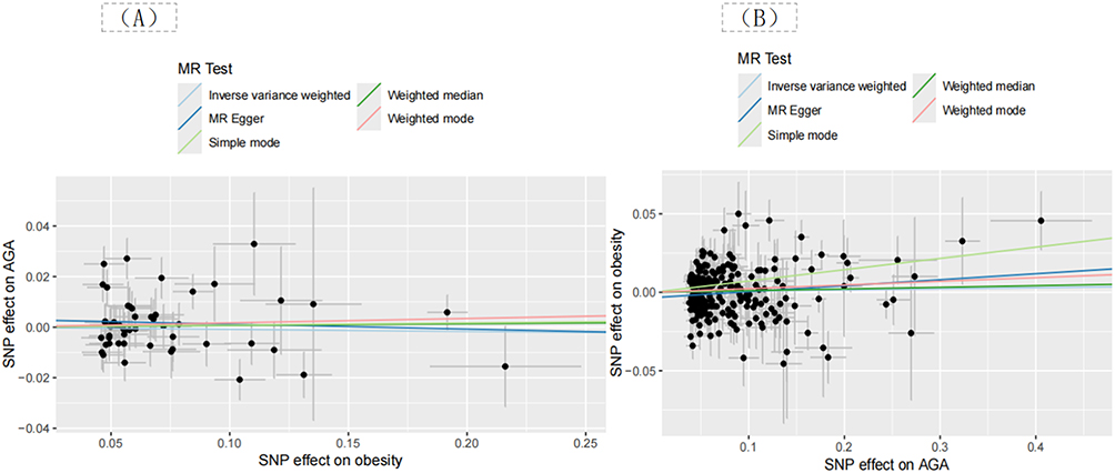 |
Figure 3 Scatter plots of MR analysis (A) and reverse MR analysis (B) for AGA and Obesity. |
MR-Egger intercept pleiotropy test yielded P=0.354>0.05, eliminating significant directional pleiotropic bias (Table 1). Leave-one-out sequential deletion analysis confirmed no single outlier SNP could drastically alter pooled MR effect size, validating result stability (Figure 4A).
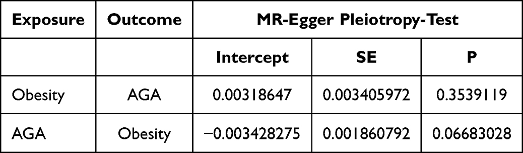 |
Table 1 Pleiotropy Test Results of MR and Reverse MR Analyses for AGA and Obesity |
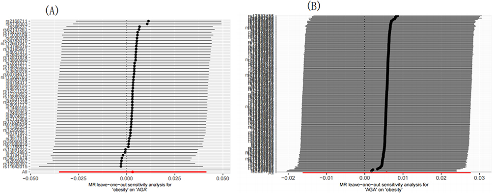 |
Figure 4 Leave-one-out plots of MR analysis (A) and reverse MR analysis (B) for AGA and Obesity. |
Reverse Mendelian Randomization Analysis (AGA as Exposure, Obesity as Outcome)
A total of 212 genome-wide significant SNPs were selected as AGA instrumental variants for reverse MR analysis (Figure 2). Heterogeneity testing indicated statistically significant between-variant dispersion (P<0.05), random-effects IVW was accordingly adopted for causal calculation, which excluded causal influence of AGA on obesity (OR=1.00,95% CI 0.99–1.01,P=0.83). The other four complementary MR methods generated consistent non-significant results (Figure 3B).
MR-Egger pleiotropy intercept P=0.067>0.05 without prominent horizontal pleiotropy (Table 1). Leave-one-out sensitivity analysis verified robust pooled estimation free from single-SNP bias (Figure 4B).
Discussion
The present research is the first bidirectional two-sample MR focusing on causal association between obesity and AGA based on large-scale publicly available GWAS summary data. Multiple complementary MR approaches uniformly verified the absence of bidirectional causal links between obesity and AGA, solving the conflicting conclusions from previous observational epidemiological investigations.
AGA follicular pathological change mainly originates from elevated local scalp 5α-reductase and androgen receptor expression within balding-region hair follicle dermal papilla cells. Excess androgen binding accelerates hair follicle miniaturization via shortening the anagen hair cycle and prolonging the telogen resting phase.2 Multiple cross-sectional clinical researches have reported an epidemiological link between AGA and metabolic spectrum diseases, including metabolic syndrome, hypertension and dyslipidaemia.3–5
Obesity is a multifactorial chronic metabolic disorder largely modulated by polygenic heredity and acquired lifestyle factors.17–19 Multiple observational surveys found higher BMI and waist circumference among AGA male patients compared with non-alopecia controls,20 and several mechanistic hypotheses were previously proposed to explain potential obesity-related AGA pathogenesis: (1) adipose tissue excess promotes peripheral hyperandrogenism to aggravate scalp follicular androgen-dependent atrophy;13,21–23 (2) obesity-induced insulin resistance impairs hair follicle stem cell homeostasis and disrupts normal hair cycling;24–29 (3) decreased circulating vitamin D concentration in the obese population accelerates follicular degeneration;30–33 (4) lipid metabolic disorder damages hair follicle stem cell survival directly under obese status.34
Nevertheless, multiple independent population-based cross-sectional researches failed to replicate positive obesity-AGA correlation: Chinese college student population survey and an early-onset AGA cohort based on Han Chinese all demonstrated irrelevant BMI and AGA incidence.35–37 Such inconsistent observational outcomes are inevitably confounded by lifestyle, socioeconomic status and undetected confounding covariates, which highlights the necessity of MR-based causal inference to minimize confounder and reverse‑causation bias. Current MR results exclude a direct causal effect between obesity and AGA, implying that the previously observed positive epidemiological correlation is induced by shared confounding factors rather than intrinsic biological causation.
Our study possesses several limitations. Primarily, source GWAS data exclusively covered European ancestry cohorts, restricting extrapolation of research conclusion to non-Caucasian ethnic groups; subsequent multi-ancestry GWAS-based MR researches are required to validate our findings across diverse populations. Second, although random-effects IVW was used to offset statistical heterogeneity from instrumental variants, potential residual cryptic pleiotropy cannot be fully eliminated; an advanced multi-variable MR design can further optimize relevant limitation in future exploration.
Conclusion
By utilizing large-scale publicly available GWAS summary statistics and bidirectional two-sample MR design with multiple complementary statistical algorithms and comprehensive sensitivity verification, this study provides robust genetic evidence rejecting bidirectional causal relationships between obesity and AGA. Correlative epidemiological associations reported by prior observational studies are likely driven by shared confounding factors instead of direct biological causation. Future translational researches should continuously explore molecular mechanisms linking metabolic dysregulation and AGA pathogenesis, and replicate present findings among multi-ethnic cohorts with diverse genetic backgrounds.
Acknowledgments
We want to acknowledge the participants and investigators of the Finnish database and the GWAS catalog database. This study was funded by Tianjin Key Medical Discipline Construction Project (Grant No:TJYXZDXK-3-010C).
Disclosure
The authors report no conflicts of interest in this work.
References
1. Otberg N, Finner AM, Shapiro J. Androgenetic alopecia. Endocrinol Metab Clin North Am. 2007;36(2):379–7. doi:10.1016/j.ecl.2007.03.004
2. Lolli F, Pallotti F, Rossi A, et al. Androgenetic alopecia: a review. Endocrine. 2017;57(1):9–17. doi:10.1007/s12020-017-1280-y
3. Qiu Y, Zhou X, Fu S, Luo S, Li Y. Systematic review and meta-analysis of the association between metabolic syndrome and androgenetic alopecia. Acta Derm Venereol. 2022;102:adv00645. doi:10.2340/actadv.v101.1012
4. Hu Y, Zhu Y, Lian N, Chen M, Bartke A, Yuan R. Metabolic syndrome and skin diseases. Front Endocrinol. 2019;10:788. doi:10.3389/fendo.2019.00788
5. Gopinath H, Upadya GM. Metabolic syndrome in androgenic alopecia. Indian J Dermatol Venereol Leprol. 2016;82(4):404–408. doi:10.4103/0378-6323.174421
6. Ng M, Fleming T, Robinson M, et al. Global, regional, and national prevalence of overweight and obesity in children and adults during 1980-2013: a systematic analysis for the Global Burden of Disease Study 2013. Lancet. 2014;384(9945):766–781. doi:10.1016/S0140-6736(14)60460-8
7. Williams EP, Mesidor M, Winters K, Dubbert PM, Wyatt SB. Overweight and obesity: prevalence, consequences, and causes of a growing public health problem. Curr Obes Rep. 2015;4(3):363–370. doi:10.1007/s13679-015-0169-4
8. Guh DP, Zhang W, Bansback N, Amarsi Z, Birmingham CL, Anis AH. The incidence of co-morbidities related to obesity and overweight: a systematic review and meta-analysis. BMC Public Health. 2009;9:88. doi:10.1186/1471-2458-9-88
9. Lauby-Secretan B, Scoccianti C, Loomis D, Grosse Y, Bianchini F, Straif K. Body fatness and cancer--viewpoint of the IARC working group. N Engl J Med. 2016;375(8):794–798. doi:10.1056/NEJMsr1606602
10. Puig L. Obesity and psoriasis: body weight and body mass index influence the response to biological treatment. J Eur Acad Dermatol Venereol. 2011;25(9):1007–1011. doi:10.1111/j.1468-3083.2011.04065.x
11. Budu-Aggrey A, Brumpton B, Tyrrell J, et al. Evidence of a causal relationship between body mass index and psoriasis: a Mendelian randomization study. PLoS Med. 2019;16(1):e1002739. doi:10.1371/journal.pmed.1002739
12. Li Z, Zhao Y. Evidence of a causal relationship between body mass index and immune-mediated and inflammatory skin diseases and biomarkers: a Mendelian randomization study. Clin Cosmet Invest Dermatol. 2024;17:2659–2667. doi:10.2147/CCID.S496066
13. Darlenski R, Mihaylova V, Handjieva-Darlenska T. The link between obesity and the skin. Front Nutr. 2022;9:855573. doi:10.3389/fnut.2022.855573
14. Spiga F, Gibson M, Dawson S, et al. Tools for assessing quality and risk of bias in Mendelian randomization studies: a systematic review. Int J Epidemiol. 2023;52(1):227–249. doi:10.1093/ije/dyac149
15. Grover S, Del Greco MF, Stein CM, Ziegler A. Mendelian randomization. Methods Mol Biol. 2017;1666:581–628.
16. Bowden J, Davey Smith G, Haycock PC, Burgess S. Consistent estimation in Mendelian randomization with some invalid instruments using a weighted median estimator. Genet Epidemiol. 2016;40(4):304–314. doi:10.1002/gepi.21965
17. Fall T, Ingelsson E. Genome-wide association studies of obesity and metabolic syndrome. Mol Cell Endocrinol. 2014;382(1):740–757. doi:10.1016/j.mce.2012.08.018
18. Després JP, Lemieux I. Abdominal obesity and metabolic syndrome. Nature. 2006;444(7121):881–887. doi:10.1038/nature05488
19. Maes HH, Neale MC, Eaves LJ. Genetic and environmental factors in relative body weight and human adiposity. Behav Genet. 1997;27(4):325–351. doi:10.1023/A:1025635913927
20. Hirsso P, Rajala U, Hiltunen L, Jokelainen J, Keinänen-Kiukaanniemi S, Näyhä S. Obesity and low-grade inflammation among young Finnish men with early-onset alopecia. Dermatology. 2007;214(2):125–129. doi:10.1159/000098570
21. Hirt PA, Castillo DE, Yosipovitch G, Keri JE. Skin changes in the obese patient. J Am Acad Dermatol. 2019;81(5):1037–1057. doi:10.1016/j.jaad.2018.12.070
22. Uzuncakmak TK, Akdeniz N, Karadag AS. Cutaneous manifestations of obesity and themetabolic syndrome. Clin Dermatol. 2018;36(1):81–88. doi:10.1016/j.clindermatol.2017.09.014
23. Abrams P, Levitt Katz LE. Metabolic effects of obesity causing disease in childhood. Curr Opin Endocrinol Diabetes Obes. 2011;18(1):23–27. doi:10.1097/MED.0b013e3283424b37
24. Ding X, Bai Z, Wang W, Gu Y, Li Z, Fan Y. Exploring the causal relationship and molecular mechanisms between fasting insulin and androgenetic alopecia: a Mendelian randomization study with bioinformatics analysis. Clin Cosmet Invest Dermatol. 2025;18:355–365. doi:10.2147/CCID.S492958
25. Mocciaro G, Gastaldelli A. Obesity-related insulin resistance: the central role of adipose tissue dysfunction. Handb Exp Pharmacol. 2022;274:145–164.
26. Polsky S, Ellis SL. Obesity, insulin resistance, and type 1 diabetes mellitus. Curr Opin Endocrinol Diabetes Obes. 2015;22(4):277–282. doi:10.1097/MED.0000000000000170
27. Ruze R, Liu T, Zou X, et al. Obesity and type 2 diabetes mellitus: connections in epidemiology, pathogenesis, and treatments. Front Endocrinol. 2023;14:1161521. doi:10.3389/fendo.2023.1161521
28. Aoki M, Murase T. Obesity-associated insulin resistance adversely affects skin function. PLoS One. 2019;14(10):e0223528. doi:10.1371/journal.pone.0223528
29. Nabaie L, Kavand S, Robati RM, et al. Androgenic alopecia and insulin resistance: are they really related? Clin Exp Dermatol. 2009;34(6):694–697. doi:10.1111/j.1365-2230.2008.03118.x
30. Wu Y, Hui Y, Liu F, et al. The association of serum adipokines, insulin resistance and Vitamin D status in male patients with androgenetic alopecia. Clin Cosmet Invest Dermatol. 2023;16:419–427. doi:10.2147/CCID.S396697
31. Zubair Z, Kantamaneni K, Jalla K, et al. Prevalence of low serum Vitamin D levels in patients presenting with androgenetic alopecia: a review. Cureus. 2021;13(12):e20431. doi:10.7759/cureus.20431
32. Vimaleswaran KS, Berry DJ, Lu C, et al. Causal relationship between obesity and vitamin D status: bi-directional Mendelian randomization analysis of multiple cohorts. PLoS Med. 2013;10(2):e1001383. doi:10.1371/journal.pmed.1001383
33. Wortsman J, Matsuoka LY, Chen TC, Lu Z, Holick MF. Decreased bioavailability of vitamin D in obesity. Am J Clin Nutr. 2000;72(3):690–693. doi:10.1093/ajcn/72.3.690
34. Morinaga H, Mohri Y, Grachtchouk M, et al. Obesity accelerates hair thinning by stem cell-centric converging mechanisms. Nature. 2021;595(7866):266–271. doi:10.1038/s41586-021-03624-x
35. Vora RV, Kota R, Singhal RR, Anjaneyan G. Clinical profile of androgenic alopecia and its association with cardiovascular risk factors. Indian J Dermatol. 2019;64(1):19–22. doi:10.4103/ijd.IJD_526_16
36. He F, Shen M, Zhao Z, et al. Epidemiology and disease burden of androgenetic alopecia in college freshmen in China: a population-based study. PLoS One. 2022;17(2):e0263912. doi:10.1371/journal.pone.0263912
37. Davies NM, Holmes MV, Davey Smith G. Reading Mendelian randomisation studies: a guide, glossary, and checklist for clinicians. BMJ. 2018;362:k601. doi:10.1136/bmj.k601
 © 2026 The Author(s). This work is published and licensed by Dove Medical Press Limited. The
full terms of this license are available at https://www.dovepress.com/terms
and incorporate the Creative Commons Attribution
- Non Commercial (unported, 4.0) License.
By accessing the work you hereby accept the Terms. Non-commercial uses of the work are permitted
without any further permission from Dove Medical Press Limited, provided the work is properly
attributed. For permission for commercial use of this work, please see paragraphs 4.2 and 5 of our Terms.
© 2026 The Author(s). This work is published and licensed by Dove Medical Press Limited. The
full terms of this license are available at https://www.dovepress.com/terms
and incorporate the Creative Commons Attribution
- Non Commercial (unported, 4.0) License.
By accessing the work you hereby accept the Terms. Non-commercial uses of the work are permitted
without any further permission from Dove Medical Press Limited, provided the work is properly
attributed. For permission for commercial use of this work, please see paragraphs 4.2 and 5 of our Terms.