Back to Journals » Clinical Ophthalmology » Volume 12
Posterior capsular complication rates with femtosecond laser-assisted cataract surgery: a consecutive comparative cohort and literature review
Authors Levitz L ![]() , Reich J, Hodge C
, Reich J, Hodge C ![]()
Received 4 May 2018
Accepted for publication 13 July 2018
Published 6 September 2018 Volume 2018:12 Pages 1701—1706
DOI https://doi.org/10.2147/OPTH.S173089
Checked for plagiarism Yes
Review by Single anonymous peer review
Peer reviewer comments 4
Editor who approved publication: Dr Scott Fraser
Lewis Levitz,1 Joseph Reich,1 Chris Hodge1,2
1Vision Eye Institute Camberwell, Hawthorn East, VIC, Australia; 2Graduate School of Health, University of Technology Sydney, Sydney, NSW, Australia
Purpose: The aim of the study was to determine whether femtosecond-assisted laser cataract surgery (FLACS) reduces the posterior capsular complication (PCC) rate compared to manual cataract surgery when performed by an experienced surgeon.
Patients and methods: We reviewed 2,021 consecutive FLACS procedures between 1 June 2012 and 30 August 2017. All cases of posterior capsular rupture (PCR) with or without vitreous prolapse or zonular dialysis (ZD) that prevented an in-the-bag placement of the intraocular lens were included. Risk factors were noted and outcomes documented.
Results: Six eyes of 2,021 (0.3%) who underwent FLACS had either a PCR or ZD. One eye (0.25%) of 403 eyes that had manual cataract surgery had a PCR. There was no significant difference in outcomes. Risk factors included advanced age, dense nuclei, pseudoexfoliation and small pupil. Only a single case in the FLACS series may have been directly attributed to the FLACS procedure.
Conclusion: This study provides evidence that there is no significant difference in the PCC rate between FLACS and manual cataract surgery in the hand of an experienced surgeon who performs >350 cases annually. This low rate of complications may be achieved by less experienced surgeons adopting FLACS.
Keywords: cataract surgery, phacoemulsification complications, femtosecond laser-assisted cataract surgery, safety, posterior capsule rupture, zonular dehiscence
Introduction
Posterior capsular complications (PCCs) have a significant effect on patient outcome due to the increased risk of additional complications such as cystoid macular edema, retinal detachment and endophthalmitis.1,2 The published incidence of posterior capsular rupture (PCR) varies considerably within the literature from 0.18% to 23.3%.3,4 Ocular risk factors include miosis, zonulopathy, axial length, previous surgery and concurrent ocular procedures.5,6 The learning curve remains a further variable with studies indicating an increased incidence among inexperienced surgeons or those surgeons with relatively lower surgical volumes.4,7–9
Femtosecond laser-assisted cataract surgery (FLACS) remains a new technology that purports to increase both the safety and accuracy of cataract surgery.10 The literature has supported statistical improvements over conventional techniques albeit with significant variation across cohorts.11,12 With respect to PCC, early reports indicated an increase in incidence of both anterior and posterior capsular tears in FLACS cohorts.3,10 The strength and morphology of the femtosecond laser-created capsule were considered the contributing factors although clinical evidence now suggests no significant difference.13,14
Benchmark data remain key to improving surgical outcomes.15 The existence of several large databases has provided significant epidemiological data and serves to improve our understanding of the contribution of relative risk factors.6,16,17 Although a clear benefit of clinical registries is the power through sample size and the correlation to “real-world” results, the use of data from a significant variety of sources may not necessarily indicate the optimal level of surgical outcomes achievable. We present a surgical audit of a single surgeon using both FLACS and manual techniques in a private ambulatory theater. This review was undertaken to support existing literature and provide an additional benchmark for surgeons considering FLACS.
Patients and methods
Consecutive procedures from July 2012 to August 2017 were included in a retrospective audit of PCC in both FLACS and manual patients from a single, experienced surgeon. The audit start date corresponded with the initial procedures using the femtosecond laser at the respective clinics. Patient files were reviewed for an intraoperative diagnosis of a PCC, which included PCR (with or without vitreous prolapse) or zonular dehiscence of the capsular bag (zonular dialysis [ZD]). To avoid the risk of underreporting, clinical findings were supported by a review of concurrent theater records. Theater records identified the use of vitrectomy probes or capsular tension rings and the diagnosis of an unplanned vitrectomy. Video recordings were available for all cases and reviewed to confirm the diagnosis and management of PCR or ZD.
Patients with trabeculectomies, previous refractive surgery and floppy iris syndrome were not excluded from undertaking FLACS pretreatment. The only condition that precluded FLACS was the presence of posterior synechia resulting in suboptimal pupil dilation (<4.5 mm). This occurred in five patients who were removed from consideration within both cohorts also to avoid selection bias.
Surgery occurred at two ambulatory theaters, with FLACS pretreatment conducted using the Alcon LenSx machine (Alcon Laboratories, Inc., Fort Worth, TX, USA) as available. Surgery was completed with either the Alcon Infiniti or later the Centurion phacoemulsification units (Alcon Laboratories, Inc.). Both femtosecond and phacoemulsification units maintained similar settings across the duration of review. A four-quadrant laser pattern was used in all patients. The laser settings did not change significantly across the time period of the study.
The patients were pretreated with ketorolac (Acular; Allergan, Inc., Irvine, CA, USA) drops just prior to and after the laser procedure. Pupils were dilated with cyclopentolate (Alcon Laboratories, Inc.), tropicamide 1% (Mydriacyl; Alcon Laboratories, Inc.) and phenylephrine 2.5% (Minims; Chauvin Pharmaceuticals, London, UK) prior to the laser being performed and again after laser. Manual cases received a single round of dilating drops prior to surgery.
Statistical analysis was performed using SPSS software (version 24.0; IBM Corporation, Armonk, NY, USA). Descriptive statistical methods were used to report the basic demographic details. The ratio of PCC between laser-assisted and manual cohorts was compared using chi-square test. A P-value of <0.05 was considered statistically significant.
The Vision Eye Institute’s Low and Negligible Risk (LNR) Research Committee reviewed the retrospective research request and granted approval for the conduct of the review. Patients had previously signed a privacy form indicating their consent to de-identified information to be used for audit and research purposes.
Results
Six eyes of 2,021 consecutive procedures (six patients) had a posterior capsular tear or zonular dehiscence during surgery for an overall PCC rate of 0.3%. One of 403 consecutive manual procedures had a PCC with an incidence of 0.25%. There was no difference between the rate of PCC cases between cohorts (P=0.868).
The age range of patients who had PCC varied from 67 to 98 years although 83.3% (5/6) of FLACS patients were 84 years or older. The single manual patient was 82 years old. Preoperative patient details and existing risk factors are included in Table 1. One patient had a history of tamsulosin for benign prostatic hyperplasia. The patient with a small pupil required intraoperative dilation with an I-Ring (BVI Visitec, Waltham, MA, USA).
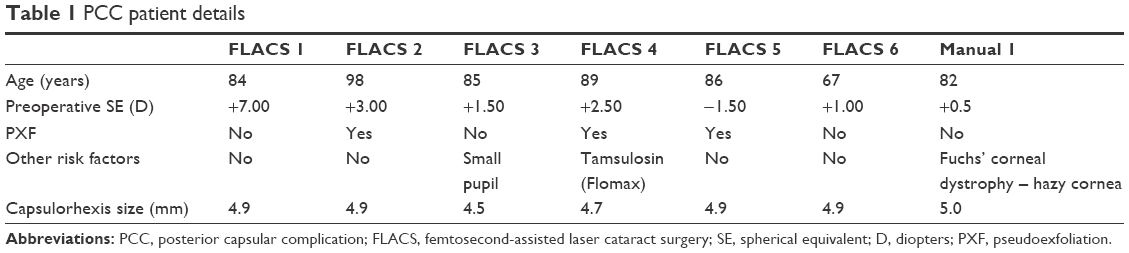 | Table 1 PCC patient details |
Surgical complication and postoperative treatments are listed in Table 2.
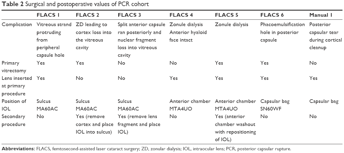 | Table 2 Surgical and postoperative values of PCR cohort |
With respect to the FLACS cases, three patients were diagnosed as having zonular dehiscence. This occurred as the last quadrant was removed (one eye) or during cortical “cleanup” (two eyes). Two of the three eyes had previously documented pseudoexfoliation (PXF). Three FLACS eyes had PCR, which occurred at different points across surgery. One occurred attempting to lift an epi-cortical plate on the posterior capsule, which led to the phaco probe puncturing the posterior capsule. A further case occurred during phacoemulsification following a post-occlusion surge, which led to a “punch through” hole. The final FLACS PCR case occurred when an anterior capsule tear extended to the posterior pole during sculpting. The patient was an 85-year-old male with a history of poorly dilated (4.5 mm) and dense brunescent cataract. During laser application, an air bubble was noticed in the periphery of the patient interface. Prior to the cataract removal, the capsulotomy was seen to be incomplete. During aspiration of the nucleus, a capsular tear developed ~180° from the section, which was manually completed. This extended to the posterior pole. A video review indicated the entire nucleus moving forward immediately prior to the tear. It was not clear if the phaco tip then inadvertently caught the capsule during this movement or whether the tear occurred as a result of the forward pressure of the nucleus itself.
Of interest, no cases occurred during the surgeon’s initial 200 FLACS cases, suggesting that a learning curve effect was not apparent.
Discussion
PCR is a significant potential complication of cataract surgery with both short- and long-term financial and safety considerations. Qatarneh et al18 identified that patients with PCR required more follow-up visits over a statistically longer duration compared to a control cohort, reflecting in a sixfold increase in costs to the patients for the visits alone. Furthermore, PCC increases the risk of complications, requiring additional surgery. Day et al19 found that the risk of retinal detachment within 3 months of surgery was 42 times higher in patients with PCR. The rate of endophthalmitis was eight times greater than that in controls, confirming PCR as a legitimate concern.
Ocular risk factors for PCC have been identified and include axial length, zonulopathy, miosis, cataract grade, previous surgery and concurrent ocular procedures. Day et al16 found that eyes with a short axial length (<20.0 mm) were more likely to have PCR (3.6% vs 1.95% for all eyes); however, further studies have failed to identify a consistent correlation between axial length and PCR.20 In a recent meta-analysis, Vazquez-Ferreiro et al found that PXF continues to represent an additional significant risk factor for complications. The authors found a pooled OR of 2.14 for PXF patients, leading to posterior rupture or ZD during cataract surgery.21 The influence of previous surgery is also a consideration. Literature suggests that patients with a history of intravitreal injections are associated with an increased risk of PCR, presumably as a result of iatrogenic lens trauma following the injection process.22 Intraoperatively, Carifi et al23 found that almost one-quarter of eyes with anterior capsular tears proceeded to posterior tears, highlighting the importance of early recognition of warning signs.
There has been a significant variation in the incidence of PCC in cataract studies. This is influenced by both the sample cohort and the respective experience of surgeons. Reported “vitreous loss” rates have been shown to increase fourfold to 1.8% when intraoperative theater nursing logs, postoperative clinical discharge summaries and clinical letters are reviewed, suggesting significant recall bias as a contributing factor in the historically large variation in literature findings.24 Less experienced surgeons have been identified as a considerable risk factor for PCR. Fathallah et al4 described an incidence of PCR as high as 23.3%. In their study, >40% of surgeons were junior staff who disproportionally contributed to the complication rate. These findings are replicated elsewhere with trainee surgeons, albeit with reduced incidence (Table 3). Turnbull and Lash25 found a cumulative PCR rate of 2.1% in a survey of ophthalmology trainees, a value that approaches general registry findings.
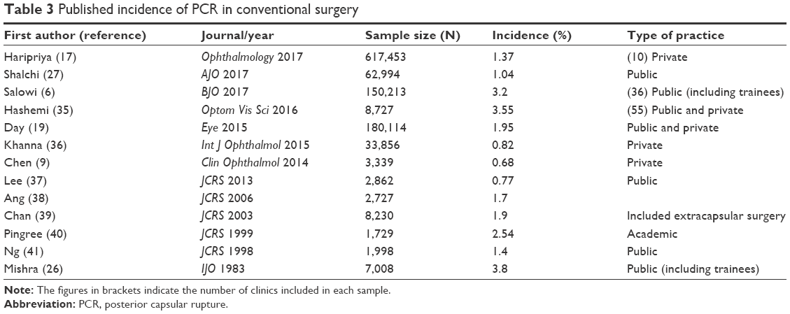 | Table 3 Published incidence of PCR in conventional surgery |
As expected, the incidence of PCC is reduced in audits including experienced surgeons with rates ranging from 0.68% to 3.8%25,26 (Table 3). It would appear that within a large study, a finding of ≤2.0% would represent a realistic goal.19,27 This however may not represent the true benchmark. More recently, Abell et al3 described a rate of 0.18% in patients undergoing conventional surgery in a comparative cohort study between conventional and FLACS techniques, which remains the lowest published mark within PCC literature within studies with reasonable sample sizes. Our results in both FLACS and manual cohorts remain broadly equivalent to this finding and indicate no difference between cohorts.
Consistency may also represent a key issue. Previously, Habib et al28 found a significant difference in the rate of complications in surgeons completing >400 surgeries each year as compared to those doing less. Chen et al9 more recently supported this assumption, albeit with a rate of 274 procedures per year as a differentiating factor in their audit of a small private clinic. These results may have significance when determining the potential benefits of converting from manual to FLACS techniques. For the purposes of this study, an “experienced surgeon” was defined as one who does >350 cases per year, as this number has been validated both in a public and private hospital settings.9,27 Scott et al14 indicated that all surgeons improved the PCC rate following conversion to FLACS surgery; however, results suggest that the surgeons performing the most annual procedures experienced the least improvement in PCC rates. Further analysis is required from additional cohorts; however, we believe that our results support the authors’ findings that consistency and volume serve to further reduce the risk of complications. Of note, the surgeon (LL) in this audit performs ~400 procedures per year.
The initial report of PCC in FLACS literature found an incidence of 3.5%.10 The cohort represented the initial learning curve among surgeons experienced in conventional techniques. The patient interface and software used by the first reporting groups are now obsolete, and thereof, these results cannot be extrapolated to the software and patient interface used today. Subsequently, debate increased regarding the possibility of an inherently weaker laser-created capsule, leading to a greater risk of capsule-related complications.3 The published literature suggests however that this does not reflect clinical practice with published rates between 0.27% and 0.87%3,28 (Table 4). Our study provides an additional benchmark in a standard cataract population undergoing FLACS technique.43
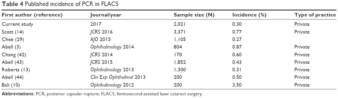 | Table 4 Published incidence of PCR in FLACS |
Several case reports indeed suggest that in cases with increased risk of capsule or zonular deficiency, FLACS may provide some additional benefit over conventional techniques; however, further numbers are required to confirm this potential.30,31 The findings do reflect some internal bias as the early studies, at least initially, removed complex cases or eyes with risk factors from consideration from FLACS, thereby reducing the potential for PCC. Similarly, the use of femtosecond laser was primarily available to experienced surgeons rather than trainees in a public or teaching hospital. More recently, the use of FLACS by residents has been shown to be well tolerated, albeit possibly less efficient than conventional techniques at the same stage of training.32–34 Our study found a PCC rate of ~0.3% in both manual and laser cohorts, which is comparable to the best found in the current literature. Of importance is that the FLACS cohort represented all patients with minimal exclusions highlighting the potential use as a revised current benchmark target. That the majority of our PCC patients had one or more potential risk factors highlights the need for continued awareness with either FLACS or conventional procedures.
Conclusion
The PCC of 0.3% has been validated as a benchmark for FLACS. This study also provides primary evidence that there is no significant difference in the PCC rate between FLACS and manual cataract surgery undertaken by an experienced surgeon who performs >350 cases annually. The rate of PCC with FLACS of 0.3% is well below that usually reported for manual cataract surgery.
Author contributions
All authors contributed toward data analysis, drafting and critically revising the paper and agree to be accountable for all aspects of the work.
Disclosure
The author reports no conflicts of interest in this work.
References
Johansson B, Lundström M, Montan P, Stenevi U, Behndig A. Capsule complication during cataract surgery: Long-term outcomes: Swedish Capsule Rupture Study Group report 3. J Cataract Refract Surg. 2009;35(10):1694–1698. | ||
Day AC, Donachie PH, Sparrow JM, Johnston RL. Royal College of Ophthalmologists’ National Ophthalmology Database. United Kingdom National Ophthalmology Database Study of Cataract Surgery: Report 3: Pseudophakic Retinal Detachment. Ophthalmology. 2016;123(8):1711–1715. | ||
Abell RG, Davies PEJ, Phelan D, Goemann K, Mcpherson ZE, Vote BJ. Anterior capsulotomy integrity after femtosecond laser-assisted cataract surgery. Ophthalmology. 2014;121(1):17–24. | ||
Fathallah M, Eltanamly RM, Saadeldin H, Elnahry GH. Causes of suboptimal corrected visual acuity following phacoemulsification in a teaching university hospital. Eur J Ophthalmol. 2017;27(2):169–173. | ||
Lundström M, Behndig A, Montan P, et al. Capsule complication during cataract surgery: background, study design, and required additional care: Swedish Capsule Rupture Study Group report 1. J Cataract Refract Surg. 2009;35(10):1679–1687. | ||
Salowi MA, Chew FLM, Adnan TH, King C, Ismail M, Goh PP. The Malaysian Cataract Surgery Registry: risk Indicators for posterior capsular rupture. Br J Ophthalmol. 2017;101(11):1466–1470. | ||
Hashemi H, Mohammadpour M, Jabbarvand M, Nezamdoost Z, Ghadimi H. Incidence of and risk factors for vitreous loss in resident-performed phacoemulsification surgery. J Cataract Refract Surg. 2013;39(9):1377–1382. | ||
Ti SE, Yang YN, Lang SS, Chee SP. A 5-year audit of cataract surgery outcomes after posterior capsule rupture and risk factors affecting visual acuity. Am J Ophthalmol. 2014;157(1):180–185. | ||
Chen M, Lamattina KC, Patrianakos T, Dwarakanathan S. Complication rate of posterior capsule rupture with vitreous loss during phacoemulsification at a Hawaiian cataract surgical center: a clinical audit. Clin Ophthalmol. 2014;8:375–378. | ||
Bali SJ, Hodge C, Lawless M, Roberts TV, Sutton G. Early experience with the femtosecond laser for cataract surgery. Ophthalmology. 2012;119(5):891–899. | ||
Roberts TV, Lawless M, Chan CC, et al. Femtosecond laser cataract surgery: technology and clinical practice. Clin Exp Ophthalmol. 2013;41(2):180–186. | ||
Nagy ZZ, Takacs AI, Filkorn T, et al. Complications of femtosecond laser-assisted cataract surgery. J Cataract Refract Surg. 2014;40(1):20–28. | ||
Roberts TV, Lawless M, Bali SJ, Hodge C, Sutton G. Surgical outcomes and safety of femtosecond laser cataract surgery: a prospective study of 1500 consecutive cases. Ophthalmology. 2013;120(2):227–233. | ||
Scott WJ, Tauber S, Gessler JA, Ohly JG, Owsiak RR, Eck CD. Comparison of vitreous loss rates between manual phacoemulsification and femtosecond laser-assisted cataract surgery. J Cataract Refract Surg. 2016;42(7):1003–1008. | ||
Ashwin PT, Mohamed SR. Representative data remains the key for national benchmarks. Eye. 2010;24(7):1298. | ||
Day AC, Donachie PH, Sparrow JM, Johnston RL, Royal College of Ophthalmologists’ National Ophthalmology Database. The Royal College of Ophthalmologists’ National Ophthalmology Database Study of cataract surgery: report 2, relationships of axial length with ocular copathology, preoperative visual acuity, and posterior capsule rupture. Eye. 2015;29(12):1528–1537. | ||
Haripriya A, Chang DF, Ravindran RD. Endophthalmitis reduction with intracameral moxifloxacin prophylaxis: analysis of 600000 surgeries. Ophthalmology. 2017;124(6):768–775. | ||
Qatarneh D, Mathew RG, Palmer S, Bunce C, Tuft S. The economic cost of posterior capsule tear at cataract surgery. Br J Ophthalmol. 2012;96(1):114–117. | ||
Day AC, Donachie PH, Sparrow JM, Johnston RL. Royal College of Ophthalmologists’ National Ophthalmology Database. The Royal College of Ophthalmologists’ National Ophthalmology Database study of cataract surgery: report 1, visual outcomes and complications. Eye. 2015;29(4):552–560. | ||
Lam JK, Chan TC, Ng AL, Chow VW, Wong VW, Jhanji V. Outcomes of cataract operations in extreme high axial myopia. Graefes Arch Clin Exp Ophthalmol. 2016;254(9):1811–1817. | ||
Vazquez-Ferreiro P, Carrera-Hueso FJ, Poquet Jornet JE, et al. Intraoperative complications of phacoemulsification in pseudoexfoliation:Metaanalysis. J Cataract Refract Surg. 2016;42(11):1666–1675. | ||
Lee AY, Day AC, Egan C, et al. United Kingdom age-related macular degeneration and diabetic retinopathy electronic medical records users group. Previous intravitreal therapy is associated with increased risk of posterior capsule rupture during cataract surgery. Ophthalmology. 2016;123(6):1252–1256. | ||
Carifi G, Miller MH, Pitsas C, et al. Complications and outcomes of phacoemulsification cataract surgery complicated by anterior capsule tear. Am J Ophthalmol. 2015;159(3):463–469. | ||
Kim BZ, Patel DV, Mcghee CN. Auckland cataract study 2: clinical outcomes of phacoemulsification cataract surgery in a public teaching hospital. Clin Exp Ophthalmol. 2017;45(6):584–591. | ||
Turnbull AM, Lash SC. Confidence of ophthalmology specialist trainees in the management of posterior capsule rupture and vitreous loss. Eye. 2016;30(7):943–948. | ||
Mishra RK. Anterior vitrectomy. Indian J Ophthalmol. 1983;31(3):288–289. | ||
Shalchi Z, Okada M, Whiting C, Hamilton R. Risk of posterior capsule rupture during cataract surgery in eyes with previous intravitreal injections. Am J Ophthalmol. 2017;177:77–80. | ||
Habib M, Mandal K, Bunce CV, Fraser SG. The relation of volume with outcome in phacoemulsification surgery. Br J Ophthalmol. 2004;88(5):643–646. | ||
Chee SP, Yang Y, Ti SE, Se T. Clinical outcomes in the first two years of femtosecond laser-assisted cataract surgery. Am J Ophthalmol. 2015;159(4):714–719. | ||
Martin AI, Hodge C, Lawless M, Roberts T, Hughes P, Sutton G. Femtosecond laser cataract surgery: challenging cases. Curr Opin Ophthalmol. 2014;25(1):71–80. | ||
Taravella MJ, Meghpara B, Frank G, Gensheimer W, Davidson R. Femtosecond laser-assisted cataract surgery in complex cases. J Cataract Refract Surg. 2016;42(6):813–816. | ||
Day AC, Dhallu SK, Maurino V, Wilkins MR. Initial experience using a femtosecond laser cataract surgery system at a UK National Health Service cataract surgery day care centre. BMJ Open. 2016;6(7):e012078. | ||
Pittner AC, Sullivan BR. Resident surgeon efficiency in femtosecond laser-assisted cataract surgery. Clin Ophthalmol. 2017;11:291–297. | ||
Cohen MN, Intili A, Ni N, Blecher MH. Femtosecond laser-assisted cataract surgery in residency training. Curr Opin Ophthalmol. 2015;26(1):56–60. | ||
Hashemi H, Rezvan F, Etemad K, et al. Intraoperative complications of cataract surgery in Tehran Province, Iran. Optom Vis Sci. 2016;93(3):266–271. | ||
Khanna RC, Ray VP, Latha M, Cassard SD, Mathai A, Sekhar GC. Risk factors for endophthalmitis following cataract surgery-our experience at a tertiary eye care centre in India. Int J Ophthalmol. 2015;8(6):1184–118918. | ||
Lee RM, Foot B, Eke T. Posterior capsule rupture rate with akinetic and kinetic block anesthetic techniques. J Cataract Refract Surg. 2013;39(1):128–131. | ||
Ang GS, Whyte IF. Effect and outcomes of posterior capsule rupture in a district general hospital setting. J Cataract Refract Surg. 2006;32(4):623–627. | ||
Chan FM, Mathur R, Ku JJ, Jj K, et al. Short-term outcomes in eyes with posterior capsule rupture during cataract surgery. J Cataract Refract Surg. 2003;29(3):537–541. | ||
Pingree MF, Crandall AS, Olson RJ. Cataract surgery complications in 1 year at an academic institution. J Cataract Refract Surg. 1999;25(5):705–708. | ||
Ng DT, Rowe NA, Francis IC, et al. Intraoperative complications of 1000 phacoemulsification procedures: a prospective study. J Cataract Refract Surg. 1998;24(10):1390–1395. | ||
Chang JS, Chen IN, Chan WM, Ng JC, Chan VK, Law AK. Initial evaluation of a femtosecond laser system in cataract surgery. J Cataract Refract Surg. 2014;40(1):29–36. | ||
Abell RG, Darian-Smith E, Kan JB, Allen PL, Ewe SY, Vote BJ. Femtosecond laser-assisted cataract surgery versus standard phacoemulsification cataract surgery: outcomes and safety in more than 4000 cases at a single center. J Cataract Refract Surg. 2015;41(1):47–52. | ||
Abell RG, Kerr NM, Vote BJ. Femtosecond laser-assisted cataract surgery compared with conventional cataract surgery. Clin Exp Ophthalmol. 2013;41(5):455–462. |
 © 2018 The Author(s). This work is published and licensed by Dove Medical Press Limited. The
full terms of this license are available at https://www.dovepress.com/terms
and incorporate the Creative Commons Attribution
- Non Commercial (unported, 3.0) License.
By accessing the work you hereby accept the Terms. Non-commercial uses of the work are permitted
without any further permission from Dove Medical Press Limited, provided the work is properly
attributed. For permission for commercial use of this work, please see paragraphs 4.2 and 5 of our Terms.
© 2018 The Author(s). This work is published and licensed by Dove Medical Press Limited. The
full terms of this license are available at https://www.dovepress.com/terms
and incorporate the Creative Commons Attribution
- Non Commercial (unported, 3.0) License.
By accessing the work you hereby accept the Terms. Non-commercial uses of the work are permitted
without any further permission from Dove Medical Press Limited, provided the work is properly
attributed. For permission for commercial use of this work, please see paragraphs 4.2 and 5 of our Terms.