Back to Journals » Clinical Ophthalmology » Volume 14
Minimally Invasive Technique for Choroidal Fluid Drainage
Authors Safuri S, Bar-David L, Barak Y
Received 15 March 2020
Accepted for publication 2 June 2020
Published 9 July 2020 Volume 2020:14 Pages 1955—1958
DOI https://doi.org/10.2147/OPTH.S253989
Checked for plagiarism Yes
Review by Single anonymous peer review
Peer reviewer comments 2
Editor who approved publication: Dr Scott Fraser
Supplementary video of "Minimally invasive technique for choroidal fluid drainage" [ID 253989].
Views: 2619
Shadi Safuri, Laura Bar-David, Yoreh Barak
Department of Ophthalmology, Rambam Health Care Campus, Haifa, Israel
Correspondence: Yoreh Barak
Department of Ophthalmology, Rambam Health Care Campus, P.O.Box 9602, Haifa 31096, Israel
Tel +972-4-777-2668
Fax +972-4-777-2142
Email [email protected]
Background: This study describes a simple technique for the treatment of kissing choroidal detachment. In contrast to the commonly used technique, this technique is minimally invasive, fast, sutureless, and does not require access to the vitreous space.
Methods: A maintainer is inserted into the anterior chamber. A 25G trocar is inserted at the pars plana into the suprachoroidal space. Drainage is evident by the clear yellowish fluid freely emerging through the trocar, accompanied with deepening of the anterior chamber and an increase in the red reflex.
Results: Follow-up ultrasound 1 week after the surgery demonstrated resolution of the choroidal detachment. Net surgery time is about 10 minutes. No complications were noted.
Discussion: This is the first report of the technique performed in phakic eye, with video description of the steps and real-time clues for successful drainage even with reduced posterior segment visibility due to lens opacities.
Keywords: choroidal, drainage, kissing-detachment, minimally invasive, sutureless, trocar
Introduction
The suprachoroidal space is normally non-existent. Choroidal detachment, also termed choroidal effusion, results from several pathological states manifesting in abnormal accumulation of fluid in this space.1 The pathophysiology of these states can be understood using Starling’s law. An increase in the choroid capillary permeability, or abnormal balance of hydrostatic and osmotic gradients between the choroidal capillaries and interstitial space of the eye (ie, the intraocular pressure), may lead to accumulation of fluid in the suprachoroidal space.1 The most common etiology of choroidal effusion is glaucoma surgery resulting in hypotony.1,2 Other etiologies include hypotony due to other causes (such as leaking wound), inflammation resulting in increased permeability, and systemic hypertension via increase of the hydrostatic drive.1,2 A distinction must be made between serous and hemorrhagic choroidal effusion, the latter resulting from capillary rupture and entitling a worst prognosis. Choroidal detachment may be asymptomatic, but larger effusions are accompanied with shallowing of the anterior chamber and a decrease in vision. Pain often accompanies hemorrhagic effusion.1,3
Most choroidal effusions are managed conservatively. Most resolve spontaneously or after treating the inciting event such as leaking wound or inflammation. However, some cases demand surgery. Indications for surgical drainage include: flat anterior chamber, appositional (kissing) choroidal detachment, and non-resolving effusion.2,4
The classic and commonly performed drainage procedure1,2 involves conjunctival peritomy followed by a 3-mm radial incision in the sclera, about 3 to 4 mm posterior to the limbus. During the surgery the eye is kept pressurized by a recurrent injections of Balanced Salt Solution (BSS), or the use of a maintainer. Usually the sclerotomy site is left open with closure of overlying conjunctiva by suturing.
A debate exists where some experts advocate for routine simultaneous vitrectomy in order to reduce the risk of subsequent retinal detachment, giant retinal tear, and proliferative vitreoretinopathy, while others perform vitrectomy only following the development of such sequelle.1,2,5
Since choroidal effusion can be encountered by any ophthalmologist and can have a drastic effect if left untreated, there is a need for a simple technique that does not require expertise in posterior segment surgery. Here we describe a minimally invasive technique to treat choroidal effusion, which is fast, easy to learn and perform, sutureless, and independent of posterior segment visibility.
Methods
The patient gave signed informed consent for both the surgery and for publishing any case details, images, and videos. The case was presented to the Institutional Review Board (IRB) of Rambam Health Care Campus (Haifa, Israel) and was granted a waiver.
This simple technique is composed of two cardinal steps. The first being insertion of a maintainer into the anterior chamber using a paracentesis incision in the cornea. The infusion line is opened at 65 mmHg. The second step requires no exposure of the conjunctiva, diathermy, nor scleral cutting. A 25G non-valved trocar (25G Constellation Vitrectomy Pak, Alcon, Switzerland) is inserted at the pars plana at 15° into the suprachoroidal space.
Thereafter, successful drainage can be followed intra-operatively with three signs: fluid emerging from the trocar port, deepening of the anterior chamber, and the increase in red reflex (evaluated by periodically switching the light mode of the microscope, as can be seen in Video 1).
In case the lumen is suspected to be blocked by tissue, the trocar can be slightly pulled out. If required, a soft tool (eg, Weck-Cel) can be used to apply pressure on the eye globe posteriorly in order to facilitate drainage.
When fluid ceases to drain despite the above measures, and given the red reflex has improved, the trocar can be removed. No sutures are needed. Post-operative care includes topical steroid qid and topical antibiotics qid. B-mode ultrasound performed 1 week after the operation should confirm resolution of the choroidal fluid.
Results
The described technique is demonstrated in Video 1. A 67 years old female with a history of uncontrolled hypertension and no history of diabetes mellitus first presented with vitreous hemorrhage in her left eye, most probably due to uncontrolled hypertension. B-mode ultrasound demonstrated an attached retina with no flaps observed. She had undergone an uneventful 25G Pars Plana Vitrectomy, with 25% SF6 tamponade. No sutures were used. No immediate post-operative complications were noted and she was discharged on POD1. One week later she presented with appositional serous choroidal detachment (Figure 1B) on the operated eye, for which she underwent choroidal drainage. At the beginning of surgery the red reflux was dim (Figure 1A). The choroidal fluid was drained as described above. Specific to the patient described in this video an additional step was performed: posterior adhesions were released via gentle injection of viscoelastic material, thus ensuring free flow of BSS from the anterior chamber to the posterior chamber and vitreous space. Once the trocar is inserted, a clear yellow fluid drains freely, accompanied by deepening of the anterior chamber. A counter of the Weck-Cels used to absorb the drained fluid is presented at the bottom right corner of the video to allow tracking. Snapshots of the red reflex status at discrete stages of the surgery are presented on the right side. For this aim the light mode of the microscope was periodically switched for a brief time. The video runs at 3X speed. Net time of surgery (speculum to speculum) was ~10 minutes. At the end of surgery a bright reflex was observed (Figure 1C). One week after surgery b-mode ultrasound confirmed resolution of choroidal fluid (Figure 1D). No complication were noted. Visual acuity improved from hand motion at presentation to counting fingers from 2 meters 1 week after surgery, and is expected to improve more after the cataract is treated.
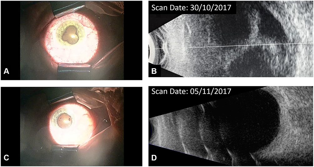 |
Figure 1 (A) Red reflex at the beginning of surgery, (B) B-mode ultrasound scan prior to surgery, (C) Red reflex at the end of the surgery, (D) B-mode ultrasound scan 1 week after surgery. |
Discussion
We described a simple, minimally invasive, fast, and easy to perform technique for choroidal fluid drainage. Choroidal detachment can result from several etiologies. However, it is most commonly encountered following hypotony due to glaucoma surgery.1,2 In this context, avoidance of scarring of the conjunctival tissue is of utmost importance for future interventions that might be required.
Using the classic approach, complications such as endophthalmitis and progression of cataract were reported.1,2 Due to the minimalistic perturbation of the eye globe and lack of access to the vitreous space, the risk of endophthalmitis is expected to be lower using this technique, but a large scale study is required to confirm this claim.
In the case described in the video, a 25G was used to drain serous fluid. Drainage of hemorrhagic choroidal detachment, after liquefaction was confirmed by ultrasound (usually occurring in 7–14 days6,8), was previously performed successfully via 20G trocar.4 However, Rezende et al4 advocate that the trocar should be inserted 7 mm posterior to the limbus, rather than at the pars plana, to avoid damage to the retina. While this may provide an increased safety margin, in the case of appositional choroidal detachment, we think it is safe to insert the trocar at the pars plana provided it is inserted at a 15° angle. At the end of the drainage of choroidal fluid, routine and careful inspection of the site of drainage with wide angle viewing system or indirect ophthalmoscope is recommended if the visibility allows, as to ensure the absence of any retinal tissue perforation or incarceration. In the attached video, the canula which was inserted at the pars plana later assumes vertical inclination. Nevertheless, the inclination of the canula should be kept angulated throughout the procedure as to minimize the risk of injury to the retina and further hemorrhage.
There are several reports of the technique with minor modifications,9,11 However, this is the first report of this technique performed on phakic eye with poor posterior segment visibility. A detailed video demonstrating both the technique and clues of successful drainage are provided here for the first time. This technique is independent of good visibility of the posterior segment, as in the case described in the video where cataract hindered visualization of the fundus. To follow the efficacy of the drainage, in addition to the free flow of fluid via the trocar, the deepening of the anterior chamber and enhancement of the red reflex provide additional clues for successful drainage.
Disclosure
The authors have no conflicts of interest to disclose.
References
1. Bellows AR, Chylack LT, Hutchinson BT. Choroidal detachment: clinical manifestation, therapy and mechanism of formation. Ophthalmology. 1981;88(11):1107–1115. doi:10.1016/S0161-6420(81)34897-0
2. WuDunn D, Ryser D, Cantor LB. Surgical drainage of choroidal effusions following glaucoma surgery. J Glaucoma. 2005;14(2):103–108. doi:10.1097/01.ijg.0000146370.28625.fc
3. Becquet F, Caputo G, Mashhour B, Chauvaud D, Pouliquen Y. Management of delayed massive suprachoroidal hemorrhage: a clinical retrospective study. Eur J Ophthalmol. 1996;6(4):393–397. doi:10.1177/112067219600600409
4. Rezende FA, Kickinger MC, Li G, Prado RF, Regis LGT. Transconjunctival drainage of serous and hemorrhagic choroidal detachment. Retina. 2012;32(2):242–249. doi:10.1097/IAE.0b013e31821c4087
5. Lee SC, Lee I, Koh HJ, Kim SH, Kwon OW. Massive suprachoroidal hemorrhage with retinal and vitreous incarceration; a vitreoretinal surgical approach. Korean J Ophthalmol. 2000;14(1):41–44. doi:10.3341/kjo.2000.14.1.41
6. Healey PR, Herndon L, Smiddy W. Management of suprachoroidal hemorrhage. J Glaucoma. 2007;16(6):577. AAO OPHTHALMIC PEARLS. doi:10.1097/IJG.0b013e318156a5a9
7. Lavinsky F, Moisseiev J, Levkovitch-Verbin H. The surgical management of massive intraoperative and postoperative suprachoroidal hemorrhage: anatomic and functional outcomes. Arq Bras Oftalmol. 2013;76(4):212–214. doi:10.1590/s0004-27492013000400003
8. Lakhanpal V. Experimental and clinical observations on massive suprachoroidal hemorrhage. Trans Am Ophthalmol Soc. 1993;91:545–652.
9. Hu ZZ, Liu QH, Ding YZ, Su Y, Ji JD. A modified method for suprachoroidal fluid drainage in kissing choroidal detachment. Int J Ophthalmol. 2020;13(2):346–348. doi:10.18240/ijo.2020.02.20
10. Rossi T, Boccassini B, Iossa M, Lesnoni G, Tamburrelli C. Choroidal hemorrhage drainage through 23-gauge vitrectomy cannulas. Retina. 2010;30(1):174–176. doi:10.1097/IAE.0b013e3181bdfa5e
11. Snyder LL, Kitchens JW, Patel SN. External choroidal drainage using direct visualization. Ophthalmic Surg Lasers Imaging Retina. 2019;50(8):529–531. doi:10.3928/23258160-20190806-11
 © 2020 The Author(s). This work is published and licensed by Dove Medical Press Limited. The
full terms of this license are available at https://www.dovepress.com/terms
and incorporate the Creative Commons Attribution
- Non Commercial (unported, 3.0) License.
By accessing the work you hereby accept the Terms. Non-commercial uses of the work are permitted
without any further permission from Dove Medical Press Limited, provided the work is properly
attributed. For permission for commercial use of this work, please see paragraphs 4.2 and 5 of our Terms.
© 2020 The Author(s). This work is published and licensed by Dove Medical Press Limited. The
full terms of this license are available at https://www.dovepress.com/terms
and incorporate the Creative Commons Attribution
- Non Commercial (unported, 3.0) License.
By accessing the work you hereby accept the Terms. Non-commercial uses of the work are permitted
without any further permission from Dove Medical Press Limited, provided the work is properly
attributed. For permission for commercial use of this work, please see paragraphs 4.2 and 5 of our Terms.