")
Back to Journals » OncoTargets and Therapy » Volume 17
Uterine Burkitt Lymphoma with Rare Extranodal Deposits in the Bone, Breast, and Sacral Canal: A Case Report
Authors Liu XS , Liu C, Chen YJ, Zeng DB, Wang YL, Pei ZJ
Received 14 September 2023
Accepted for publication 24 January 2024
Published 26 January 2024 Volume 2024:17 Pages 41—44
DOI https://doi.org/10.2147/OTT.S440228
Checked for plagiarism Yes
Review by Single anonymous peer review
Peer reviewer comments 2
Editor who approved publication: Prof. Dr. Daniel Neureiter
Xu-Sheng Liu,1 Chao Liu,2 Yi-Jia Chen,1 Dao-Bing Zeng,1 Ya-Lan Wang,1 Zhi-Jun Pei1
1Department of Nuclear Medicine, Taihe Hospital, Hubei University of Medicine, Shiyan, People’s Republic of China; 2Medical Imaging Center, Taihe Hospital, Hubei University of Medicine, Shiyan, People’s Republic of China
Correspondence: Zhi-Jun Pei, Department of Nuclear Medicine, Taihe Hospital, Hubei University of Medicine, Shiyan, People’s Republic of China, Email [email protected]
Abstract: Burkitt lymphoma is a highly invasive non-Hodgkin lymphoma. Sporadic Burkitt’s lymphoma is commonly found in the abdomen. However, Burkitt lymphoma infiltrating the uterus is uncommon in occurrence. We report the results of 18F-FDG PET/CT examination of a 36-year-old woman. The report indicates that in addition to the strong uptake of FDG imaging agent in the uterus, bilateral cervical and abdominal lymph nodes also have strong activity. At the same time, it was also found that bilateral small breast nodules, sacral canal and multiple bones in the whole body showed a radiation uptake pattern similar to that of the uterus. 18F-FDG PET/CT imaging can help determine the extent of the disease and the affected body area, which is helpful to guide the treatment decision. This case report shows the application of 18F-FDG PET/CT imaging in the diagnosis, staging and post-treatment evaluation of Burkitt lymphoma of the uterus. It provides very useful information for clinicians and helps to improve the accuracy of diagnosis and treatment effect.
Keywords: uterine, Burkitt lymphoma, case report, PET/CT, bone involvement
Introduction
Burkitt lymphoma is a highly invasive non-Hodgkin lymphoma, known for its rapid growth and invasion of the body’s lymphatic system.1,2 Sporadic Burkitt’s lymphoma is commonly found in the abdomen.3 However, there have been few reported cases of Burkitt lymphoma involving the uterus. Uterine Burkitt lymphoma is a rare and challenging diagnosis, as it can present with non-specific symptoms and can often be confused with other gynecological malignancies.4,5 The use of imaging modalities such as 18F-FDG PET/CT has greatly improved the detection and staging of this disease, particularly in cases with multi-focal disease.6–8 Early diagnosis and active treatment are crucial to improve the prognosis of patients. However, there are few reports on PET/CT imaging of uterine Burkitt lymphoma.
Case Presentation
In January 2013, Shiyan Taihe Hospital accepted a 36-year-old female referral patient. The patient had abnormal uterine bleeding a month prior to presentation, which was characterized by increased menstrual volume, prolonged menstrual period and occasional dizziness. after the onset of the disease, the patient was treated in an external hospital and underwent hysteroscopy and uterine tissue aspiration. The examination suggested the likelihood of: (uterine) Burkitt lymphoma. Immunohistochemical results showed LCA (+++), CD38 (+++), CD138 (-), CKpan (-), EMA (-), Vimentin (+), CD10 (+), CD3 (+), CD5 (+), CD20 (+++), CD79α (+++), Ki-67 (positive rate 95%), MUM-1 (+), CyclinD1 (-), SOX-10 (-), CD23 (-), Pax-5 (+++), Bcl-2 (-), Bcl-6 (+), TdT (-), κ (+), λ (-), CD43 (+), SMA (-), h-Caldesmon (-), Desmin (-), ER (-), PR (-). In order to further evaluate the situation and determine the hidden metastasis, she was referred to the PET center for 18F-FDG PET/CT lymphoma staging.
18F-FDG PET/CT results showed that there was abnormal imaging agent concentration in the uterine body and cervix of the patient (Figure 1), suggesting that the possibility of malignant tumor was high, which supported the diagnosis of immunohistochemistry.
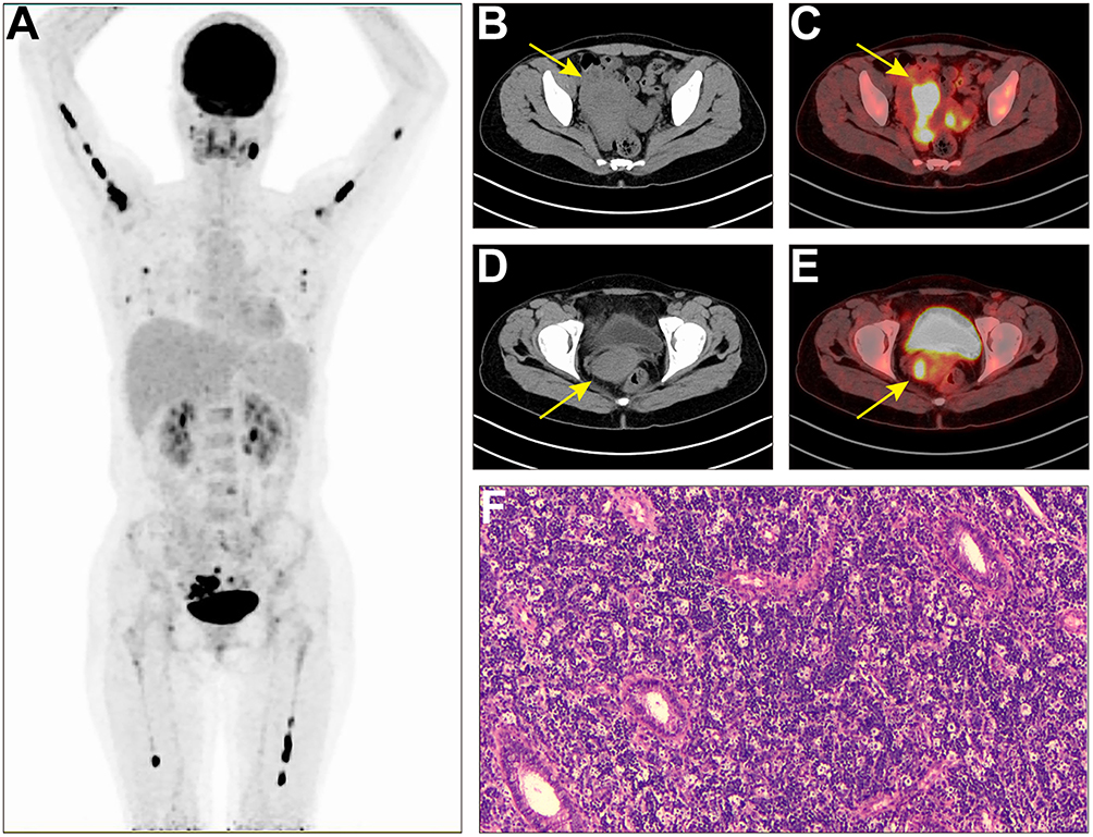 |
Figure 1 MIP on 18F-FDG scan (A) showed hypermetabolic lesions in the whole body. Increased tracer uptake occurred in the pelvic area, and the most significant abnormal activity of the uterine body was located in the axial image (B), CT; (C), PET/CT fusion image; (arrow, SUVmax, 19.5). The most significant abnormal activity of the cervix is located in the axial image (D), CT; (E), PET/CT fusion image; (arrow, SUVmax, 9.5). (F) Hematoxylin-eosin stain, original magnification ×40. Histopathological reports support Burkitt’s lymphoma. |
At the same time, PET/CT scan also found abnormal uptake of imaging agents in multiple lymph nodes, including left cervical lymph nodes and abdominal lymph nodes. In addition, several extranodal parts of the body, including breast, sacral canal and bone, have metabolic abnormalities of different degrees. These findings support the diagnosis of Burkitt lymphoma and suggest that the case has widespread invasion. Based on the PET/CT scan results, the clinician diagnosed the patient as stage IIIa of Burkitt lymphoma and had lost the opportunity for surgery. The patient received CD20 monoclonal antibodies combined with chemotherapy.
The patient underwent a PET/CT scan after the completion of the fourth round of chemotherapy. The post-treatment PET/CT scan revealed a restored uterine morphology (decreased size and disappearance of hypermetabolic lesions). The size and metabolism of the lymph nodes in the left neck and abdominal cavity, and the small nodules scattered on both sides of the breast glands decreased compared with before. The bone density of many bones in the whole body was uneven and the uptake of imaging agents was lower than before. After the above consideration, the activity of tumor cells was significantly suppressed after comprehensive treatment (Figure 2).
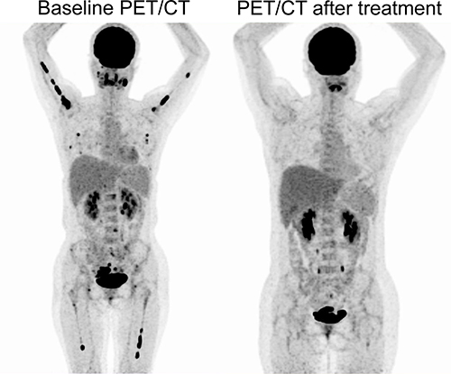 |
Figure 2 18F-FDG PET/CT imaging after and before treatment. |
Discussion
Burkitt lymphoma is a dangerous malignant lymphoma, which usually occurs in children and young people. It is characterized by rapid tumor growth and rapidly deteriorating symptoms. Although the treatment of Burkitt lymphoma has made significant progress in the past decades, the treatment of advanced or recurrent cases still faces challenges.1,2 18F-FDG PET/CT imaging has very high diagnostic accuracy, can provide systemic metabolic information, and help guide treatment and monitor the efficacy.9,10
In previous reports, Burkitt lymphoma often involved lymph nodes and extranodal sites, such as peritoneal lymph nodes, spleen and bone. Wang et al6 reported that a 7-year-old boy had peritoneal Burkitt lymphoma. PET/CT showed peritoneal, mesenteric and omental masses, accompanied by obvious ascites, mesenteric nodules and kidney involvement. Wen et al7 reported the results of FDG PET/CT examination of a 21-year-old man. His Burkitt lymphoma involved not only lymph nodes, spleen, brain and bone, but also four organs of the endocrine system, including thyroid gland, right adrenal gland, pancreas and right testicle. Previous studies reported the first well-documented case of Burkitt lymphoma in the uterus. A 40-year-old woman presented with vaginal bleeding. Endometrial scrapes revealed diffuse infiltration of medium-sized lymphocytes with the characteristic morphological and immunophenotypic characteristics of Burkitt lymphoma. There was no evidence of extrauterine disease.11 However, there are few reports of PET/CT scan involving uterine Burkitt lymphoma. In the treatment of Burkitt lymphoma, chemotherapy and immunotherapy are the main treatment methods.1,2 For Burkitt lymphoma of the uterus, depending on the patient’s age, condition and range of disease, other treatment methods such as surgery or chemotherapy may be required.4,5 In our report, not only lymph node diseases are involved, but also a large number of extranodal sites, including breast, bone and sacral canal. In particular, the appearance of lesions involving bones throughout the body is a manifestation of its deterioration, which usually occurs in patients with advanced disease. There are relatively few clinical studies on Burkitt lymphoma with bone involvement throughout the body, and no standard treatment scheme has been established for this subtype. Burkitt lymphoma with bone involvement throughout the body is a rare and challenging disease. Clinicians need to consider multiple treatment methods according to the patient’s condition to improve the treatment effect. However, 18F-FDG PET/CT imaging can help determine the extent of the disease and the affected body area, which is helpful to guide the treatment decision.9
Conclusion
In conclusion, this case report shows the application of 18F-FDG PET/CT imaging in the diagnosis, staging and post-treatment evaluation of Burkitt lymphoma of the uterus. It provides very useful information for clinicians and helps to improve the accuracy of diagnosis and treatment effect.
Ethics Approval and Informed Consent
The studies involving human participants were reviewed and approved by The Ethics Committee of Taihe Hospital Affiliated of Hubei University of Medicine. Written informed consent was obtained from the patient for publication of this report.
Acknowledgments
This work was supported by the Shiyan Taihe Hospital hospital-level project (2022JJXM007) and the Key Discipline Project of Hubei University of Medicine.
Disclosure
The authors report no conflicts of interest in this work.
Reference
1. López C, Burkhardt B, Chan JKC, et al. Burkitt lymphoma. Nat Rev Dis Primers. 2022;8(1):78. doi:10.1038/s41572-022-00404-3
2. Roschewski M, Staudt LM, Wilson WH. Burkitt’s Lymphoma. N Engl J Med. 2022;387(12):1111–1122. doi:10.1056/NEJMra2025746
3. Zeng W, Lechowicz MJ, Winton E, Cho SM, Galt JR, Halkar R. Spectrum of FDG PET/CT findings in Burkitt lymphoma. Clin Nucl Med. 2009;34(6):355–358. doi:10.1097/RLU.0b013e3181a34552
4. Horowitz NA, Benyamini N, Wohlfart K, Brenner B, Avivi I. Reproductive organ involvement in non-Hodgkin lymphoma during pregnancy: a systematic review. Lancet Oncol. 2013;14(7):e275–e282. doi:10.1016/S1470-2045(12)70589-2
5. Khan WA, Deshpande KA, Kurdukar M, Patil P, Mahure H, Pawar V. Primary Burkitts’s lymphoma of endometrium and bilateral ovaries in a 6-year-old female: report of a rare entity and review of the published work. J Obstet Gynaecol Res. 2013;39(10):1484–1487. doi:10.1111/jog.12085
6. Wang X, Chen Z, Tang G, Zhang X, Child With A. Burkitt lymphoma with pleural, peritoneal, mesenteric, omental, and renal involvement. Clin Nucl Med. 2011;36(7):612–615. doi:10.1097/RLU.0b013e318217af84
7. Wen Z, Zhuang H. Burkitt’s lymphoma involving multiple hormone-producing organs on FDG PET/CT. Clin Nucl Med. 2019;44(12):995–997. doi:10.1097/RLU.0000000000002807
8. Chen Z, Xue Q, Huang C, Yao S, Miao W. Burkitt lymphoma/ leukemia presented on 68Ga-Pentixafor and 18F-FDG PET/CT. Clin Nucl Med. 2022;47(1):98–100. doi:10.1097/RLU.0000000000003750
9. Milgrom SA, Rechner L, Berthelsen A. The optimal use of PET/CT in the management of lymphoma patients. Br J Radiol. 2021;94(1127):20210470. doi:10.1259/bjr.20210470
10. Juweid ME, Mueller M, Alhouri A, A‐Risheq MZ, Mottaghy FM. Positron emission tomography/computed tomography in the management of Hodgkin and B-cell non-Hodgkin lymphoma: an update. Cancer. 2021;127(20):3727–3741. doi:10.1002/cncr.33772
11. Keller C, Savage DG, Rusta-Villa M, Bhagat G, Alobeid B. Primary Burkitt lymphoma of the uterine corpus. Leuk Lymphoma. 2006;47(1):141–145. doi:10.1080/10428190500144821
 © 2024 The Author(s). This work is published and licensed by Dove Medical Press Limited. The full terms of this license are available at https://www.dovepress.com/terms.php and incorporate the Creative Commons Attribution - Non Commercial (unported, v3.0) License.
By accessing the work you hereby accept the Terms. Non-commercial uses of the work are permitted without any further permission from Dove Medical Press Limited, provided the work is properly attributed. For permission for commercial use of this work, please see paragraphs 4.2 and 5 of our Terms.
© 2024 The Author(s). This work is published and licensed by Dove Medical Press Limited. The full terms of this license are available at https://www.dovepress.com/terms.php and incorporate the Creative Commons Attribution - Non Commercial (unported, v3.0) License.
By accessing the work you hereby accept the Terms. Non-commercial uses of the work are permitted without any further permission from Dove Medical Press Limited, provided the work is properly attributed. For permission for commercial use of this work, please see paragraphs 4.2 and 5 of our Terms.