Back to Journals » International Journal of Nephrology and Renovascular Disease » Volume 13
Calciphylaxis: An Analysis of Concomitant Factors, Treatment Effectiveness and Prognosis in 30 Patients
Authors Panchal S, Holtermann K, Trivedi N, Regunath H , Yerram P
Received 7 December 2019
Accepted for publication 20 March 2020
Published 5 April 2020 Volume 2020:13 Pages 65—71
DOI https://doi.org/10.2147/IJNRD.S241422
Checked for plagiarism Yes
Review by Single anonymous peer review
Peer reviewer comments 2
Editor who approved publication: Professor Pravin Singhal
Sarju Panchal,1 Kirstie Holtermann,2 Namrita Trivedi,1 Hariharan Regunath,3 Preethi Yerram4
1Department of Internal Medicine, Hospital of University of Pennsylvania, Philadelphia, PA 19146, USA; 2Department of Medicine, University of Missouri School of Medicine, Columbia, MO 65212, USA; 3Department of Medicine – Divisions of Pulmonary, Critical Care and Infectious Diseases, University of Missouri School of Medicine, Columbia, MO 65212, USA; 4Department of Medicine – Division of Nephrology, University of Missouri School of Medicine, Columbia, MO 65212, USA
Correspondence: Preethi Yerram
Department of Medicine - Division of Nephrology, University of Missouri School of Medicine, One Hospital Drive, Columbia, MO 65212, USA
Tel +1 573 882 7992
Email [email protected]
Background: Calciphylaxis is a rare but severe complication mostly affecting patients with end-stage renal disease (ESRD) and is associated with high morbidity and mortality. The natural history, concomitant factors, pathogenesis, and treatment for calciphylaxis remain equivocal.
Methods: We conducted a retrospective study on patients diagnosed with calciphylaxis in a tertiary care center between January 1, 2012, and December 31, 2017. We describe demographics, co-morbidities, laboratory parameters, effectiveness of sodium thiosulfate treatment and outcomes.
Results: Of the 30 patients (age 65.6 ± 12.79 years, male:female = 8:22), 23 (76.67%) had ESRD and were either on hemodialysis (15 [65.22%], median duration 22.5 months [range 0.2– 96 months]) or peritoneal dialysis (8 [34.78%], duration 29± 10 months). Predisposing home medications: 8 (28%) had calcium supplements, 10 (36%) had warfarin, 16 (57%) had vitamin D and 5 (18%) had iron supplements. The median parathyroid hormone (PTH) level was 239.8 pg/mL (range 4.7– 2922). Calciphylaxis was found on extremities in 21 (70%) and on torso in 6 (20%) patients. Sodium thiosulfate (STS) was given for treatment in 20 (67%) patients and 3 were cured in < 2.25 months. One-year survival for all patients with calciphylaxis was 26% (29% for STS group and 20% for those that did not receive STS) and following any surgical treatment regardless of STS use was 14%.
Limitations: Retrospective design, absence of a control group and low power.
Conclusion: Calciphylaxis was more common among females with a predilection for extremities over the torso. Elevations in PTH and inflammatory markers were common. Treatment with STS did not show a statistically significant improvement in survival. Those who were cured, were treated with STS up to three months.
Keywords: calciphylaxis, sodium thiosulfate, calcific uremic arteriolopathy, calcification, non-uremic calciphylaxis
Background
Calciphylaxis is a serious condition with high morbidity and mortality commonly affecting the ESRD population. However, it can also be seen in patients without kidney disease or those with earlier stages of kidney disease and is termed Non-Uremic Calciphylaxis.1 The condition is rare with an annual incidence estimated to be around 0.4% in the United States and increasing, perhaps due to increased awareness of the condition.1,2 Unfortunately, the pathogenesis of calciphylaxis is not fully understood which makes it extremely challenging to treat. Histologic analysis and animal models suggest that calciphylaxis occurs due to calcification of small arterioles in the dermis and hypodermis layers of the skin. The calcification begins in the media of small arterioles and is associated with thrombosis and hyperplasia of the intimal layer. Occlusion of microvasculature results in ischemia and necrosis of the overlying skin and soft tissue.1,3
The current leading hypothesis is that there is an imbalance of promoters and inhibitors of calcification that lead to calciphylaxis. The three main inhibitors of calcification studied are Matrix G1a-Protein (MGP), Fetuin-A and inorganic pyrophosphate (PPi). The levels of these proteins are stipulated to be low in patients with calciphylaxis. MGP is an extracellular protein released by endothelial cells and vascular smooth muscle cells which must undergo vitamin K mediated carboxylation to be active. Active MGP is thought to inhibit precipitation of calcium phosphate based on animal models. Fetuin-A is a major carrier protein like albumin that is synthesized by the liver. Fetuin-A becomes downregulated in inflammatory states such as chronic kidney disease. Role of PPi is less clear; however, studies in murine models have shown that upregulating levels of PPi will prevent cardiovascular calcifications.1,3 Upregulation of pro-calcification factors, genes and cytokines is thought to potentiate calciphylaxis. Inflammatory cytokines damage the endothelial lining of blood vessels which promotes differentiation of endothelial cells into a type of cell that enables calcification. The precise cellular pathway remains elusive.1,3,4
Much remains to be understood about the pathogenesis of calciphylaxis before a sound treatment can be discovered. There is limited convincing literature on the epidemiology, risk factors, treatment and prognosis of calciphylaxis. Current postulated risk factors include ESRD, female gender, obesity, elevations in calcium (Ca), phosphate (PO4), parathyroid hormone (PTH), and comorbid conditions. Select medications are stipulated to increase risk of calciphylaxis, while few have been postulated to reduce risk. Medications thought to increase risk include, vitamin K antagonists like warfarin, calcium, phosphate, iron and vitamin D supplements and corticosteroids. Cinacalcet, a calcimimetic agent, is thought to reduce risk by reducing PTH levels and reducing calcium phosphate precipitation. Bisphosphonates are pyrophosphate analogs, and may be protective based on data from murine models, but limited data exist in humans. Additional factors with limited to no data in the literature include race, frequency, duration, and type of hemodialysis before diagnosis, protective medications, treatment, and prognosis data. To better comprehend these factors, we analyzed a cohort of patients at a single institution who presented with calciphylaxis over a five-year period.
Methods
The study is a retrospective chart review of patients diagnosed with calciphylaxis between January 1, 2012, to December 31, 2017, at the University of Missouri Hospital, Columbia, Missouri. The study was approved by the University of Missouri’s Institutional Review Board (IRB) as an expedited review. The IRB approved a “waiver of consent” for the study based on the retrospective nature of the study which would involve use of only de-identified information for data analysis and publication. The IRB also took into consideration the fact that the study could not practicably be conducted without a waiver of consent as many of the patients were either dead, or out of reach making it improbable to get individual consents. Once the data were collected, it was de-identified (medical record number was the only patient identifier we collected) prior to data analysis. All the collected data were stored securely per IRB approved methods, and patient data confidentiality was maintained in compliance with the Declaration of Helsinki.
After obtaining IRB approval, our electronic medical record system (Cerner PowerChart®) was queried through a request to Cerner support with the following inclusion criteria: age >18 years and terms “Calciphylaxis” or “Calcific Uremic Arteriolopathy.” The initial list consisted of 52 patients, which was scrutinized case-by-case by two authors to eliminate duplicates, and to determine if a biopsy was obtained, and whether the biopsy showed a definite or probable diagnosis of calciphylaxis. Thirty patients with biopsy-proven calciphylaxis were included from the original cohort and their records analyzed. Of note, the reported data were preferably collected at the time of diagnosis, if not within a month of diagnosis.
Statistics
Numbers, mean ± standard deviation (SD), median and range were used as appropriate based on the distribution of values for each parameter. Data were gathered and tabulated using Microsoft® Excel 2016 and imported into IBM® SPSS Statistics version 25 software for analysis. The survival estimates for the two groups were compared using a Kaplan–Meier curve.
Results
Among the 30 patients diagnosed with calciphylaxis, the mean age was 65.6 ± 12.79 years with a female preponderance (22, 73%). Twenty-seven patients (90%) were Caucasian, and only one patient (3%) was African American. Twenty-three patients (76.67%) had ESRD of which 15 (65.22%) were on hemodialysis (HD), and 8 (34.78%) were on peritoneal dialysis (PD) at the time of diagnosis. One had ESRD but was not on dialysis. The frequency of dialysis in the HD group was three times per week in 71% of the patients, less than three times per week in 19% of patients and more than three times per week in 10% of patients. Information on the duration on dialysis (any form of dialysis) before the diagnosis of calciphylaxis was available for 20 patients. The mean duration on dialysis before diagnosis was 42 months (3.5 years) with a range of 5 days to 18 years. Nine patients (45%) had been diagnosed with calciphylaxis within two years of starting any form of dialysis. Five patients (20%) developed ESRD secondary to nephritic or nephrotic syndromes including focal segmental glomerulosclerosis (FSGS), Alport syndrome and Membranous glomerulonephritis. Non-uremic calciphylaxis was seen in 6 patients (20%) with 2/6 patients having a glomerular filtration rate (GFR) <30mL/min at the time of diagnosis.
In 28 patients, medication history and co-morbidities at the time of diagnosis were available. Eight patients (28%) were found to be on a calcium supplement (either calcium acetate or carbonate), 16 (57%) were on vitamin D supplements (cholecalciferol, ergocalciferol or calcitriol) and 10 (36%) were on warfarin (8 at time of diagnosis, 2 prior use). One patient (4%) had been using glucocorticoids for >1 month, five (18%) were on iron supplements, three (11%) were on cinacalcet and only one (4%) was taking a bisphosphonate at the time of diagnosis. Ten patients (36%) were using a statin at the time of diagnosis.
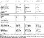
When analyzing co-morbidities in patients with calciphylaxis, we found that 1 patient (3%) had end-stage liver disease (cirrhosis), 20 patients (67%) had diabetes mellitus type 1 or 2, 17 patients (57%) had atherosclerotic disease (coronary artery disease or peripheral arterial disease), 14 patients (47%) were currently using tobacco or had a history of tobacco use, and 4 (13%) were using illicit drugs or had a history of drug use. We found that 17 patients (57%) were obese with a BMI of >30kg/m2 and 14 (47%) were morbidly obese with BMI of >35kg/m2. Table 1 lists the baseline characteristics including demographic, comorbidities and laboratory parameters of all patients and also provides a comparison of these parameters between the two groups.
 |
Table 1 Baseline Characteristics Between Two Groups Based on the Use of Sodium Thiosulfate |
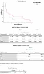
Six patients (20%) had calciphylaxis centrally, 21 (70%) had a lesion on extremities, and 3 (10%) had a combination of central and peripheral involvement. Out of the 30 patients, 20 received sodium thiosulfate (STS) and 10 did not. Out of the patients that received STS, three patients were cured within 2.25 months of use. Out of the 17 patients that were not healed, only two remained alive at the time of data collection. The overall one-year survival rate for patients with calciphylaxis in our study was 26%. The one-year survival rate for patients who receive STS was 29%, and those that did not receive STS was 20% (Figure 1). The mean survival time for patients who received STS was 529 days, 95% CI [238–821] versus patients who did not receive STS was 255 days, 95% CI [98–414] and there was no statistical significance (p- 0.218). Eight patients from the group had some form of surgery for calciphylaxis (excision, debridement or above the knee amputation). The one-year survival rate in patients who received surgery regardless of STS use was 14%.
 |
Figure 1 Kaplan–Meier survival estimates between those who received sodium thiosulfate and those who did not. |
Discussion
Our study has a moderate size cohort of patients diagnosed with calciphylaxis from a single institution. Due to verification of biopsies by two authors of the study, we are confident that our findings are specific to patients with calciphylaxis. The mean age in our study was ~66 years with a preponderance of female sex (73%) and Caucasians (90%), which were consistent with other studies.2,4,5 McCarthy et al studied a cohort of 101 patients with a mean age of 60 years and 80% being female.2 Nigwekar et al studied a cohort of 172 patients with a mean age of 55 years, 74% being female5 Bleyer et al studied a cohort of 19 cases in a referral center with the population being 50% African Americans, and they found that all patients with calciphylaxis were Caucasian.6
Majority of the patients in our study had CKD stage 5 (80%) with 65.22% being on HD and 34.78% being on PD. Only one patient had healthy kidney function, and 17% had CKD stage 2–4. McCarthy et al reported similar findings in a cohort of 101 patients with 62% having CKD stage 5 and 19% had CKD stage 3 or 4. PD has recently been implicated as a risk factor for calciphylaxis; however, data are lacking.7 According to the National Institute of Health, as of 2013, approximately 7% of patients in the United States are being treated with PD vs 64% with HD. Our findings support PD being a risk factor with 33% of patients being on PD at the time of diagnosis of calciphylaxis. Longer duration on dialysis is also an emerging risk factor. However, to our knowledge, there are no large prospective or retrospective studies to date that support or refute this as a risk factor. A cross-sectional survey of Angelis et al found that younger patients who had been on dialysis for an average of 80 months were at increased risk.8 Our findings differed from the results of this study because 50% of our patients were diagnosed within 2.4 years (29 months) of starting any form of dialysis, the range of 5 days to 18 years.
There are data supporting that elevated calcium, phosphate, and parathyroid hormone levels are seen in patients with calciphylaxis but they are not highly specific.1 Analysis of our cohort revealed a mean corrected calcium of 9.26mg/dl with 75% of patients have serum calcium <9.9mg/dL and all of them having values <11.3mg/dL. Mean phosphate was 5.57mg/dl with 50% of patients having values <4.4mg/dL. The mean PTH in our study was 481 pg/mL with 75% having values >140pg/mL. Normal values of PTH were only seen in patients with CKD stage less than 3. Studies in animal models have shown cutaneous calcification with the administration of megadoses of vitamin D and high phosphate diet. Ingesting calcium and vitamin D analogs, therefore, are deemed to be a risk factor.9 Our findings were consistent with this as 28% of patients were taking either calcium carbonate or calcium acetate at the time of diagnosis, and 57% of patients were on a vitamin D analog. Out of the patients taking vitamin D, 35% were taking calcitriol – the activated form of the vitamin D. Other medications implicated as risk factors include warfarin, iron supplements, and chronic glucocorticoid use. Proteins such as MGP that inhibit precipitation of calcium phosphate rely upon carboxylation by vitamin K to be active. Hence, warfarin, a vitamin K antagonist, is now known to be a risk factor based on multiple studies.4,10,11 Galloway et al directed a case–control study explicitly looking at the odds of developing calciphylaxis in patients taking warfarin. The study cohort was 2234 patients receiving dialysis from which 142 patients were using warfarin, and five total cases of calciphylaxis were found. Four out of the five patients with calciphylaxis were on warfarin (OR: 61, p=0.0001).12 We found that 36% of patients were exposed to warfarin at some time before diagnosis and 29% of patients were on warfarin at the time of diagnosis of calciphylaxis. Iron supplementation has been proposed as a risk factor because Farah et al and Amuluru et al found that tissue iron content is increased in calciphylaxis.13,14 Our study found that 18% of patients were on some form of iron supplement, but all the patients had normal or low serum iron. Ferritin was elevated above the upper limit of normal (150ng/mL for females and 300ng/mL for males) in over 75% of patients. Systemic glucocorticoid use is a possible emerging risk factor. To our knowledge, no specific studies have been conducted looking at this risk factor. Nigwekar et al conducted a systematic review of nonuremic calciphylaxis and found preceding corticosteroid use in 61% of patients (N=36). In our study, only one patient (4%) had been on corticosteroids for more than one month at the time of diagnosis.
There are limited data on medications that may protect against calciphylaxis. Cinacalcet, a calcimimetic used to treat secondary hyperparathyroidism, has been shown to reduce the incidence of calciphylaxis in hemodialysis patients. Three patients (11%) in our study were on cinacalcet at the time of diagnosis. Statins are known to have anti-inflammatory and anti-thrombotic properties. To our knowledge, only one previous study has looked at statin use in calciphylaxis.10 The study had 62 cases, 124 controls and found statin use was more common among controls than cases (39 vs 19%, p<0.01). In our study, 36% of patients were on statins at the time of diagnosis. There are scarce data behind bisphosphonates preventing calciphylaxis. Bisphosphonates are pyrophosphate (PPi) analogs, and studies in murine models have shown that raising levels of PPi’s decreases cardiovascular calcification. A study by Price et al found that Ibandronate prevents calciphylaxis in rat models. Only one patient (4%) in our study was on a bisphosphonate at the time of diagnosis.
The co-morbidities that have been strongly linked to calciphylaxis include obesity, diabetes, inflammatory conditions and malnutrition.11,15 Our findings are consistent with this as we found most of our patients were obese (60%), had diabetes mellitus type 1 or 2 (67%), and had albumin values <3.1 (greater than 50%). Among the markers for inflammation, we found that ESR was consistently elevated, whereas most CRP levels were within normal range indicating that such inflammatory markers may not be necessarily elevated in calciphylaxis. Among atherosclerotic diseases, 57% of our patients had a history of CAD or PAD, and ~50% had a history of smoking. Because there have been many risk factors involving the liver, such as vitamin K carboxylation, hypoalbuminemia, deficiencies of Fetuin-A,1 we analyzed the prevalence of the end-stage liver disease in patients with calciphylaxis. We found only one patient (3%) with cirrhosis.
A large retrospective review found that the majority of the calciphylaxis lesions tend to occur in the lower extremities rather than abdomen or buttocks (60% vs 30%, respectively).16 Our findings were consistent in that, 70% of the patients had calciphylaxis lesions in their extremities (upper or lower), and 20% had lesions on their central torso. The optimal treatment or treatment duration for calciphylaxis is not known. Sodium thiosulfate (STS) is currently the standard treatment based upon retrospective reviews. One case–control study of 16 cases reported that one-year survival of patients treated with STS is 65% vs 45% for patients not treated with STS.4 In our study, 20 patients (66%) received STS, of which, only three were cured and all three received STS for <2.25 months. Sodium thiosulfate use improved survival by 3% in the patients that were treated, but was not statistically significant probably from the low power of our study. Larger, prospective studies are needed to evaluate the efficacy, safety, and duration of STS use. In our study, eight patients received some form of debridement and the one-year survival among them was 14%. However, it is possible that patients who received debridement had a more severe case of calciphylaxis correlating to worse outcomes. Our results are discordant with the only other retrospective study of 64 cases which looked at mortality in patients with calciphylaxis undergoing debridement. This study reported improved survival in those who underwent surgical debridement (17 vs 46 cases [61% vs 27%]).17
In conclusion, calciphylaxis is a complex condition that is usually fatal. Our findings suggest that patients on dialysis may be predisposed to calciphylaxis as soon as one to two years. Having an elevated parathyroid hormone, ESR, ferritin, and low albumin seems to be a common occurrence. Levels of calcium, phosphate, and inflammatory markers may vary in patients with calciphylaxis and do not aid in the diagnosis. Warfarin and vitamin D analogs have a noticeably significant association with calciphylaxis. Further studies are needed to determine whether cinacalcet and/or bisphosphonates are indeed protective for calciphylaxis. There was no statistically significant difference in survival among patients who received STS compared to those that did not. The optimal duration of using STS is unknown, but our data suggest that in those that were cured of calciphylaxis, the average treatment duration was around 3 months. With this retrospective review, we hope to contribute to the existing literature on calciphylaxis and potentially spark new ideas for further research. Given the lack of definitive/effective treatment options for calciphylaxis, rigorous trials are desperately needed to test the efficacy of currently available treatments as well as novel options such as compound SNF472.17
Disclosure
Dr. Regunath has received honoraria from ©Getinge AB and ©Hamilton Medical; none were relevant to this study. Dr. Yerram has received honoraria from Reata Pharmaceuticals and CareDx; none were relevant to this study. The authors report no other potential conflicts of interest.
References
1. Nigwekar SU, Thadhani R, Brandenburg VM. Calciphylaxis. N Engl J Med. 2018;378(18):1704–1714. doi:10.1056/NEJMra1505292
2. Mccarthy JT, El-azhary RA, Patzelt MT, et al. Survival, risk factors, and effect of treatment in 101 patients with calciphylaxis. Mayo Clin Proc. 2016;91(10):1384–1394. doi:10.1016/j.mayocp.2016.06.025
3. Kramann R, Bradenburg VM, Schurgers IJ, et al. Novel insights into osteogenesis and matrix remodeling association with calcific uremic arteriolopathy. Nephrol Dial Transplant. 2013;28:856–868. doi:10.1093/ndt/gfs466
4. Mazhar AR, Johnson RJ, Gillen D, et al. Risk factors and mortality associated with calciphylaxis in end-stage renal disease. Kidney Int. 2001;60(1):324–332. doi:10.1046/j.1523-1755.2001.00803.x
5. Nigwekar SU, Brunelli SM, Meade D, Wang W, Hymes J, Lacson E. Sodium thiosulfate therapy for calcific uremic arteriolopathy. Clin J Am Soc Nephrol. 2013;8(7):1162–1170. doi:10.2215/CJN.09880912
6. Bleyer AJ, Choi M, Igwemezie B, De la Torre E, White WL. A case control study of proximal calciphylaxis. Am J Kidney Dis. 1998;32(3):376–383. doi:10.1053/ajkd.1998.v32.pm9740152
7. Zhang Y, Corapi KM, Luongo M, Thadhani R, Nigwekar SU. Calciphylaxis in peritoneal dialysis patients: a single center cohort study. Int J Nephrol Renovasc Dis. 2016;9:235–241. doi:10.2147/IJNRD.S115701
8. Angelis M, Wong LL, Myers SA, Wong LM. Calciphylaxis in patients on hemodialysis: a prevalence study. Surgery. 1997;122(6):1083–1089. doi:10.1016/S0039-6060(97)90212-9
9. Fine A, Zacharias J. Calciphylaxis is usually non-ulcerating: risk factors, outcome and therapy. Kidney Int. 2002;61(6):2210–2217. doi:10.1046/j.1523-1755.2002.00375.x
10. Nigwekar SU, Bhan I, Turchin A, et al. Statin use and calcific uremic arteriolopathy: a matched case-control study. Am J Nephrol. 2013;37(4):325–332. doi:10.1159/000348806
11. Hayashi M, Takamatsu I, Kanno Y, et al. A case-control study of calciphylaxis in Japanese end-stage renal disease patients. Nephrol Dial Transplant. 2012;27(4):1580–1584. doi:10.1093/ndt/gfr658
12. Galloway PA, El-damanawi R, Bardsley V, et al. Vitamin K antagonists predispose to calciphylaxis in patients with end-stage renal disease. Nephron. 2015;129(3):197–201. doi:10.1159/000371449
13. Amuluru L, High W, Hiatt KM, et al. Metal deposition in calcific uremic arteriolopathy. J Am Acad Dermatol. 2009;61(1):73–79. doi:10.1016/j.jaad.2009.01.042
14. Farah M, Crawford RI, Levin A, Chan Yan C. Calciphylaxis in the current era: emerging ‘ironic’ features? Nephrol Dial Transplant. 2011;26(1):191–195. doi:10.1093/ndt/gfq407
15. Nigwekar SU, Zhao S, Wenger J, et al. A nationally representative study of calcific uremic arteriolopathy risk factors. J Am Soc Nephrol. 2016;27(11):3421–3429. doi:10.1681/ASN.2015091065
16. Weenig RH, Sewell LD, Davis MD, Mccarthy JT, Pittelkow MR. Calciphylaxis: natural history, risk factor analysis, and outcome. J Am Acad Dermatol. 2007;56(4):569–579. doi:10.1016/j.jaad.2006.08.065
17. Brandenburg V, Sinha S, Torregrosa JV, et al. Phase 2 open label single arm repeat dose study to assess the effect of SNF472 on wound healing in uraemic calciphylaxis patients. J Am Soc Nephrol. 2017;28. doi:10.1681/ASN.2016080886
 © 2020 The Author(s). This work is published and licensed by Dove Medical Press Limited. The full terms of this license are available at https://www.dovepress.com/terms.php and incorporate the Creative Commons Attribution - Non Commercial (unported, v3.0) License.
By accessing the work you hereby accept the Terms. Non-commercial uses of the work are permitted without any further permission from Dove Medical Press Limited, provided the work is properly attributed. For permission for commercial use of this work, please see paragraphs 4.2 and 5 of our Terms.
