")
Back to Journals » Nature and Science of Sleep » Volume 15
Impact of Spina Bifida on Sleep Quality: Current Insights
Authors Gunnett M, Rocque BG , Nourani A, Beltran-Ale G
Received 16 May 2023
Accepted for publication 14 November 2023
Published 24 November 2023 Volume 2023:15 Pages 967—978
DOI https://doi.org/10.2147/NSS.S401269
Checked for plagiarism Yes
Review by Single anonymous peer review
Peer reviewer comments 3
Editor who approved publication: Prof. Dr. Ahmed BaHammam
Mohini Gunnett,1 Brandon G Rocque,2 Anis Nourani,1 Guillermo Beltran-Ale1
1Department of Pediatrics, Division of Pulmonary and Sleep Medicine, University of Alabama at Birmingham (UAB), Birmingham, AL, USA; 2Department of Neurosurgery, Division of Pediatric Neurosurgery, University of Alabama at Birmingham (UAB), Birmingham, AL, USA
Correspondence: Guillermo Beltran-Ale, University of Alabama at Birmingham, Department of Pediatrics, Division of Pulmonary and Sleep Medicine, 620 Jarman F. Lowder Building, 1600 7th Avenue South, Birmingham, AL, 35233-1711, USA, Tel +1 205 638 9593, Fax +1 205 638 2850, Email [email protected]
Abstract: Spina bifida (SB) is one of the most common birth defects in children. The care for patients with SB continues to evolve, and there has been notable improvement in survival outcomes, degree of disability and quality of life for these children. However, patients with SB continue to remain at higher risk for sleep-related breathing disorders (SRBD), unexplained sudden death, and potential alterations in their sleep chronotype. Previous studies report on abnormalities in the spinal cord, brainstem function, and dysfunction of upper airway maintenance as the likely mechanisms behind SRBD that is commonly seen in SB. Most studies looking at prevalence of SRBD in SB have been retrospective studies. A recent prospective study identified a prevalence as high as 42% when a polysomnography (PSG) was completed on all patients regardless of symptomatology. Treatment options vary depending on the type and severity of SRBD and can range widely. Despite advances in care for patients with SB and SRBD, a subset of these patients with myelomeningocele (MMC) continue to experience sudden unexplained death. Studies continue to evaluate ways to stratify which of these patients may be at higher risk of this devastating outcome. Given that SRBD is potentially treatable, early assessment and intervention could become an integral part of a multidisciplinary treatment strategy to optimize long-term medical and neurodevelopmental outcomes for this patient population. By understanding the impact that SB may have on a patient’s sleep quality, their biological chronotype and their potential of developing SRBD, a provider may help to optimize the care a patient with SB receives from birth into adulthood.
Keywords: sleep apnea, sleep-disordered breathing, myelomeningocele, central sleep apnea, hydrocephalus, Chiari 2 malformation
Sleep-Disordered Breathing in the Child with Spina Bifida
Introduction
Spina bifida (SB) is the third most common disability of childhood in the United States, with an estimate of over 166,000 people affected nationwide.1–3 It is a birth defect caused by abnormalities of caudal neurogenesis early in pregnancy resulting in a split (“bifid”) spinal column. As a result, the posterior bony elements of the vertebrae do not close completely around the contents of the spinal canal. The term “spina bifida” ranges from milder forms of caudal neuropore failure such as spina bifida occulta and meningocele, to the most severe form of myelomeningocele (MMC) or open spina bifida. In MMC, the embryonic posterior neuropore fails to close early in gestation, leaving the spinal cord as an open placode on the back without covering of dura, bone, muscle, or skin. Children born with MMC have neurologic deficits at the level of the spinal cord defect and below, resulting in varying degrees of paralysis, numbness, bladder and bowel dysfunction.3–7 Due to cerebral spinal fluid (CSF) escaping the open spinal defect, MMC is almost always accompanied by a Type II Chiari Malformation (CM-II), which is a developmental brain abnormality consisting of herniation of the hindbrain (brainstem and cerebellum) through the foramen magnum (Figure 1).8 This herniation can obstruct the proper circulation of CSF and cause hydrocephalus (HC), which is widening of the ventricles as a consequence of increased CSF accumulation.9
 |
Figure 1 (A) Normal Brain MRI of a pediatric patient; (B) Brain MRI findings of a pediatric patient with Chiari Malformation Type II (CM-II); (C) Illustration showing elongation and compression of anatomical structures of the brainstem and spinal cord. Note that both central respiratory centers and the nuclei of the vagus and glossopharyngeal nerves are involved. This leads to increased risk of central apnea and/or upper airway obstruction. |
The neurologic and medical consequence secondary to the co-morbidities of a patient with MMC have shown to affect breathing response while sleeping. This review discusses what is currently known about sleep-related breathing disorders in the SB population. As mentioned, the term SB encompasses several types of congenital abnormalities in the spinal cords, therefore we attempt to simplify the information provided by presenting evidence that pertains almost entirely to the patient population with MMC. Though patients with MMC typically will have an associated CM-II, several research articles in this review did not stratify patients with a diagnosis of MMC by the degree of Chiari malformation present. Therefore, we attempt to clarify this by noting whether a study evaluated patients with a diagnosis of MMC or with a more specific diagnosis of CM-II.
The Normal Control of Breathing
Respiration is controlled by “respiratory centers” of the brain that stimulate the contraction of the respiratory muscles.10 Areas within the medulla oblongata and pons respond to stimuli from sensory neurons within the brain to generate rhythmic nerve impulses for muscle contraction that results in inspiration and expiration. Central chemoreceptors within the medulla oblongata help to monitor the chemistry of cerebrospinal fluid, whereas peripheral chemoreceptors within the aortic and carotid bodies help to monitor the chemistry of the blood. Changes in carbon dioxide levels, pH, or oxygen levels result in these centers modifying the inspiratory rate and force of respiratory muscles to help restore physiologic baseline. A negative feedback system helps to control the stimulation of these centers that result in the normal human respiration patterns.10–13 During sleep, there are several normal changes in respiratory physiology. During the rapid eye movement (REM) stage of sleep, breathing becomes irregular, paralysis of accessory muscles of respiration develops, and breathing becomes dependent on the activity of the diaphragm. The upper respiratory dilator muscles become more hypotonic. Lastly, the respiratory center becomes less responsive to changes in arterial oxygen and carbon dioxide changes. These changes of respiration during sleep leave a person vulnerable to sleep dysfunction if there is any condition that may induce alterations in respiratory patterns or rate.
Impact of Sleep-Disordered Breathing in the Pediatric Population
Sleep-related breathing disorders (SRBD), also referred to as sleep-disordered breathing (SDB), represent a spectrum of disorders that lead to sleep disruption, including primary snoring, obstructive sleep apnea, central sleep apnea, and sleep-related hypoventilation. SDB is characterized by respiratory symptoms, such as snoring, gasping, and pauses in breathing.14,15 Regardless of its severity, untreated pediatric SDB is associated with significant adverse outcomes in multiple functional domains.11–13,16 Chronic SDB has been associated with cardiovascular changes,17,18 poor asthma control,15 metabolic disorders, growth failure,19 cognitive and neurobehavioral deficits including impairments in attention,12,15,17,20–23 behavioral regulation,12,24 and broad executive functioning skills.24,25 Studies have noted that untreated SDB is costly for patients and the healthcare system, as SDB can result in an elevation in healthcare usage and increased morbidity.13,23,26–30
SDB is common in the general pediatric population with a prevalence reported of up to 5% in children and 11% in adolescents.11,12 In comparison, prevalence of SDB among individuals with MMC has been noted to range from 42% (when a sleep study was performed as a screening examination in all patients)31 to 81% in patients with MMC who presented with symptoms suggestive of SDB.32 Kirk et al identified 996 sleep studies (including overnight in-lab polysomnography (PSG), nocturnal oximetry readings, and cardiorespiratory polygraphy) using a survey of 86 SB clinics within the United States and Canada, eliciting a 42% prevalence of SDB in this population.33 Studies with smaller cohorts have reported similar results.28–30 When looking at severe forms of SDB (apnea/hypopnea index [AHI] > 5 events/hour), around 20% in patients with MMC have been reported.31,34,35 The prevalence of SDB in infants and newborns is higher and ranges from 72% in infants to as high as 100% in newborns.36,37 These studies will be reviewed in more detail but clearly indicate that SDB is a common comorbidity in patients with MMC.
Factors Causing Abnormal Breathing Patterns in MMC
The increased risk of SDB in patients with MMC is a consequence of the abnormalities in the spinal cord, brainstem function, pulmonary function, and upper airway maintenance that are commonly appreciated in this patient population.33,34,36,38,39 In fact, commonly associated neurological conditions like CM-II and hydrocephalus are known to affect respiratory pattern and reflex response while sleeping, leading to SDB.32,40 Abnormalities of the brainstem and respiratory centers may serve as the mechanism involved in producing central apnea, whereas compression of the nuclei of cranial nerves IX and X may result in upper airway obstruction due to decreased ability to maintain upper airway patency41 MMC with CM-II results in abnormal brainstem control resulting in absent arousal responses to hypoxia and hypercapnia, and absent ventilatory responses to hypoxia and hypercapnia in these patients (Figure 1).38 Given that children with MMC are at increased risk for obesity,4 and that obesity is an independent risk factor for SDB in all children,12 the combination of higher obesity rates and brainstem dysfunction present in patients with MMC may place this population at increased risk for SDB and life-threatening complications, such as cardiorespiratory arrest and sudden death.42–46 Further studies on the prevalence and severity of SDB in a patient with MMC and obesity could help in determining if frequent screenings or earlier interventions are recommended for this subset of patients.
Despite the known increased prevalence of SDB in patients with MMC, more specifically CM-II, there remains no clear signs or symptoms of SDB that may indicate which patient remains at highest risk of developing it. While some studies observed clinical symptoms or MRI findings that may have been associated with SDB in that specific cohort of patients,31,34,38 others did not observe any correlations between clinical symptoms or MRI findings and likelihood of developing SDB or SDB severity.47
Current Knowledge of the Prevalence of SDB in Patients with MMC
The types of SDB described in patients with MMC include central sleep apnea (CSA), periodic breathing, obstructive sleep apnea (OSA), and central hypoventilation (Table 1).34,36,39,48–50 SDB of central origin has been the most common type of SDB associated to patients with MMC, particularly those with brainstem involvement secondary to CM-II (Table 2). Filho and Pratesi evaluated 24 patients with CM-II and revealed a SDB prevalence of 50%, with all patients having CSA.23 Shellhaas et al compared 19 newborns with MMC against 19 healthy control infants with polysomnography. They found that infants with MMC had significantly higher AHI compared to control patients (34 events/hour vs 19 events/hour, respectively), with majority demonstrating a more predominant CSA index (10 events/hour vs 4 events/hour) and hypopnea index (21 events/hour vs 12 events/hour), versus OSA index (3 events/hour vs 2.5 events/hour).51
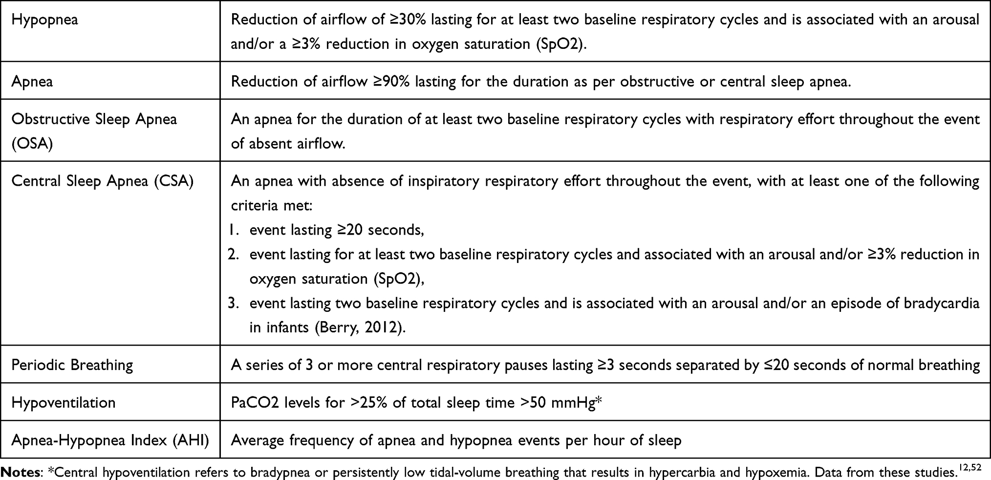 |
Table 1 Definition of Respiratory Events as per the AASM |
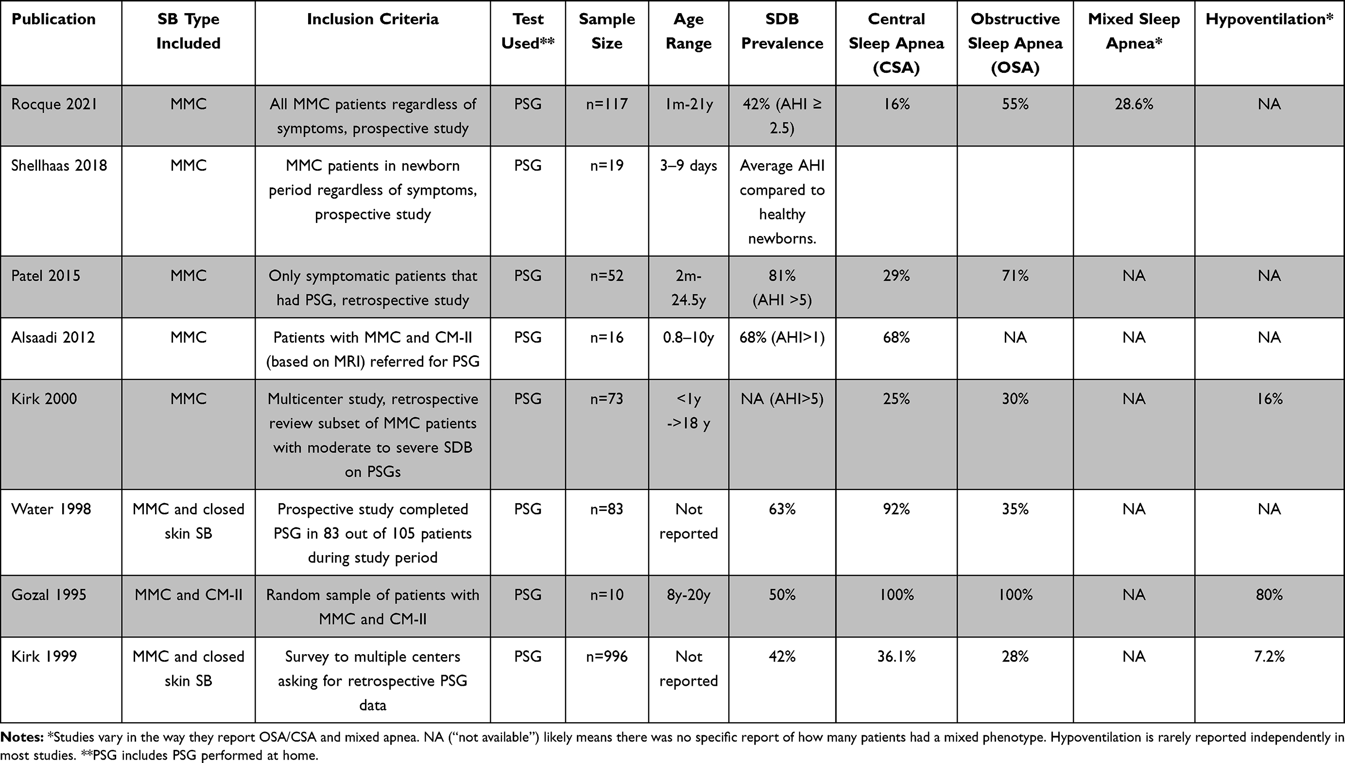 |
Table 2 Summary of Publications Reporting SDB Prevalence in Patients with SB |
The reported prevalence of obstructive sleep apnea is more variable. Published studies on the prevalence of OSA in this patient population report a wide range of ages. Commonly, patients have associated comorbidities for OSA such as adenoid or tonsillar hypertrophy. The degree of obstruction may also be dependent on pharyngeal dilator muscles dysfunction secondary to CM-II involvement of cranial nerves. Some studies have reported a high prevalence of mild OSA in up to 40% and moderate-to-severe OSA in up to 31% of this patient population.11,32,34,53–55 Other studies did not report any OSA in their cohort.23,29 Several studies did not find elevated body mass index (BMI), a known risk factor for OSA, to predict SDB in patients with CM-II, suggesting that SDB in patients with CM-II may more likely be a consequence of ventilatory control instability.
Clinical Symptoms of SDB in MMC
Reported symptoms of SDB in this population are typically similar to that of the general pediatric population and are frequently reported as snoring, apnea, blue spells, shortness of breath, irritability, choking or gasping with sleep, or fragmented sleep.32,56 Excessive daytime sleepiness is less common in children than in adults.57 In infants up to a year of age with MMC, the most common presentations include stridor, apnea, and feeding difficulties.4 Waters et al evaluated for symptoms predictive of SDB and found that snoring, witnessed apnea, dysphagia, and enlarged tonsils were symptoms associated with OSA, whereas witnessed cyanosis and vocal cord dysfunction were symptoms correlated with CSA.34 For children under the age of 2 years with MMC, the presence of SDB can result in rapid progression of neurological deterioration and can result in cardiorespiratory arrest and sudden unexplained death during sleep.33,43,56–59
Several studies have reported a higher prevalence of SDB in patients with MMC and CM-II by evaluating patients with the signs or symptoms suggestive of respiratory abnormalities,36,39,53 or after retrospectively evaluating patients with MMC based on having any previously completed polysomnography.32 Despite these findings, no clear predictors of SDB have been identified in patients with MMC and CM-II. Additionally, patients are at risk of SDB despite the absence of clinical symptoms, as was demonstrated by Rocque et al when a prevalence of SDB of 42% (diagnosed by AHI >2.5 events/h) was identified in a cohort of patients with CM-II that underwent screening with PSG regardless of the presence of symptoms suggestive of SDB.31 Given that SDB is potentially treatable, early assessment and intervention could become an integral part of a multidisciplinary treatment strategy to optimize long-term medical and neurodevelopmental outcomes.
Screening for SDB in the MMC Population
In children, clinical evaluation along with routine exam and existing sleep questionnaires are often not accurate or sensitive enough to establish a diagnosis of SDB in patients with MMC.33,50,56,60–62 The AAP currently recommends that each child be questioned regarding snoring and other signs and symptoms of SDB.3 Several questionnaires designed to screen for SDB are available, such as the Children’s Sleep Habits Questionnaire or the Pediatric Sleep Questionnaire (PSQ) are available. However, these instruments are not specific to patients with MMC. Beltran et al evaluated the efficacy of the PSQ in predicting OSA in patients with MMC and any CM, and they found the questionnaire to be a poor tool for screening, with a sensitivity of 73.58%, specificity of 20.83%, PPV of 33.91%, and NPV of 58.82%.37 Clinical evaluation is also not a reliable marker in determining which patients with MMC are at increased risk of SDB.17,31,34,38,42,61 For an infant with MMC with a history of stridor, dysphagia, apnea, cyanosis or a higher spinal lesion, the accuracy and sensitivity in predicting SDB based on clinical evaluation alone is estimated to be only 83% and 65%, respectively.38 Due to the low sensitivity of clinical evaluation alone and the notable morbidity and mortality risk associated with SDB in patients with MMC, it is recommended that all patients diagnosed with MMC undergo overnight observed polysomnography (PSG) evaluation regardless of symptom burden present.12,32,56
Overnight PSG is currently the “gold standard” in identifying SDB. A full pediatric PSG in an American Academy of Sleep Medicine-accredited laboratory includes monitoring of several physiologic parameters throughout the course of a night that help characterize a patient’s sleep architecture and respiratory patterns. The combination of electroencephalogram (EEG), electro-oculogram (EOG) for eye movements, and sub-mental electromyogram (EMG) for muscle tone allow for monitoring wake state, various sleep stages and arousals. Respiratory function is assessed using an airflow cannula at the nose and mouth, belts at the chest and abdomen monitoring movements, oximetry, and either end-tidal or transcutaneous CO2 monitoring. Heart rate is monitored with an electrocardiogram (ECG) and limb movements are monitored using limb EMG sensors. Video recordings with audio are generally present to detect sounds and movements during sleep. The data is scored by a trained sleep technologist and then interpreted by a sleep physician, who provides a summary report that outlines a patient’s overall quality and architecture of sleep and their respiratory patterns as observed in that overnight timeframe. Respiratory apneas are reported as an apnea–hypopnea index (AHI), which provides a general reflection on all obstructive events, central apneas, and hypopneas that were present overnight as an average number of events per hour. Given that a developing child is very sensitive to untreated sleep apnea, the AHI value required to classify sleep apnea severity is lower than that of adults (Table 3).52
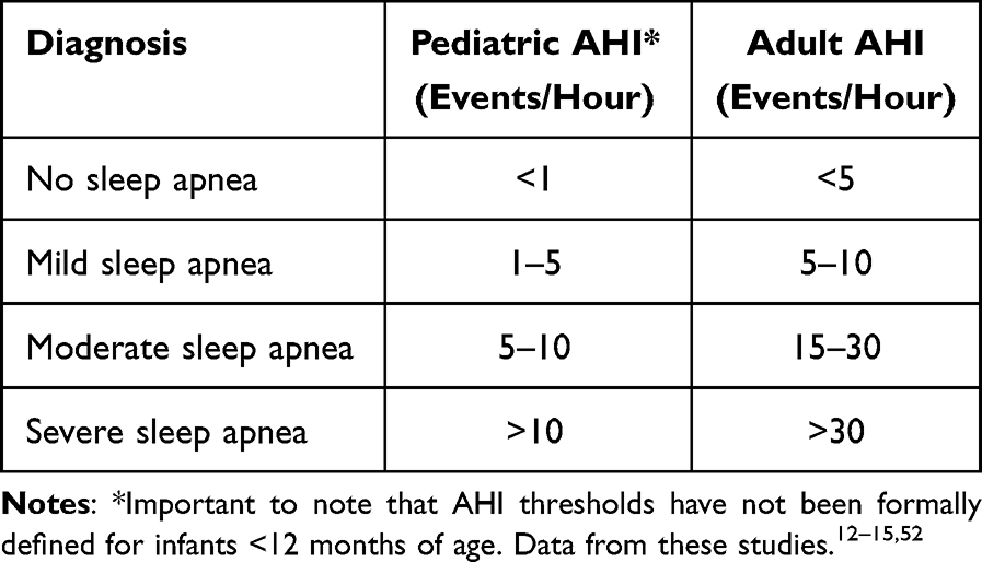 |
Table 3 Classification of Sleep Apnea by AHI |
Screening with PSG may help identify SDB in patients with MMC earlier. Given the known prevalence of SDB in the MMC population and the potential therapeutic options available from infancy, there is a need for improved standardization of protocols evaluating for SDB in individuals with MMC at institutions with specialized MMC programs. Given that patients with MMC remain at increased risk of SDB throughout their lifetime,63 even after neurosurgical interventions, this is a patient population that likely warrants regular screening with PSG starting in infancy or at time of initial diagnosis into adulthood.33,62,64 Several large centers are shifting towards providing coordinated clinical care pathways that help to optimize routine screening in the ion with MMC.4 Screening methods remain an area for potential improvement in management and care for this patient population.
Treatment for SDB in MMC
Individuals with MMC are likely to undergo multiple surgical procedures over the course of their lives.36,39,50 Many of these patients undergo fetal surgical closure of the open MMC prior to birth. Otherwise, the remainder of children undergo this closure within 48 hours of birth.31,32 Overall, about 80% of children with MMC also develop hydrocephalus that requires surgical treatment.9 While the anatomic findings associated with CM-II are present in nearly all individuals with MMC, fewer than 10% of children undergo CM-II decompression surgery in the modern era.9,36,39,50,61 Prenatal surgery has been shown to decrease the need for hydrocephalus surgery to as low as 40%, and decrease the anatomic severity of CM-II in children treated with prenatal closure compared to standard post-natal repair.65 However, little is known about the effect of neurosurgical intervention on SDB in patients with MMC. In infants who have not had treatment for hydrocephalus, the presence of stridor is considered to be an indication for hydrocephalus treatment, and treatment often leads to reduction or elimination of stridor.51 However, no studies have consistently demonstrated an improvement in SDB after neurosurgical treatment in any children older than infancy.4,51
As previously mentioned, SDB encompasses OSA, CSA, periodic breathing, and central hypoventilation. Treatment options for SDB in patients with SB vary by age, co-morbidities and complications associated with the individual patient, as well as the type and severity of the SDB present. Respiratory support recommended for patients with CSA vary depending on the age and concomitant hypoventilation. Even if a neurosurgical intervention is warranted for a patient, respiratory support will often be initiated for interval management given that CSA resolution will unlikely be immediate. Oxygen supplementation is the most common recommendation for infants.32,38,51 Older children who can tolerate positive pressure are started on non-invasive bi-level positive pressure instead.38 In cases with severe CSA, regardless of age and concomitant hypoventilation, bi-level positive pressure may be indicated. Invasive ventilation (via tracheostomy) may be the safest way to provide this in young children and infants or severe cases.10,32,38
Treatment options for OSA will also vary based on age and co-morbidities and will depend on the presence of surgically reversible airway obstruction. As with CSA, there is little data on the effectiveness of neurosurgical interventions on resolving OSA. These interventions, often discussed as improvement in pharyngeal and/or laryngeal tone, could in theory improve OSA severity and subsequently the degree of support required. Surgically reversible airway obstruction includes laryngomalacia, palatine tonsils hypertrophy, adenoid enlargement, and other less common upper airway pathologies.
While tonsillectomy/adenoidectomy remains a common treatment choice for OSA in the general pediatric patient population, it has not shown to resolve the upper airway obstruction of patients with MMC and CM-II.34,38 The decision to perform any of these interventions often warrants a multidisciplinary discussion to determine if the benefits of surgery outweigh the outcomes that may be exhibited in these patients given their complex underlying neurological pathology and multifactorial or multilevel airway obstruction. Evaluation for interventions start with an airway evaluation that can be completed with rigid and/or flexible airway endoscopy and in some centers with specialized airway imaging like airway MRIs. These evaluations are more dependable when they are done while mimicking physiologic sleep. No studies have been conducted looking into more reliable tests in patients with MMC.
As with respiratory support for CSA, respiratory support options for OSA start with oxygen supplementation. This is commonly used in infants and young children with OSA and associated hypoxia. There is controversy in the sleep medicine community about the benefits of supplemental oxygen in patients with OSA without significant hypoxia. However, this discussion is beyond the focus of this review. In older children, noninvasive use of CPAP is the first option for OSA. In severe cases, non-invasive bi-level positive pressure is trialed in older children. Infants and young children, on the other hand, may not tolerate non-invasive positive pressure nor are they safe with this management and so may require tracheostomy placement as a bypass for the areas of airway obstruction. Older children may also require tracheostomy placement if non-invasive bi-level positive pressure fails to manage severe OSA. The need for concomitant positive pressure would depend on the presence of CSA or hypoventilation in these cases. There is not widespread use of alternative options like lingual nerve stimulation for glossoptosis, and/or high flow nasal cannula support in the pediatric population.
Studies have reported that more than half of infants with MMC will continue to demonstrate SDB often with CSA, periodic breathing, and hypoventilation despite relief of upper airway obstruction through CM decompression, shunt placement, and/or tracheostomy tube placement.4,14,21,22,32,36,39,40,46,50 In fact, most studies reporting a high prevalence of SDB in CM-II patients were performed on patients after they had undergone neurosurgery, suggesting that SDB may develop regardless of neurosurgical intervention. Patel et al included repeat overnight polysomnography on nine children that underwent neurosurgical intervention (6 CM-II decompression, 4 HC shunt placement, 1 with both procedures), with repeat polysomnography indices indicating interval improvement in the severity of their SDB, though none of these improvements met statistical significance. Future studies should look more closely at the role hydrocephalus may be playing in the development or exacerbation of SDB in this patient population.
Finally, behavioral interventions and treatment of concomitant obesity likely play a role in ameliorating SRDB severity or its effects in the MMC population. To our knowledge, there are no published studies that have looked specifically into these interventions in MMC.
Adult Care and Sudden Death
The Spina Bifida Association has encouraged the development of organized multidisciplinary teams with expertise to be more readily available for adults.34,56,66 Coordinated, patient-centered programs that incorporate the patient, family, school and workplace personnel, social work services, and healthcare providers for several years prior to transition of care from childhood to adulthood have shown promising support of the adult population with MMC.13,26,27
Symptoms of SDB in adults with MMC are typically similar to the general population and includes snoring, witnessed cessation of breathing, gasping or choking at night, excessive daytime sleepiness, impaired cognition, and mood changes.13,17,64 For adults, validated questionnaires to predict the presence of sleep apnea include the STOP-BANG (Snoring, Tiredness, Observed apnea, High blood Pressure-Body Mass Index (BMI), Age, Neck Circumference, Gender), Berlin, Epworth Sleepiness Scale (ESS), and OSA 50 (Obesity-Snoring-Apnea-Age > 50).20,43 As with children, the “gold standard” evaluation for SDB in an adult with MMC remains overnight observed PSG. Considering that the risk of SDB remains with a patient with MMC throughout their lifetime, the recent US Preventive Services Task Force (USPSTF) recommends that patients with MMC be considered an exception to the recommendation of not routinely screening for SDB in asymptomatic adults.17 More importantly, clinicians who maintain healthy vigilance of sleep problems in this patient population will likely identify opportunities to improve the overall function and health of the patient as well as decrease stress for caregivers.
Despite preventive, medical, and surgical successes in the last several decades resulting in most patients with MMC reaching into adulthood,32,67 these individuals remain at risk for sudden unexplained death. Jernigan et al evaluated 106 patients with MMC with ages between 19 and 30 years, with 5.6% (6/106) of them experiencing sudden death. Case studies have reported on the potential correlation between sudden death in adults with MMC in the presence of chronic tonsillar herniation and hydrocephalus. While these co-morbidities have known associations with SDB, studies have not yet evaluated the impact of these specific conditions on the prevalence or severity of SDB in patients with MMC. Further research is needed to determine which characteristics may place a patient with MMC at increased risk of SDB and sudden death.4,33
Other Etiologies Impacting Sleep Quality in the Child with Spina Bifida
Sleep Quality in the Child with Spina Bifida
Only few studies have been able to evaluate sleep quality in the patient population with any severity of SB. To better understand sleep effects in individuals with SB, Murray et al assessed the sleep of 37 adolescents with SB against 37 adolescents without SB using sleep questionnaires, actigraphy, and sleep diaries.63 They found that adolescents with SB experienced poorer sleep quality, reduced sleep, increased insomnia, and increased daytime fatigue compared to otherwise typically developing adolescents. This is similar to evidence that exists showing that patients with chronic illnesses have poor sleep quality.8,24,66 Pathological and nighttime sleep deprivations have substantial adverse effects on cognition, which further worsens the burden of chronic illness.8,23,28,29 Therefore, prompt identification and treatment of co-existing sleep problems among patients with chronic illness, such as SB, is an important step in improving the overall outcomes for these individuals.
Chronotype
Chronotype is an individual’s preferred timing of sleep, activity, and cognitive performance and is often referred to as a later chronotype (evening-type) or an earlier chronotype (morning-type). Across the lifespan, chronotype is associated with psychological wellbeing, environmental and endogenous factors.68,69 Studies have previously validated that chronotype changes during life.69–71 Infants and children experience earlier chronotypes. In adolescence, chronotype progresses towards later hours resulting in an evening-type chronotype for adolescents and young adults. Typically, this evening-type chronotype peaks around 20 years of age, after which chronotype typically will begin to shift back towards an earlier chronotype. Therefore, elderly have a morning-type chronotype.
Several studies have suggested that evening chronotype is associated with adverse health effects including increased risk of cardiovascular and metabolic disorders.35,68–71 Moreover, any disruption to the normal circadian rhythm can lead to circadian misalignment producing immediate consequences of the sleep-wake cycle and results in sleep disturbances.69 Edelstein et al explored the potential of sleep disorders due to anomalies in brain regions of MMC individuals that may influence circadian rhythmicity.8 They measured chronotype and the presence of sleep problems in 202 individuals with MMC and 62 typically developing, healthy age peers. They found that while individuals with MMC showed the characteristic progression towards a later chronotype in adolescent and young adulthood, they peaked at an older age in comparison to controls (29.2 years versus 23.4 years). This means that at the age when later chronotype peaks for individuals with MMC, the controls were already returning to earlier sleep-wake times. Additionally, children and adults with MMC endorsed sleep problems more often than controls. Interestingly, the problems endorsed by adults appear to involve sleep timing and quality. In fact, they found that in adults with MMC endorsed problems involving sleep timing and quality, there was a correlation between chronotype and the different sleep problems.8 The impact of this shift in circadian timing, contributing to sleep difficulties in an individual with MMC, still remains unclear but does present an important consideration in the chronic management of other sleep-related disorders in this patient population.
Conclusions
It is understood that SDB conditions such as obstructive sleep apnea or central sleep apnea are associated with developmental concerns, cognitive consequences, and risk of sudden death in the patient population with MMC.72 Current evidence indicates a high prevalence of SDB in patients with MMC and CM-II, even when asymptomatic. Therefore, screening with PSG should occur promptly at regular intervals to avoid potentially serious complications such as sudden death. Early identification of SDB may lead to earlier assessment and treatment interventions that could help optimize long-term medical and neurodevelopmental outcomes for this vulnerable patient population. Nonetheless, there remains a paucity of data to help understand the extent to which SB and its associated co-morbidities effect sleep continuity or exacerbate other sleeping conditions. Further prospective studies that include a large number of participants will be necessary to further evaluate the potential impact of early PSG screening, surgical interventions, and/or other treatment interventions on managing SDB in patients with MMC. Future research will also hopefully provide insight on which interventions for SDB may help alleviate the risk of sudden death for this patient population.
Acknowledgments
The work for this study was performed at the University of Alabama at Birmingham. Figure 1 was produces by the National Center for Advancing Translational Sciences of the National Institutes of Health under award number UL1TR003096. The content is solely the responsibility of the authors and does not necessarily represent the official views of the National Institutes of Health.
Author Contributions
All authors made a significant contribution to the work reported, whether that is in the conception, study design, execution, acquisition of data, analysis and interpretation, or in all these areas; took part in drafting, revising or critically reviewing the article; gave final approval of the version to be published; have agreed on the journal to which the article has been submitted; and agree to be accountable for all aspects of the work.
Funding
No financial support was provided for development of this study.
Disclosure
None of the authors have any conflicts of interest to disclose for this work.
References
1. Copp AJ, Adzick NS, Chitty LS, Fletcher JM, Holmbeck GN, Shaw GM. Spina bifida. Nat Rev Dis Primers. 2015;1(1):15007. doi:10.1038/nrdp.2015.7
2. Hopson B, Rocque BG, Joseph DB, et al. The development of a lifetime care model in comprehensive spina bifida care. J Pediatr Rehabil Med. 2018;11(4):323–334. doi:10.3233/PRM-180548
3. Phillips LA, Burton JM, Evans SH. Spina Bifida Management. Curr Probl Pediatr Adolesc Health Care. 2017;47(7):173–177. doi:10.1016/j.cppeds.2017.06.007
4. Spina Bifida Association. What is Spina Bifida? Resources and Prevention; 2021.
5. National Center on Birth Defects and Developmental Disabilities, C.f.D.C.a.P. Spina Bifida; 2020.
6. Sullivan AM, Herdt M. Characteristics and first-year mortality, by lesion level, among infants with spina bifida in the New York State Birth Defects Registry, 2008–2017. Birth Defects Res. 2022;114(2):62–68.
7. Mai CT, Isenburg JL, Canfield MA, et al. National population-based estimates for major birth defects, 2010–2014. Birth Defects Res. 2019;111(18):1420–1435. doi:10.1002/bdr2.1589
8. Edelstein K, Cirino PT, Hasher L, Fletcher JM, Dennis M. Sleep problems, chronotype, and diurnal preferences in children and adults with spina bifida. J Biol Rhythms. 2012;27(2):172–175. doi:10.1177/0748730411435209
9. Kim I, Hopson B, Aban I, et al. Treated hydrocephalus in individuals with myelomeningocele in the National Spina Bifida Patient Registry. J Neurosurg Pediatr. 2018;22(6):646–651. doi:10.3171/2018.5.PEDS18161
10. Mizuguchi K, Morota N, Kubota M. [Respiratory complications in children with Chiari malformation type II associated with myelomeningocele]. No to Hattatsu. 2016;48(1):25–28. Japanese.
11. Kaditis AG, Alonso Alvarez ML, Boudewyns A, et al. Obstructive sleep disordered breathing in 2- to 18-year-old children: diagnosis and management. Eur Respir J. 2016;47(1):69–94. doi:10.1183/13993003.00385-2015
12. Section on Pediatric Pulmonology, S.o.O.S.A.S.A.A.o.P. Clinical practice guideline: diagnosis and management of childhood obstructive sleep apnea syndrome. Pediatrics. 2002;109(4):704–712. doi:10.1542/peds.109.4.704
13. Lurie A. Obstructive sleep apnea in adults: epidemiology, clinical presentation, and treatment options. Obstr Sleep Apnea Adults. 2011;46:1–42.
14. Leu RM. Sleep-related breathing disorders and the Chiari 1 malformation. Chest. 2015;148(5):1346–1352. doi:10.1378/chest.14-3090
15. Tan HL, Alonso Alvarez ML, Tsaoussoglou M, Weber S, Kaditis AG. When and why to treat the child who snores? Pediatr Pulmonol. 2017;52(3):399–412. doi:10.1002/ppul.23658
16. Di Carlo G, Zara F, Rocchetti M, et al. Prevalence of sleep-disordered breathing in children referring for first dental examination. A multicenter cross-sectional study using pediatric sleep questionnaire. Int J Environ Res Public Health. 2020;17(22):8460. doi:10.3390/ijerph17228460
17. Bibbins-Domingo K, Grossman DC, Curry SJ et al; Force, U.S.P.S.T. Screening for obstructive sleep apnea in adults: US preventive services task force recommendation statement. JAMA. 2017;317(4):407–414. doi:10.1001/jama.2016.20325
18. O’Driscoll DM, Foster AM, Ng ML, et al. Acute cardiovascular changes with obstructive events in children with sleep disordered breathing. Sleep. 2009;32(10):1265–1271. doi:10.1093/sleep/32.10.1265
19. Bonuck K, Parikh S, Bassila M. Growth failure and sleep disordered breathing: a review of the literature. Int J Pediatr Otorhinolaryngol. 2006;70(5):769–778. doi:10.1016/j.ijporl.2005.11.012
20. Lindquist B, Jacobsson H, Strinnholm M, Peny-Dahlstrand M. A scoping review of cognition in spina bifida and its consequences for activity and participation throughout life. Acta Paediatr. 2022;111(9):1682–1694. doi:10.1111/apa.16420
21. Del Bigio MR. Neuropathological changes caused by hydrocephalus. Acta Neuropathol. 1993;85(6):573–585. doi:10.1007/BF00334666
22. Lindquist B, Carlsson G, Persson EK, Uvebrant P. Learning disabilities in a population-based group of children with hydrocephalus. Acta Paediatr. 2005;94(7):878–883. doi:10.1111/j.1651-2227.2005.tb02005.x
23. Henriques Filho PS, Pratesi R. Sleep disorder: a possible cause of attention deficit in children and adolescents with Chiari malformation type II. Arq Neuropsiquiatr. 2009;67(1):29–34. doi:10.1590/S0004-282X2009000100008
24. Halbower AC, Degaonkar M, Barker PB, et al. Childhood obstructive sleep apnea associates with neuropsychological deficits and neuronal brain injury. PLoS Med. 2006;3(8):e301. doi:10.1371/journal.pmed.0030301
25. Trosman I, Trosman SJ. Cognitive and behavioral consequences of sleep disordered breathing in children. Med Sci. 2017;5(4):30. doi:10.3390/medsci5040030
26. Le JT, Mukherjee S. Transition to adult care for patients with spina bifida. Phys Med Rehabil Clin N Am. 2015;26(1):29–38. doi:10.1016/j.pmr.2014.09.007
27. Hopson B, Alford EN, Zimmerman K, Blount JP, Rocque BG. Development of an evidence-based individualized transition plan for spina bifida. Neurosurg Focus. 2019;47(4):E17. doi:10.3171/2019.7.FOCUS19425
28. McDowell MM, Blatt JE, Deibert CP, Zwagerman NT, Tempel ZJ, Greene S. Predictors of mortality in children with myelomeningocele and symptomatic Chiari type II malformation. J Neurosurg Pediatr. 2018;21(6):587–596. doi:10.3171/2018.1.PEDS17496
29. Dauvilliers Y, Stal V, Abril B, et al. Chiari malformation and sleep related breathing disorders. J Neurol Neurosurg Psychiatry. 2007;78(12):1344–1348. doi:10.1136/jnnp.2006.108779
30. Gozal D, Arens R, Omlin KJ, Jacobs RA, Keens TG. Peripheral chemoreceptor function in children with myelomeningocele and Arnold-Chiari malformation type 2. Chest. 1995;108(2):425–431. doi:10.1378/chest.108.2.425
31. Rocque BG, Maddox MH, Hopson BD, et al. Prevalence of sleep disordered breathing in children with myelomeningocele. Neurosurgery. 2021;88(4):785–790. doi:10.1093/neuros/nyaa507
32. Patel DM, Rocque BG, Hopson B, et al. Sleep-disordered breathing in patients with myelomeningocele. J Neurosurg Pediatr. 2015;16(1):30–35. doi:10.3171/2014.11.PEDS14314
33. Kirk VG, Morielli A, Brouillette RT. Sleep-disordered breathing in patients with myelomeningocele: the missed diagnosis. Dev Med Child Neurol. 1999;41(1):40–43. doi:10.1017/s0012162299000079
34. Waters KA, Forbes P, Morielli A, et al. Sleep-disordered breathing in children with myelomeningocele. J Pediatr. 1998;132(4):672–681. doi:10.1016/S0022-3476(98)70359-2
35. Lazzareschi I, Curatola A, Massimi L, et al. Sleep-disordered breathing in patients with Chiari malformation type II: a case-control study and review of the literature. J Clin Sleep Med. 2022;18(9):2143–2154. doi:10.5664/jcsm.10062
36. Ward SL, Jacobs RA, Gates EP, Hart LD, Keens TG. Abnormal ventilatory patterns during sleep in infants with myelomeningocele. J Pediatr. 1986;109(4):631–634. doi:10.1016/S0022-3476(86)80226-8
37. Beltran Ale G, Pascoe J, George A, et al. Validation of pediatric sleep questionnaire in children with Chiari malformation and/or spina bifida with or without myelomeningocele. Sleep Med. 2021;84:93–97. doi:10.1016/j.sleep.2021.05.030
38. Kirk VG, Morielli A, Gozal D, et al. Treatment of sleep-disordered breathing in children with myelomeningocele. Pediatr Pulmonol. 2000;30(6):445–452. doi:10.1002/1099-0496(200012)30:6<445::AID-PPUL2>3.0.CO;2-C
39. Ward SL, Nickerson BG, van der Hal A, Rodriguez AM, Jacobs RA, Keens TG. Absent hypoxic and hypercapneic arousal responses in children with myelomeningocele and apnea. Pediatrics. 1986;78(1):44–50. doi:10.1542/peds.78.1.44
40. National Institute of Neurological Disorders and Stroke. Spina Bifida Fact Sheet. National Institute of Neurological Disorders and Stroke; 2021.
41. Pasterkamp H, Cardoso ER, Booth FA. Obstructive sleep apnea leading to increased intracranial pressure in a patient with hydrocephalus and syringomyelia. Chest. 1989;95(5):1064–1067. doi:10.1378/chest.95.5.1064
42. Ely EW, McCall WV, Haponik EF. Multifactorial obstructive sleep apnea in a patient with Chiari malformation. J Neurol Sci. 1994;126(2):232–236. doi:10.1016/0022-510X(94)90280-1
43. Jernigan SC, Berry JG, Graham DA, et al. Risk factors of sudden death in young adult patients with myelomeningocele. J Neurosurg Pediatr. 2012;9(2):149–155. doi:10.3171/2011.11.PEDS11269
44. Omer S, al-Kawi MZ, Bohlega S, Bouchama A, McLean D. Respiratory arrest: a complication of Arnold-Chiari malformation in adults. Eur Neurol. 1996;36(1):36–38. doi:10.1159/000117197
45. Yumer MH, Nachev SS, Dzhendov TY, Kalev OK. Chiari type II malformation: a case report and review of literature. Folia Med. 2006;48:55–59.
46. Zolty P, Sanders MH, Pollack IF. Chiari malformation and sleep-disordered breathing: a review of diagnostic and management issues. Sleep. 2000;23(5):637–643. doi:10.1093/sleep/23.5.1f
47. Wealthall SR, Whittaker GE, Greenwood N. The relationship of apnoea and stridor in spina bifida to other unexplained infant deaths. Dev Med Child Neurol. 1974;16(s32):107–116. doi:10.1111/j.1469-8749.1974.tb03458.x
48. Cochrane DD, Adderley R, White CP, Norman M, Steinbok P. Apnea in patients with myelomeningocele. Pediatr Neurosurg. 1990;16(4–5):232–239. doi:10.1159/000120533
49. Chung F, Yegneswaran B, Liao P, et al. STOP questionnaire: a tool to screen patients for obstructive sleep apnea. Anesthesiology. 2008;108(5):812–821. doi:10.1097/ALN.0b013e31816d83e4
50. Holinger PC, Holinger LD, Reichert TJ, Holinger PH. Respiratory obstruction and apnea in infants with bilateral abductor vocal cord paralysis, meningomyelocele, hydrocephalus, and Arnold-Chiari malformation. J Pediatr. 1978;92(3):368–373. doi:10.1016/S0022-3476(78)80421-1
51. Shellhaas RA, Kenia PV, Hassan F, Barks JDE, Kaciroti N, Chervin RD. Sleep-disordered breathing among newborns with myelomeningocele. J Pediatr. 2018;194:244–247 e241. doi:10.1016/j.jpeds.2017.10.070
52. Berry RB, Brooks R, Gamaldo CE, et al. The AASM manual for the scoring of sleep and associated events: rules, terminology and technical specifications, version 2.0. Am Acad Sleep Med. 2012;176:2012.
53. Alsaadi MM, Iqbal SM, Elgamal EA, Gozal D. Sleep-disordered breathing in children with Chiari malformation type II and myelomeningocele. Pediatr Int. 2012;54(5):623–626. doi:10.1111/j.1442-200X.2012.03660.x
54. Losurdo A, Dittoni S, Testani E, et al. Sleep disordered breathing in children and adolescents with Chiari malformation type I. J Clin Sleep Med. 2013;9(04):371–377. doi:10.5664/jcsm.2592
55. Ferre A, Poca MA, de la Calzada MD, et al. Sleep-related breathing disorders in Chiari malformation type 1: a prospective study of 90 patients. Sleep. 2017;40(6). doi:10.1093/sleep/zsx069
56. Spina Bifida Association. Guidelines for the Care of People with Spina Bifida. Spina Bifida Association; 2018.
57. Gozal D, Wang M, Pope DW. Objective sleepiness measures in pediatric obstructive sleep apnea. Pediatrics. 2001;108(3):693–697. doi:10.1542/peds.108.3.693
58. Hays RM, Jordan RA, McLaughlin JF, Nickel RE, Fisher LD. Central ventilatory dysfunction in myelodysplasia: an independent determinant of survival. Dev Med Child Neurol. 1989;31(3):366–370. doi:10.1111/j.1469-8749.1989.tb04005.x
59. Oakeshott P, Hunt GM, Poulton A, Reid F. Expectation of life and unexpected death in open spina bifida: a 40-year complete, non-selective, longitudinal cohort study. Dev Med Child Neurol. 2010;52(8):749–753. doi:10.1111/j.1469-8749.2009.03543.x
60. Netzer NC, Stoohs RA, Netzer CM, Clark K, Strohl KP. Using the Berlin Questionnaire to identify patients at risk for the sleep apnea syndrome. Ann Intern Med. 1999;131(7):485–491. doi:10.7326/0003-4819-131-7-199910050-00002
61. Rosen CL, Wang R, Taylor HG, et al. Utility of symptoms to predict treatment outcomes in obstructive sleep apnea syndrome. Pediatrics. 2015;135(3):e662–e671. doi:10.1542/peds.2014-3099
62. Kenia PV, Lester SG, Rau SM, Shellhaas RA. 0778 sleep-disordered breathing among children with myelomeningocele: an opportunity to improve clinical practice. Sleep. 2018;41:A289–A289.
63. Murray CB, Palermo TM, Holmbeck GN. A multimethod, case-controlled study of sleep-wake disturbances in adolescents with Spina Bifida. J Pediatr Psychol. 2018;43(6):601–612. doi:10.1093/jpepsy/jsx150
64. Murray CB, Kirsch AC, Palermo TM, et al. Developmental Course and determinants of sleep disturbances in adolescents with Spina Bifida. J Pediatr Psychol. 2016;41(6):631–642. doi:10.1093/jpepsy/jsw021
65. Adzick NS, Thom EA, Spong CY, et al. A randomized trial of prenatal versus postnatal repair of myelomeningocele. N Engl J Med. 2011;364(11):993–1004. doi:10.1056/NEJMoa1014379
66. Domino JS, Lundy P, Glynn EF, Partington M. Estimating the prevalence of neurosurgical interventions in adults with spina bifida using the Health Facts data set: implications for transition planning and the development of adult clinics. J Neurosurg Pediatr. 2022;29(4):371–378. doi:10.3171/2021.10.PEDS21293
67. Wolujewicz P, Steele JW, Kaltschmidt JA, Finnell RH, Ross ME. Unraveling the complex genetics of neural tube defects: from biological models to human genomics and back. Genesis. 2021;59(11):e23459. doi:10.1002/dvg.23459
68. Roenneberg T, Kuehnle T, Juda M, et al. Epidemiology of the human circadian clock. Sleep Med Rev. 2007;11(6):429–438. doi:10.1016/j.smrv.2007.07.005
69. Allebrandt KV, Teder-Laving M, Kantermann T, et al. Chronotype and sleep duration: the influence of season of assessment. Chronobiol Int. 2014;31(5):731–740. doi:10.3109/07420528.2014.901347
70. Dijk DJ, Duffy JF, Czeisler CA. Contribution of circadian physiology and sleep homeostasis to age-related changes in human sleep. Chronobiol Int. 2000;17(3):285–311. doi:10.1081/CBI-100101049
71. Fischer D, Lombardi DA, Marucci-Wellman H, Roenneberg T, Tosini G. Chronotypes in the US - influence of age and sex. PLoS One. 2017;12(6):e0178782. doi:10.1371/journal.pone.0178782
72. Eisenberg A, Hobart-Porter L, Jambhekar S, et al. Sleep related breathing disorders in the spina bifida population ages 1–20 years: a retrospective study in Arkansas. J Pediatr Rehabil Med. 2022;15(4):581–586. doi:10.3233/PRM-210129
 © 2023 The Author(s). This work is published and licensed by Dove Medical Press Limited. The full terms of this license are available at https://www.dovepress.com/terms.php and incorporate the Creative Commons Attribution - Non Commercial (unported, v3.0) License.
By accessing the work you hereby accept the Terms. Non-commercial uses of the work are permitted without any further permission from Dove Medical Press Limited, provided the work is properly attributed. For permission for commercial use of this work, please see paragraphs 4.2 and 5 of our Terms.
© 2023 The Author(s). This work is published and licensed by Dove Medical Press Limited. The full terms of this license are available at https://www.dovepress.com/terms.php and incorporate the Creative Commons Attribution - Non Commercial (unported, v3.0) License.
By accessing the work you hereby accept the Terms. Non-commercial uses of the work are permitted without any further permission from Dove Medical Press Limited, provided the work is properly attributed. For permission for commercial use of this work, please see paragraphs 4.2 and 5 of our Terms.