")
Back to Journals » Clinical Interventions in Aging » Volume 13
Compression of the right coronary artery by an aortic pseudoaneurysm after infective endocarditis: an unusual case of myocardial ischemia
Authors Lacalzada-Almeida J , De la Rosa-Hernández A, Izquierdo-Gómez MM, García-Niebla J, Hernández-Betancor I, Bonilla-Arjona JA, Barragán-Acea A, Laynez-Cerdeña I
Received 25 June 2017
Accepted for publication 11 November 2017
Published 21 December 2017 Volume 2018:13 Pages 9—11
DOI https://doi.org/10.2147/CIA.S144840
Checked for plagiarism Yes
Review by Single anonymous peer review
Peer reviewer comments 3
Editor who approved publication: Dr Richard Walker
Juan Lacalzada-Almeida,1 Alejandro De la Rosa-Hernández,1 María Manuela Izquierdo-Gómez,1 Javier García-Niebla,2 Iván Hernández-Betancor,1 Juan Alfonso Bonilla-Arjona,3 Antonio Barragán-Acea,1 Ignacio Laynez-Cerdeña1
1Cardiology Department, Hospital Universitario de Canarias, Tenerife, 2Health services from the Health Area of El Hierro, Valle del Golfo Health Center, El Hierro, 3Radiology Department, Hospital Universitario de Canarias, Tenerife, Spain
Abstract: A 61-year-old male with a prosthetic St Jude aortic valve size 24 presented with heart failure symptoms and minimal-effort angina. Eleven months earlier, the patient had undergone cardiac surgery because of an aortic root dilatation and bicuspid aortic valve with severe regurgitation secondary to infectious endocarditis by Coxiela burnetii and coronary artery disease in the left circumflex coronary artery. Then, a prosthesis valve and a saphenous bypass graft to the left circumflex coronary artery were placed. The patient was admitted to the Cardiology Department of Hospital Universitario de Canarias, Tenerife, Spain and a transthoracic echocardiography was performed that showed severe paraprosthetic aortic regurgitation and an aortic pseudoaneurysm. The 64-slice multidetector computed tomography confirmed the pseudoaneurysm, originating from the right sinus of Valsalva, with a compression of the native right coronary artery and a normal saphenous bypass graft. On the basis of these findings, we performed surgical treatment with a favorable postoperative evolution. In our case, results from complementary cardiac imaging techniques were crucial for patient management. The multidetector computed tomography allowed for a confident diagnosis of an unusual mechanism of coronary ischemia.
Keywords: pseudoaneurysm, infective endocarditis, myocardial ischemia, aortic valve prosthesis
Photo essays
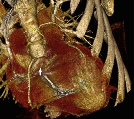
A 61-year-old male patient was admitted to our hospital with heart failure because of a bicuspid aortic valve with severe regurgitation, secondary to Q-fever infective endocarditis (IE). Prior to cardiac surgery, invasive coronary angiography was performed, which showed a severe proximal lesion in the left circumflex coronary artery (LCX). A prosthetic St Jude aortic valve size 24 and a saphenous bypass graft to LCX were placed, with no postoperative complications. Eleven months later, the patient was readmitted with heart failure and minimal-effort angina. A transthoracic echocardiography (TTE) was performed, and an image of an aortic pseudoaneurysm was detected in TTE (Figure 1A, C and D). A severe paraprosthetic aortic regurgitation was observed (Figure 1B), following which a 64-slice multidetector computed tomography (MDCT) was performed. The presence of an aortic pseudoaneurysm originating from the right sinus of Valsalva was successfully shown in the volume rendering images (Figure 2). In the MDCT axial views, the pseudoaneurysm was localized in the anterior wall of the thoracic aorta, showing contrast enhancement within it (Figure 3A and C), as well as an ascending aortic aneurysm, up to the aortic arch (Figure 3B). The MDCT revealed compression of the native right coronary artery (RCA) due to the pseudoaneurysm (Figure 3D), this being the cause for the probable ischemia in this area. The LCX had a chronic occlusion and the venous grafting was functioning properly (Figure 3E). MDCT oblique reconstruction also showed the aortic prosthesis valve (Figure 3F). The Bentall–De Bono procedure was performed, with resection of the pathological section of the root and ascending aorta and replacement using composite valve-graft prosthesis, using a bioprosthetic valve. The coronary ostiums and the venous grafting to LCX were reimplanted.
 | Figure 1 Echocardiography study. |
 | Figure 2 MDCT volume rendering image shows compression of the RCA by the aortic pseudoaneurysm (white arrow) and the saphenous venous bypass graft (yellow arrow). |
 | Figure 3 Multidetector computed tomography study. |
Pseudoaneurysm may be located in the mitral–aortic intervalvular fibrosa due to perivalvular complications of IE1 or secondary to aortic valve surgery complications.2 It can also be located in relation with the aortic root.2 Other authors described systolic compression by pseudoaneurysm of the left main coronary artery,1 of the left anterior descending,3 and of the RCA.2 In our case, the complementary use of cardiac imaging techniques was crucial for the management of the patient. Even having a prosthetic mechanic aortic valve and coronary bypass surgery, the MDCT allowed a confident diagnosis of an unusual mechanism of probable coronary ischemia, since it showed the compression of the RCA by the pseudoaneurysm, guiding the surgical treatment, after which the patient recovered adequately.
Consent
The patient provided written informed consent for the case and accompanying images to be published.
Disclosure
The authors report no conflicts of interest in this work.
References
Parashara DK, Jacobs LE, Kotler MN, et al. Angina caused by systolic compression of the left coronary artery as a result of pseudoaneurysm of the mitral-aortic intervalvular fibrosa. Am Heart J. 1995;129(2):417–421. | ||
Zientara A, Häussler A, Genoni M, Dzemali O. 41 Years after Björk–Shiley valve implantation: advanced preparation of a giant root pseudoaneurysm entrapping the right coronary artery. Eur J Cardiothorac Surg. 2015;48(3):512–513. | ||
Bouabdallaoui N, Achouh P, Lacaze-gadonneix J, Ennezat PV. Unusual cause of acute coronary syndrome: dynamic coronary compression by an aortic pseudoaneurysm. Eur Heart J Cardiovasc Imaging. 2014;15(4):468–469. |
 © 2017 The Author(s). This work is published and licensed by Dove Medical Press Limited. The full terms of this license are available at https://www.dovepress.com/terms.php and incorporate the Creative Commons Attribution - Non Commercial (unported, v3.0) License.
By accessing the work you hereby accept the Terms. Non-commercial uses of the work are permitted without any further permission from Dove Medical Press Limited, provided the work is properly attributed. For permission for commercial use of this work, please see paragraphs 4.2 and 5 of our Terms.
© 2017 The Author(s). This work is published and licensed by Dove Medical Press Limited. The full terms of this license are available at https://www.dovepress.com/terms.php and incorporate the Creative Commons Attribution - Non Commercial (unported, v3.0) License.
By accessing the work you hereby accept the Terms. Non-commercial uses of the work are permitted without any further permission from Dove Medical Press Limited, provided the work is properly attributed. For permission for commercial use of this work, please see paragraphs 4.2 and 5 of our Terms.