")
Back to Journals » Clinical, Cosmetic and Investigational Dentistry » Volume 15
Validity and Reliability of Polarized vs Non-Polarized Digital Images for Measuring Gingival Melanin Pigmentation
Received 20 May 2023
Accepted for publication 7 September 2023
Published 12 September 2023 Volume 2023:15 Pages 189—197
DOI https://doi.org/10.2147/CCIDE.S422139
Checked for plagiarism Yes
Review by Single anonymous peer review
Peer reviewer comments 2
Editor who approved publication: Professor Christopher E. Okunseri
Talal M Zahid,1 Zuhair S Natto2
1Periodontology Department, King Abdulaziz University, Jeddah, Saudi Arabia; 2Dental Public Health Department, King Abdulaziz University, Jeddah, Saudi Arabia
Correspondence: Talal M Zahid, Periodontology Department, King Abdulaziz University, Jeddah, Saudi Arabia, Email [email protected]
Aim: This study aimed to compare the validity and reliability of polarized and non-polarized intraoral photography for the measurement of gingival melanin pigmentation.
Materials and Methods: A case series study was conducted on ten patients scheduled for gingival depigmentation. A total of 976 polarized and non-polarized image samples were collected, capturing two rows above the gingival margin, for analysis. These images were taken both before and one year after the depigmentation procedure. Three independent evaluators assessed the photographs (an orthodontist, a general dentist, and a layperson). The Dummett Oral Pigmentation Index (DOPI) and Gingival Melanosis Record (GMR) indices were used to measure the level of gingival pigmentation.
Results: The study found no significant differences between polarized and non-polarized images taken before and after depigmentation. Both methods of imaging received similar scores from the evaluators. The orthodontist identified more pigmented slides than the layperson and the general dentist.
Conclusion: Both polarized and non-polarized photographic methods may be used for assessing gingival pigmentation. However, further research is warranted to confirm this finding and examine additional factors.
Keywords: intraoral photography, polarized, non-polarized, gingival melanin pigmentation, Dummett Oral Pigmentation Index, Gingival Melanosis Record, dental photography, image analysis
Introduction
Over the past few decades, photography has become an integral part of the regular practice of dentistry. It is considered a critical component of medical documentation and dental education. Dentists are required to take pictures of the oral cavity before, during, and after treatment for further examination of the teeth. These images need to be of good quality, with correct depth and true color for the accurate assessment of soft and hard tissues.1–3 Consequently, the use of intraoral and extraoral photographs has grown exponentially over the years, owing to their ability to provide detailed information about the oral cavity and aesthetic appearance. Intraoral photography captures photos of the oral cavity for diagnosis, treatment planning, and patient education, while extraoral photography shows how procedures carried out inside the mouth impact the aesthetic look of the face. However, the image quality largely depends on the type of camera and the photography technique used.1,4,5
Intraoral photography is a popular method for assessing gingival pigmentation. Non-polarized and polarized photography are two commonly used techniques for capturing intraoral photographs.6,7 Non-polarized photography involves taking photos of the oral cavity without polarizing filters. This implies that when a photo is taken using the non-polarized technique, the light waves generated from the camera flash will vibrate internally at random angles without any plane. This technique captures images of the gingiva as it appears to the naked eye, making it easier to assess the color correctly. However, glare and reflection from the flash are two main downsides of non-polarized photography, which make it challenging to capture in-depth pictures of glossy surfaces and assess how opaque the teeth’s whiteness is.4,6,8
Polarized photography, on the other hand, uses a special filter to minimize glare and remove surface reflections, allowing the light waves to vibrate in a single direction. This polarization of the light waves makes surface details more visible.4,5,9 This technique is particularly useful for capturing images of dental implants and reflective surfaces (eg, enamel and ceramic restorations). However, the use of a polarized filter can create color distortion, making it difficult to assess the gingiva’s true color. In addition, excessive polarization can radically darken the images.7,8,10
Studies suggest that polarized intraoral photography is probably more reliable for capturing images of enamel and ceramic restorations,11–13 while non-polarized intraoral photography may be more appropriate for photographing the gingiva.1,4,5,8 Some authors also suggested that cross-polarized photography is probably the best method for visualizing hyperpigmentation while concealing hypopigmentation and subsurface features like vascularity.7,10,14 However, more research is needed to establish the clinical relevance of these techniques. Although much research has been done in the last decade to study tooth color photography techniques, there is a knowledge gap regarding gingival color photography. This study, therefore, aimed to compare the validity and reliability of polarized and non-polarized intraoral photography for the measurement of gingival melanin pigmentation.
Materials and Methods
Study Design and Patient Selection
The study was conducted in compliance with the Helsinki Declaration. Ethical approval (#82-04-19) was approved by the ethical research committee at Faculty of Dentistry, King Abdulaziz University, Jeddah, Saudi Arabia. This is a case series study in which we included ten patients who were scheduled for gingival depigmentation. Research details were clearly explained to the participants, and each signed a written informed consent before inclusion. Patients who underwent anterior teeth restorations and had recently bleached their teeth, at least within the previous six months, were excluded from the study.
Image Capturing
A total of 976 polarized and non-polarized image samples were extracted from the 10 included patients, using different photographic equipment. We captured photographs using a Digital Single Lens Reflex (DSLR) camera (Nikon D750, Nikon Corporation, Japan) outfitted with a macro lens, ring flash, and cross-polarizing filter. The cross-polarizing filter was used to visualize hyperpigmentation and reduce unwanted reflections and glare. For each patient, we photographed the entire upper arch, which was then segmented into smaller samples. Each of these samples was coded and placed on a separate slide. These slides were then randomized. This approach resulted in the 976 image samples.
The camera settings included an exposure of 1/125 s at f/22, film speed (ISO) of 250, an ISO sensitivity of 100, an Electronic Through-The-Lens metering (E-TTL) flash, a distance of 25 cm, and a focusing 1:1 magnification ratio. Images were taken at a resolution of 1440×960 pixels and set to “Fine” image quality mode. Images were taken with the above parameters before and after the depigmentation procedure.
Both polarized and non-polarized photographic techniques were employed to capture the images. Figure 1 offers a side-by-side comparison of these two photographic techniques: (A) presenting a non-polarized image and (B) showing a polarized image. The non-polarized image (A) is with glare and reflection from the flash while the polarized image (B) shows the elimination of flash reflection, providing a clearer view of the upper arch. We used a stent with a marker to ensure consistency and accuracy in our image capturing process, especially when juxtaposing pre- and post-depigmentation images. The stent ensured that the camera was positioned at a consistent distance and angle for each photograph. The marker, placed on the stent, served as a reference point in the images. When capturing the images, its position remained consistent, acting as a guide to ensure the camera’s orientation was uniform across all photographs. This consistency was crucial when superimposing pre- and post-depigmentation images for comparison. The marker’s position enabled us to align the pre- and post-depigmentation photos and then overlay them to make sure that the anatomical landmarks matched precisely. Figure 2 shows the process of capturing standardized photos using the stent and marker, emphasizing the method’s utility in maintaining a consistent camera orientation and subject positioning.
 |
Figure 1 Comparative visualization of (A) non-polarized and (B) polarized images. Image (B) highlights the absence of flash reflection on the polarized side. |
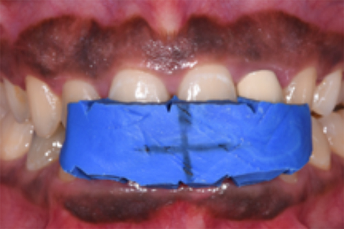 |
Figure 2 Standardized photos were taken with a stent and a marker to ensure the same position and angle when the images were overlayed over each other. |
Depigmentation was carried out using surgical blades and dental diamond burs under dental anesthesia (see Figure 3).15 One year later, post-depigmentation photos were taken with the same stent, which ensured uniformity in position and angle when superimposing the images over one another. A white balance card and a ColorChecker Passport (X-Rite Pantone, Grand Rapids, Michigan, USA) were used to ensure that all the photos were taken under the same lighting and conditions. This was done to make sure that even if the lighting changed throughout the course of the study, the colors in the picture would stay true to accuracy and consistency.
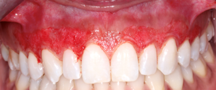 |
Figure 3 Picture of the full upper arch after gingival depigmentation. |
We captured two rows above the gingival margin, from canine to canine. Each sample was 75×75 pixels in size. Participants were asked to keep an upright posture, refrain from turning their heads, and maintain their maxillary teeth’s occlusal plane parallel to the ground. They were instructed to keep their mouth closed and maintain maximum habitual intercuspation while wearing lip retractors. They were also told to close their mouths between shots to prevent teeth dehydration.
Image Analysis
Frontal oral pictures were used to evaluate gingival pigmentation. The images were entered into a software (Adobe Photoshop 2020, Adobe Inc., San Jose, California) for analysis. The grid feature in Photoshop was utilized to ensure the use of the same area for pre- and post-depigmentation images (see Figure 4).
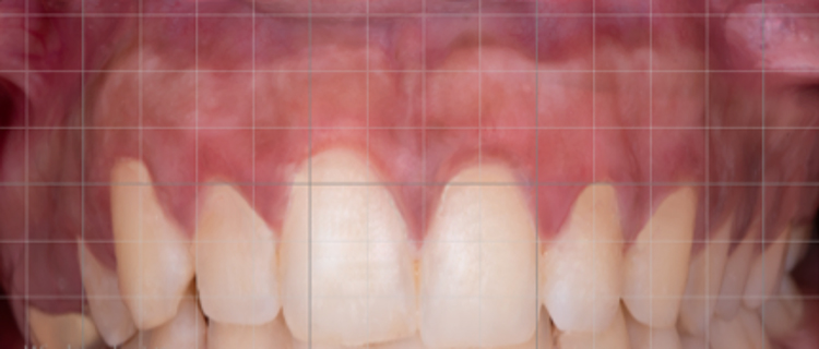 |
Figure 4 The grid feature in Photoshop 2020 was used to divide each image into numerous smaller samples. |
The Dummett Oral Pigmentation Index (DOPI)16 and Gingival Melanosis Record (GMR)17 index were utilized to measure the level of gingival pigmentation (see Figure 5). All images were split into two groups for the GMR (Yes or No) and four groups for the DOPI (0, 1, 2, and 3). A pilot study of 80 photographs was carried out to evaluate the efficacy of the DOPI and GMR.
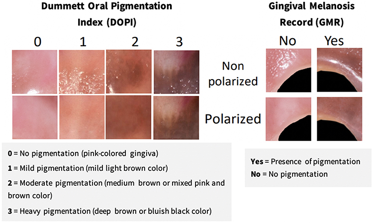 |
Figure 5 Measurement of gingival pigmentation with the Dummett Oral Pigmentation Index (DOPI) and Gingival Melanosis Record (GMR). |
Three evaluators (an orthodontist, a general dentist, and a layperson) independent of this study took part in the assessment of the photographs. For consistency in the evaluation, all images underwent complete masking and were shown to each evaluator on the same device (iPad Air, 3rd generation, Apple Inc., California, USA). Computer-generated random numbers were assigned to each image.
Each image was initially divided into smaller samples. Each sample was given a code and put into its own slide. It was then dispersed at random among other slides. Each evaluator assessed a set of 30 slides before performing the final image assessment. This was done to make sure that the evaluators understood the procedure and were ready for the image analysis. The intra- and inter-rater reliability of evaluators was then calculated using the Kappa (κ) test. All the calculated kappa were above 0.70.
Sample Size Calculation and Data Analysis
The sample size was calculated based on the assumption that the general dentist evaluating the polarized and non-polarized photos would find a 0.02 mean difference and a 0.17 standard deviation difference. The final sample size was estimated to be 976 images per group.
Data were presented using descriptive statistics (eg, mean and standard deviation for DOPI and percentage for GMR). After checking for normality, the paired t-test was used to determine within-group difference between polarized and non-polarized photos. One way ANOVA was utilized to evaluate between-group differences. A significance level of p < 0.05 was considered statistically significant. Percentage of GMR and DOPI reduction was calculated by subtracting post-gingival depigmentation from pre-depigmentation. All the analyses were conducted using the Statistical Package for the Social Sciences (SPSS) program (Version 26.0; SPSS Inc., Chicago, IL, USA).
Results
The study analyzed a total of 5856 responses. Each examiner evaluated 976 images (488 polarized and 488 non-polarized) using the DOPI and the GMR indices. This assessment was conducted two times, both before and one year after the depigmentation procedure. Photographic analysis revealed a lower GMR percentage for post-gingival depigmentation images than for pre-depigmentation images, as shown in Figure 6. In both pre- and post-depigmentation image analysis, the orthodontist had a higher GMR percentage than the other two evaluators. A similar pattern was observed in the DOPI mean of post-depigmentation images (Figure 7). The images taken after gingival depigmentation had relatively lower GMR percentage and DOPI mean than those captured before depigmentation. All three evaluators noted this reduction in GMR and DOPI values following depigmentation.
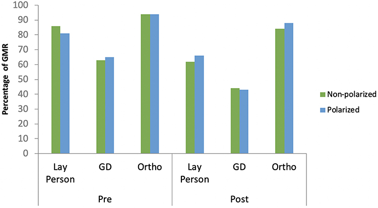 |
Figure 6 Percentage of GMR of polarized and non-polarized photos among different groups at pre- and post-depigmentation. Abbreviations: GD, general dentist; Ortho, orthodontist; GMR, Gingival Melanosis Record. |
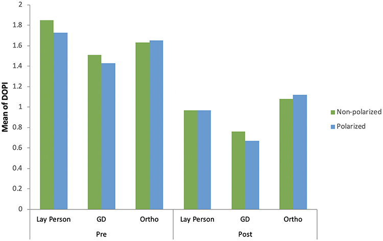 |
Figure 7 DOPI mean of polarized and non-polarized photos among different evaluators at pre-depigmentation and post-gingival depigmentation. Abbreviations: GD, general dentist; Ortho, orthodontist; DOPI, Dummett Oral Pigmentation Index. |
In both pre- and post-depigmentation image analysis, no significant difference was found in the DOPI mean and GMR percentage between polarized and non-polarized images (Table 1). The assessment of polarized images one year after depigmentation revealed 67–95% of gingival repigmentation using the GMR index (Figure 8), while the DOPI obtained a slightly lower range, 51—71% (Figure 9). A similar pattern was also noted in non-polarized images. The one-year follow-up image analysis revealed no statistically significant difference between polarized and non-polarized images in either the DOPI mean or the GMR percentage.
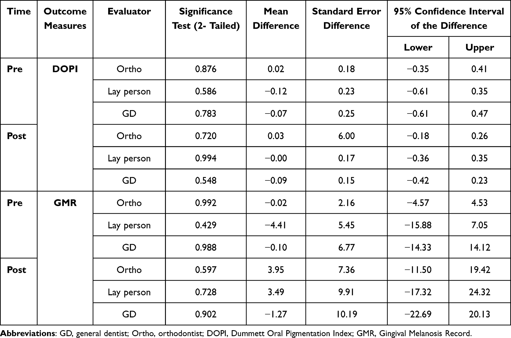 |
Table 1 Comparison of GMR and DOPI Before and After Depigmentation by Different Evaluators |
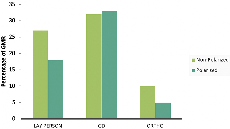 |
Figure 8 Percentage of GMR reduction of polarized and non-polarized photos among different groups. Abbreviations: GD, general dentist; Ortho, orthodontist; GMR, Gingival Melanosis Record. |
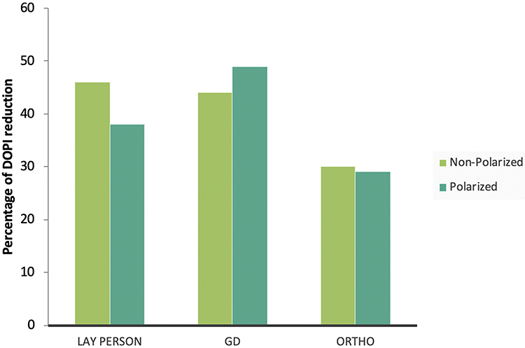 |
Figure 9 Percentage of DOPI reduction of polarized and non-polarized photos among different groups. Abbreviations: GD, general dentist; Ortho, orthodontist; DOPI, Dummett Oral Pigmentation Index. |
Discussion
In the oral cavity, the most commonly pigmented tissue is the gingiva. Dental photography is an excellent method to record and analyze these changes in gingival coloration. However, capturing images of gingival pigmentation with correct color rendition presents a challenge due to the use of a light source and its specular reflection.3,4,18 In the present study, pre- and post-depigmentation images of the gingiva were analyzed using polarized and non-polarized intraoral photography. The results of this study revealed no significant differences between polarized and non-polarized images taken before and after depigmentation, and both techniques received similar scores from the evaluators.
There is a lack of information in the current literature regarding the true color of the gingiva. In fact, since no standardized color matching or shade guides have yet been developed, gingival color is mostly determined subjectively using visual observation.19,20 The accuracy of visual technique depends on several factors including sex, training, and experience of the observer.21,22 However, the visual method has been found to be less reliable than the digital method.23 Digital images provide dental technicians with important details about a tooth’s shape, surface, and distinguishing features in addition to its color.24 Hence, a combination of both the visual and digital techniques has been suggested for color matching during cosmetic dental restoration.25
The findings of this study suggest that both polarized and non-polarized imaging modalities may be used to analyze changes in gingival pigmentation. Although gingival color photography has not been a subject of much research, our findings are in line with those published on shade selection8,25 and demineralized white lesions.12 It is important to mention that using cross-polarizing filters has reportedly been found to be the most consistent technique for tooth color assessment, because they help reduce undesirable reflections and diffuse light produced by the flash.7,9,11,12,14,26 However, the removal of specular reflections by polarizing filters can cause color distortion and result in a flawed image analysis.7,8,10 Hence, non-polarized photography is preferable to cross-polarized photography, as it is less expensive and easier to use.4,18,25 In fact, smartphone cameras are gaining popularity among dentists these days due to their affordability, software options, improved functionalities, and high-resolution images, which make them ideal for taking color references.27–29 However, both methods should be used in combination, whenever feasible.
Digital images taken with the non-polarized technique should clearly capture the gingiva’s color as seen by the eyes during an oral examination.8,26 As demonstrated in this study, when non-polarized images are captured with standardized, calibrated parameters, they can replicate gingival color in a simple, quick, and accurate manner. Several factors, however, need to be considered while taking an intraoral non-polarized picture, including white balance, color rendition, color temperature, image resolution, light source, light glare, and specular reflection.1,2,4,5,10,18,21,26 Studies have also suggested using a grey reference card to help standardize colors.24,26,30
In the present study, all three evaluators differed to some extent in their image analysis, although these differences were not statistically significant. The orthodontist comparatively identified more pigmented slides than the layperson and the general dentist. This finding may be attributed to the specialized training and experience of the orthodontist in analyzing dental irregularities, making them better equipped to identify changes in gingival pigmentation. However, because only one orthodontist was involved in the image analysis, it is difficult to make any firm conclusion at this moment. Nevertheless, numerous studies have demonstrated that trained professionals, such as dental technicians, are better at matching colors than laypeople, students, dental staff, and dentists.8,25,31
A major limitation of this study was that only three evaluators were utilized to assess a sizable sample of images. Although we were unable to add more evaluators due to logistical issues, increasing their number would have increased the validity and reliability of the results. In addition, each evaluator was at risk of having eye fatigue when analyzing those samples without a time limit. Future studies should consider giving evaluators an eye rest by watching a blue color object before continuing the evaluation. Another limitation of this study was the use of the same photography setup and a single photograph acquisition protocol. Due to these limitations, the study could be subjected to observer bias.
Therefore, one should exercise caution when generalizing the results of this study. However, more research is required to confirm these findings and investigate additional factors. Future studies should include larger image samples, an adequate of evaluators with diverse backgrounds, and varieties of equipment and parameters to gain more insights about the best standardized photography method for assessing gingival pigmentation.
Conclusion
In this study, no significant differences were found between polarized and non-polarized intraoral photography in detecting gingival pigmentation changes. Both techniques may be utilized for analyzing gingival pigmentation. The non-polarized photography, however, is preferred due to its lower cost and ease of use, but several factors must be considered to ensure accurate replication of gingival color. In addition, further research is needed to confirm these findings and investigate additional factors.
Acknowledgements
We extend our gratitude to Dr. Bashair Baqies and Dr. Shaymaa Alamoudi for their assistance in research and logistics. Their support was notably helpful throughout our research process.
Disclosure
The authors report no conflicts of interest in this work.
References
1. Desai V, Bumb D, Marwah N, Toumba KJ. Digital dental photography: a contemporary revolution. Int J Clin Pediatr Dent. 2013;6:193–196. doi:10.5005/jp-journals-10005-1217
2. Ahmad I. Digital dental photography. Part 2: purposes and uses. Br Dent J. 2009;206(9):459–464. doi:10.1038/sj.bdj.2009.366
3. Ahmad I. Digital dental photography. Part 1: an overview. Br Dent J. 2009;206(8):403–407. doi:10.1038/sj.bdj.2009.306
4. Kalpana D, Rao SJ, Joseph JK, Kurapati SKR. Digital dental photography. Indian J Dent Res. 2018;29(4):507–512. doi:10.4103/ijdr.IJDR_396_17
5. Wagner DJ. A beginning guide for dental photography: a simplified introduction for esthetic dentistry. Dent Clin North Am. 2020;64(4):669–696. doi:10.1016/j.cden.2020.07.002
6. Ahmad I. Digital dental photography. Part 5: lighting. Br Dent J. 2009;207(1):13–18. doi:10.1038/sj.bdj.2009.558
7. Lazar R, Culic B, Gasparik C, Lazar C, Dudea D. The accuracy of dental shade matching using cross-polarization photography. Int J Comput Dent. 2019;22(4):343–351.
8. Kelkar KC, Dogra ES, Bhat V, Prasad DK, Hegde C. A comparison between visual, digital photography and polarizing filter photography for shade selection. Indian J Dent Res. 2020;31(5):712–717. doi:10.4103/ijdr.IJDR_286_19
9. Kim E, Son T, Lee Y, Jung B. Development of polarization dental imaging modality and evaluation of its clinical feasibility. J Dent. 2012;40(Suppl 1):e18–e25. doi:10.1016/j.jdent.2012.04.013
10. Farah RI, Almershed AS, Albahli BF, Al-Haj Ali SN. Effect of ambient lighting conditions on tooth color quantification in cross-polarized dental photography: a clinical study. J Prosthet Dent. 2022;128(4):776–783. doi:10.1016/j.prosdent.2021.01.015
11. Robertson AJ, Toumba KJ. Cross-polarized photography in the study of enamel defects in dental paediatrics. J Audiov Media Med. 1999;22(2):63–70. doi:10.1080/014051199102179
12. Benson PE, Ali Shah A, Robert Willmot D. Polarized versus nonpolarized digital images for the measurement of demineralization surrounding orthodontic brackets. Angle Orthod. 2008;78(2):288–293. doi:10.2319/121306-511.1
13. Lakhanpal S, Neelima MS. Accuracy of three shade-matching devices in replicating the shade of metal ceramic restorations: an in vitro study. J Contemp Dent Pract. 2016;17(12):1003–1008. doi:10.5005/jp-journals-10024-1971
14. Hanlon KL. Cross-polarised and parallel-polarised light: viewing and photography for examination and documentation of biological materials in medicine and forensics. J Vis Commun Med. 2018;41(1):3–8. doi:10.1080/17453054.2018.1420418
15. Murthy MB, Kaur J, Das R. Treatment of gingival hyperpigmentation with rotary abrasive, scalpel, and laser techniques: a case series. J Indian Soc Periodontol. 2012;16(4):614–619. doi:10.4103/0972-124X.106933
16. Dummett CO, Gupta OP. Estimating the Epidemiology of Oral Pigmentation. J Natl Med Assoc. 1964;56(5):419–420.
17. Kato T, Takiuchi H, Sugiyama S, et al. Measurement of reduced gingival melanosis after smoking cessation: a novel analysis of gingival pigmentation using clinical oral photographs. Int J Environ Res Public Health. 2016;13(6):598. doi:10.3390/ijerph13060598
18. Casaglia A, P DED, Arcuri L, Gargari M, Ottria L. Dental photography today. Part 1: basic concepts. Oral Implantol. 2015;8:122–129.
19. Denissen H, Kuijkens A, Dozic A. A photographic method to measure the colour characteristics of healthy gingiva. Int J Dent Hyg. 2007;5(1):22–26. doi:10.1111/j.1601-5037.2007.00216.x
20. Gomez-Polo C, Montero J, Gomez-Polo M, Martin Casado AM. Clinical study on natural gingival color. Odontology. 2019;107(1):80–89. doi:10.1007/s10266-018-0365-2
21. Clary JA, Ontiveros JC, Cron SG, Paravina RD. Influence of light source, polarization, education, and training on shade matching quality. J Prosthet Dent. 2016;116(1):91–97. doi:10.1016/j.prosdent.2015.12.008
22. Haddad HJ, Jakstat HA, Arnetzl G, et al. Does gender and experience influence shade matching quality? J Dent. 2009;37(Suppl 1):e40–e44. doi:10.1016/j.jdent.2009.05.012
23. Preethi Suganya S, Manimaran P, Saisadan D, et al. Spectrophotometric Evaluation of Shade Selection with Digital and Visual Methods. J Pharm Bioallied Sci. 2020;12(5):S319–S323. doi:10.4103/jpbs.JPBS_95_20
24. Jorquera GJ, Atria PJ, Galan M, et al. A comparison of ceramic crown color difference between different shade selection methods: visual, digital camera, and smartphone. J Prosthet Dent. 2022;128(4):784–792. doi:10.1016/j.prosdent.2020.07.029
25. Hardan L, Bourgi R, Cuevas-Suarez CE, et al. Novel trends in dental color match using different shade selection methods: a systematic review and meta-analysis. Materials. 2022;16(1):15. doi:10.3390/ma16010015
26. Sampaio CS, Atria PJ, Hirata R, Jorquera G. Variability of color matching with different digital photography techniques and a gray reference card. J Prosthet Dent. 2019;121(2):333–339. doi:10.1016/j.prosdent.2018.03.009
27. Tam WK, Lee HJ. Accurate shade image matching by using a smartphone camera. J Prosthodont Res. 2017;61(2):168–176. doi:10.1016/j.jpor.2016.07.004
28. Moussa C, Hardan L, Kassis C, et al. Accuracy of dental photography: professional vs smartphone’s camera. Biomed Res Int. 2021;2021:3910291. doi:10.1155/2021/3910291
29. Hardan LS, Moussa C. Mobile dental photography: a simple technique for documentation and communication. Quintessence Int. 2020;51(6):510–518. doi:10.3290/j.qi.a44365
30. Hein S, Zangl M. The use of a standardized gray reference card in dental photography to correct the effects of five commonly used diffusers on the color of 40 extracted human teeth. Int J Esthet Dent. 2016;11(2):246–259.
31. Capa N, Malkondu O, Kazazoglu E, Calikkocaoglu S. Evaluating factors that affect the shade-matching ability of dentists, dental staff members and laypeople. J Am Dent Assoc. 2010;141(1):71–76. doi:10.14219/jada.archive.2010.0023
 © 2023 The Author(s). This work is published and licensed by Dove Medical Press Limited. The full terms of this license are available at https://www.dovepress.com/terms.php and incorporate the Creative Commons Attribution - Non Commercial (unported, v3.0) License.
By accessing the work you hereby accept the Terms. Non-commercial uses of the work are permitted without any further permission from Dove Medical Press Limited, provided the work is properly attributed. For permission for commercial use of this work, please see paragraphs 4.2 and 5 of our Terms.
© 2023 The Author(s). This work is published and licensed by Dove Medical Press Limited. The full terms of this license are available at https://www.dovepress.com/terms.php and incorporate the Creative Commons Attribution - Non Commercial (unported, v3.0) License.
By accessing the work you hereby accept the Terms. Non-commercial uses of the work are permitted without any further permission from Dove Medical Press Limited, provided the work is properly attributed. For permission for commercial use of this work, please see paragraphs 4.2 and 5 of our Terms.