")
Back to Journals » Journal of Inflammation Research » Volume 14
Upregulation of TMEM45A Promoted the Progression of Clear Cell Renal Cell Carcinoma in vitro
Authors Jiang H , Chen H, Wan P, Liang M, Chen N
Received 27 September 2021
Accepted for publication 22 November 2021
Published 1 December 2021 Volume 2021:14 Pages 6421—6430
DOI https://doi.org/10.2147/JIR.S341596
Checked for plagiarism Yes
Review by Single anonymous peer review
Peer reviewer comments 3
Editor who approved publication: Professor Ning Quan
Huiming Jiang,1,* Haibin Chen,2,* Pei Wan,1 Meng Liang,3 Nanhui Chen1
1Department of Urology, Meizhou People’s Hospital, Guangdong Provincial Key Laboratory of Precision Medicine and Clinical Translational Research of Hakka Population, Meizhou, People’s Republic of China; 2Department of Histology and Embryology, Shantou University Medical College, Shantou, People’s Republic of China; 3Gannan Medical University, Ganzhou, People’s Republic of China
*These authors contributed equally to this work
Correspondence: Huiming Jiang; Nanhui Chen Tel +86-13560990839
Email [email protected]; [email protected]
Background: Clear cell renal cell carcinoma (ccRCC) is the most common and aggressive type of primary kidney cancer worldwide. Transmembrane protein 45A (TMEM45A) has been reported to be closely associated with the progression of several cancers. However, the role of TMEM45A in ccRCC remains unclear. Our study intended to explore the potential role of TMEM45A in ccRCC.
Methods: Data on the expression of TMEM45A were obtained from multiple databases, including UCSC, GEPIA2, Oncomine and TIMER. Real-world samples of ccRCC and paired normal renal tissues were used to confirm the information obtained from the databases. In addition, the prognostic value of TMEM45A was evaluated. Loss-of-function assays were performed using TMEM45A-targeting lentivirus to evaluate the biological role of TMEM45A in renal cancer cells. Gene set enrichment analysis (GSEA) was performed to investigate the potential molecular mechanisms.
Results: TMEM45A was significantly overexpressed in patients with ccRCC and correlated with poor overall survival and disease-free survival. In addition, the expression of TMEM45A was closely associated with various clinicopathological parameters such as histological grade and TNM stage. Knockdown of TMEM45A inhibited the proliferation and migration and promoted the apoptosis of ccRCC cells in vitro. The results of the GSEA suggested that TMEM45A was potentially involved in the promotion of epithelial–mesenchymal transition (EMT) and inflammatory response in ccRCC.
Conclusion: TMEM45A was overexpressed and associated with poor survival and acted as a tumour promoter in ccRCC; therefore, might be a potential prognostic marker and therapeutic target.
Keywords: biomarker, clear cell renal cell carcinoma, prognosis, TMEM45A
Introduction
Renal cell carcinoma (RCC) remains a significant global health challenge. Approximately 75% of the RCC cases are clear cell renal cell carcinoma (ccRCC), which is the major histological subtype.1 According to the cancer statistics data from the United States (2020), the number of newly diagnosed cases of ccRCC was 73,750, and 14,830 patients were predicted to die of the disease.2 A contrast-enhanced computed tomography or magnetic resonance imaging remains the mainstay for the clinical diagnosis of RCC. Surgery remains the major treatment for located RCC, whereas treatment of metastatic RCC is more challenging considering the resistance of cancer cells to radiotherapy and chemotherapy.3 Nearly half of the patients progress to metastasis even undergo nephrectomy, and the 5-year survival rate of those with metastasis is less than 12%.4 Although the introduction of tyrosine kinase inhibitors and immune checkpoint inhibitors have significantly improved the survival of patients with RCC, the outcomes remain unsatisfactory.5,6 Therefore, the identification of effective prognostic biomarkers and therapeutic targets remains essential for personalised treatment and follow-up.
Transmembrane transport is one of the vital mechanisms of material transport in various biological processes. A transmembrane protein (TMEM) is a type of integral membrane protein that extends through the lipid bilayer of the plasma membrane or membrane-bound organelles. TMEMs mainly function as channels to permit the transport of specific substances across the biological membranes; however, their functions remain unknown.7 Transmembrane protein 45A (TMEM45A) is a member of the TMEM family, which is located on chromosome 3q12.2 and encodes a transmembrane protein of 275 amino acids with five or seven transmembrane domains. Several studies have reported a close relationship between TMEM45A and the progression and chemoresistance of various cancers. Downregulation of TMEM45A could inhibit the proliferation, migration and invasion of glioma cells.8 In addition, TMEM45A was found to be essential for hypoxia-induced chemoresistance in breast and liver cancer cells.9 Upregulation of TMEM45A has been reported in ccRCC.10,11 However, there is a lack of in-depth studies on the biological functions and prognostic value of TMEM45A in ccRCC. In the present study, we explored the association between TMEM45A expression and the clinicopathological characteristics and survival of patients in an integrated manner by analysing multiple databases, real-world ccRCC samples and performing loss-of-function assays in cancer cell lines. Our study demonstrates that TMEM45A may act as a tumour promoter, and its overexpression predicts an unfavourable survival outcome in patients with ccRCC.
Methods
Data Collection
The gene expression profiles and clinicopathological data of ccRCC were downloaded from the University of California Santa Cruz Xena platform (https://xena.ucsc.edu/).12 A total of 535 tumour samples and 72 normal samples were obtained. After matching the clinical information and the respective gene expression data and removing the cases with a follow-up period of less than 1 month, 518 patients were selected for the survival analysis. Several other online databases, such as GEPIA2 (http://gepia2.cancer-pku.cn/), Oncomine (https://www.oncomine.org/) and TIMER (https://cistrome.shinyapps.io/timer/) were used for external validation.13–15
Tissue Specimens
Surgical samples (paired ccRCC and normal renal tissues) were collected from 11 patients who underwent partial nephrectomy or nephrectomy at the Meizhou People’s Hospital between 2019 and 2021. The mean age of the patients was 54.9 years. Seven (63.6%) cases were histologically graded as well-differentiated (G1/2), whereas the remaining four (36.4%) cases were graded as poorly differentiated (G3/4). Most patients (n = 7, 63.6%) had localized stage I/II tumors, and other four patients (36.4%) had stage III/IV tumors. Written informed consent was obtained from all patients in accordance with the Declaration of Helsinki. The Medical Ethics Committee of the Meizhou People’s Hospital approved our study (No. 2020-CY-06).
RNA Extraction and qPCR
We extracted the total RNA using an RNA extraction kit (Haigene Biotech Co., Ltd., China). RNA was reverse transcribed into complementary DNA using the PrimeScript RT reagent kit (Takara Bio Inc., Dalian, China) following the manufacturer’s protocol. We performed quantitative polymerase chain reaction (qPCR) with the SYBR Green PCR kit (Takara Bio Inc., Dalian, China) using the ABI 7500 fluorescent quantitative PCR system (Applied Biosystems Inc., Foster City, CA, USA). The primers used were as follows: TMEM45A (forward: GTTCACTTCCTGTGTCCTTAACC; reverse: CATTTCCCGGCCATGAGTGT), and GAPDH (forward: GTCAAGGCTGAGAACGGGAA; reverse: AAATGAGCCCCAGCCTTCTC). The 2−ΔΔCt method was used to measure the expression of TMEM45A.
Cell Culture and Transfection
The ccRCC cell line ACHN was obtained from the Chinese Academy of Sciences, and cultured in Dulbecco’s Modified Eagle Medium (DMEM) containing 10% fetal bovine serum (Gibco, USA) and 1% penicillin–streptomycin at 37°C with 5% CO2 and 95% humidity. The short hairpin RNA (shRNA) target against the TMEM45A sequences (NM_001363876.2, NCBI) was 5ʹ-CCTGTGTCCTTAACCAAGTTA-3ʹ. Recombinant TMEM45A-targeting lentivirus (LV-sh-TMEM45A) or control mock lentivirus (LV-sh-NC) constructed by GenePharma (Suzhou, China) were transfected into the RCC cells. After culturing for 48–72h, puromycin was used to obtain stable clones in which the TMEM45A gene was downregulated. We confirmed the stably transfected clones by quantitative reverse transcription PCR.
Cellular Experiments of Proliferation, Migration, and Apoptosis After TMEM45A Knockdown
The cell proliferation ability was assessed by the MTT assay. The cell migration ability was evaluated by the wound-healing assay. Apoptosis of cells was evaluated using the Annexin V‐APC 7-AAD apoptosis kit (Biolegend, USA) according to the manufacturer’s instructions. All the cellular experiments were performed on three groups (control, LV-sh-NC and LV-sh-TMEM45A).
Functional Enrichment Analysis
Functional enrichment analysis was performed to explore the potential molecular mechanisms of TMEM45A in ccRCC. First, we divided the patients with ccRCC in The Cancer Genome Atlas (TCGA) into high- and low-TMEM45A groups based on the median expression of TMEM45A. Next, we performed gene set enrichment analysis (GSEA) to identify the significantly enriched pathways. The sample-wise gene set activities of the different pathways were calculated in GSVA using the “single-sample GSEA” (ssGSEA) method. Lastly, we detected the relationship between TMEM45A expression and the enriched pathways by the Spearman correlation method.
Statistics
All statistical analyses were performed using R software version 3.6.1 (the R Foundation for Statistical Computing, Vienna, Austria). Analysis of variance (ANOVA) or t-test was performed to assess the significant differences in TMEM45A expression among the different subgroups. The chi-square test or Fisher’s exact test was performed to evaluate the correlation between TMEM45A expression and the clinicopathological parameters of ccRCC. The Kaplan–Meier method and Log rank test were considered for the survival analyses. Univariate and multivariate survival analyses were performed by a Cox regression model. Pearson’s correlation test was used in the correlation analysis. A p-value < 0.05 was considered statistically significant.
Results
TMEM45A Was Significantly Upregulated in ccRCC
First, we evaluated the expression level of TMEM45A in TCGA-KIRC cohort. The results revealed a significantly higher expression of TMEM45A in ccRCC tissues than in normal kidney tissues (p <0.001) (Figure 1A). Several other online databases, including GEPIA2, Oncomine and TIMER, were analysed to validate the results. The results confirmed the upregulation of TMEM45A in ccRCC tissues (Figure 1B–D). More details regarding the patients, in addition to those presented in Figure 1D, could be found in Supplementary Table S1. An interesting finding was the significantly higher expression of TMEM45A in several other types of cancers, including breast cancer, oesophageal cancer, head and neck squamous cell carcinoma and lung squamous cell carcinoma (Figure 1D). We evaluated the expression of TMEM45A in 11 pairs of ccRCC tissues and the adjacent normal kidney tissues by qPCR to confirm the results obtained from the databases. As demonstrated in Figure 1E and F, the expression of TMEM45A in ccRCC was significantly higher than that in normal renal tissues (p = 0.038). The American Joint Committee on Cancer (AJCC) staging method (7th edition) was used to denote the TNM stage of ccRCC,16 and subsequent analysis revealed that the expression of TMEM45A increased significantly with higher histological grade, increased T stage, distant metastasis and advanced TNM stage (p <0.001, Figure 1G–J).
 |
Figure 1 Higher expression of TMEM45A in ccRCC tissues than in normal renal tissues. (A) Data of the 535 tumour samples and 72 normal renal samples from TCGA-KIRC cohort. (B) Data from the GEPIA2 (http://gepia2.cancer-pku.cn/) database. (C) Data from the Oncomine (https://www.oncomine.org/) database. (D) TMEM45A expression in the pan-cancer analysis from the TIMER (https://cistrome.shinyapps.io/timer/) database. (E) Upregulation of TMEM45A in eight of the eleven ccRCC samples compared with that in the paired normal renal samples. (F) Significant increase in TMEM45A expression in the tumour samples compared with that in the paired normal renal samples. (G–J) Significantly increased TMEM45A expression with higher histological grade and T stage, distant metastasis and advanced TNM stages. *p < 0.05, **p < 0.01, ***p < 0.001. Abbreviation: ccRCC, clear cell renal cell carcinoma. |
The Prognostic Value of TMEM45A in ccRCC
We divided patients with ccRCC into high- and low-expression groups based on the median expression of TMEM45A to determine its prognostic value and relationship with clinicopathological characteristics. As shown in Table 1, the expression of TMEM45A was closely related to various vital clinicopathological parameters in patients with ccRCC, including cancer status (p <0.001), histological grade (p <0.001), T stage (p = 0.003), M stage (p = 0.001) and TNM stage (p <0.001). The Kaplan–Meier survival analysis revealed that patients with high TMEM45A expression exhibited poorer overall survival (OS) and disease-free survival (DFS) (p <0.001, Figure 2A and B). Analysis ofthe GEPIA2 database revealed the same results (Figure 1C and D). Considering that the histological grade and TNM stage play an important role in the prognosis of patients with cancer, we conducted an additional stratified analysis. The analysis revealed that patients in the high TMEM45A expression group had significantly poorer OS than those in the low-expression group with histological grade 1/2 (p = 0.037), grade 3/4 (p = 0.043) and stage I/II (p = 0.033) but not stage III/IV (p = 0.477) disease (Figure 2E–H). The results of DFS were consistent with those of OS (Figure 2I–L).
 |
Table 1 Correlations Between the Expression of TMEM45A and Clinicopathologic Characteristics in ccRCC |
 |
Figure 2 The prognostic value of TMEM45A in ccRCC. (A and B) Overexpression of TMEM45A in patients with ccRCC correlated with poor overall survival (OS) and disease-free survival (DFS). (C and D) Validation of the association between TMEM45A expression and OS or DFS on the GEPIA2 platform. (E–H) Stratified survival analysis to determine the prognostic value of TMEM45A on OS based on the histological grade or stage. (I–L) Stratified analysis to determine the prognostic value of TMEM45A on DFS based on the histological grade or stage. Abbreviation: ccRCC, clear cell renal cell carcinoma. |
TMEM45A Knockdown Inhibited the Proliferation and Migration and Promoted the Apoptosis of ccRCC Cells in vitro
Considering the upregulation and significant correlation of TMEM45A with the prognosis and various clinicopathological features, we analysed the biological functions by knocking down TMEM45A in ccRCC cell lines. We established a stable knockdown model of TMEM45A expression in the ccRCC cell line ACHN via lentiviral shRNA-mediated infection. The qPCR results revealed that the expression of TMEM45A decreased significantly in the LV-sh-TMEM45A group compared with the LV-sh-NC group (p = 0.021, Figure 3A). The analysis revealed that knockdown of TMEM45A inhibited the migration and proliferation abilities than that observed in the LV-sh-NC group (Figure 3B and C). Furthermore, the apoptosis of ACHN cells increased significantly after knockdown in the LV-sh-TMEM45A group (Figure 3D). The results suggested that TMEM45A acted as a tumour promoter in ccRCC cells.
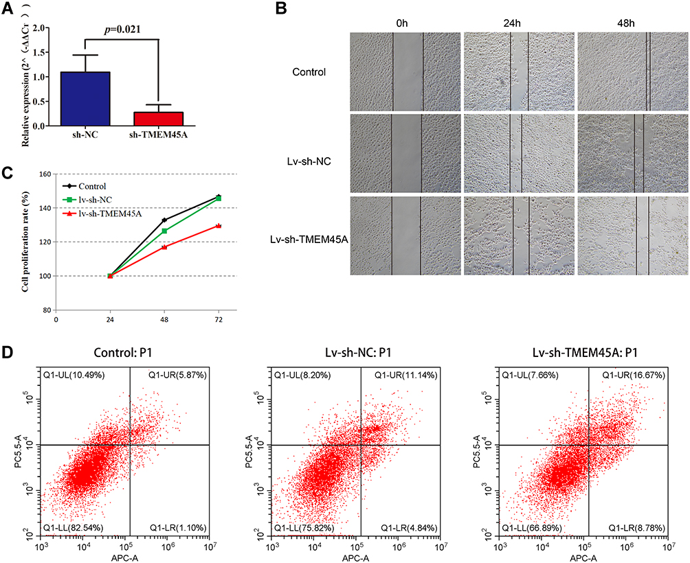 |
Figure 3 Effect of TMEM45A knockdown on the proliferation, migration, and apoptosis of renal cancer cells. (A) Confirmation of the knockdown efficiency by qPCR demonstrating significantly decreased expression of TMEM45A in the LV-sh-TMEM45A group compared with the LV-sh-NC group. (B) Wound healing assay results showing that TMEM45A knockdown significantly inhibited the migration of ACHN. (C) The results of the MTT assay showing that TMEM45A knockdown significantly inhibited the proliferation of ACHN. (D) TMEM45A knockdown significantly promoted the apoptosis of ACHN. |
Functional Enrichment Analysis
GSEA is a common method for functional enrichment analysis, which can determine significant differences between two biological statuses in a certain gene set.17 The results demonstrated that high TMEM45A expression was significantly associated with several cancer-related pathways, including epithelial–mesenchymal transition (EMT), IL-6-JAK-STAT3 signaling pathway, inflammatory response, tumour necrosis factor alpha (TNF-α) signalling pathway and interferon-γ response (Figure 4A and B). EMT is an important characteristic of tumour metastasis, whereas interleukin-6 (IL-6), TNF-α and interferon-γ are important inflammatory factors, and inflammation is closely associated with EMT.18–21 Therefore, we further analysed the relationship between the expression of TMEM45A and EMT and inflammatory response. The analysis revealed that TMEM45A had a significantly positive correlation with EMT (r = 0.472, p <0.001) and the inflammatory response pathway (r = 0.412, p <0.001) (Figure 4C and D). Moreover, the pan-cancer analysis revealed that the positive correlation between TMEM45A expression and EMT and inflammatory response was significant in most cancer types (Figure 4E and F).
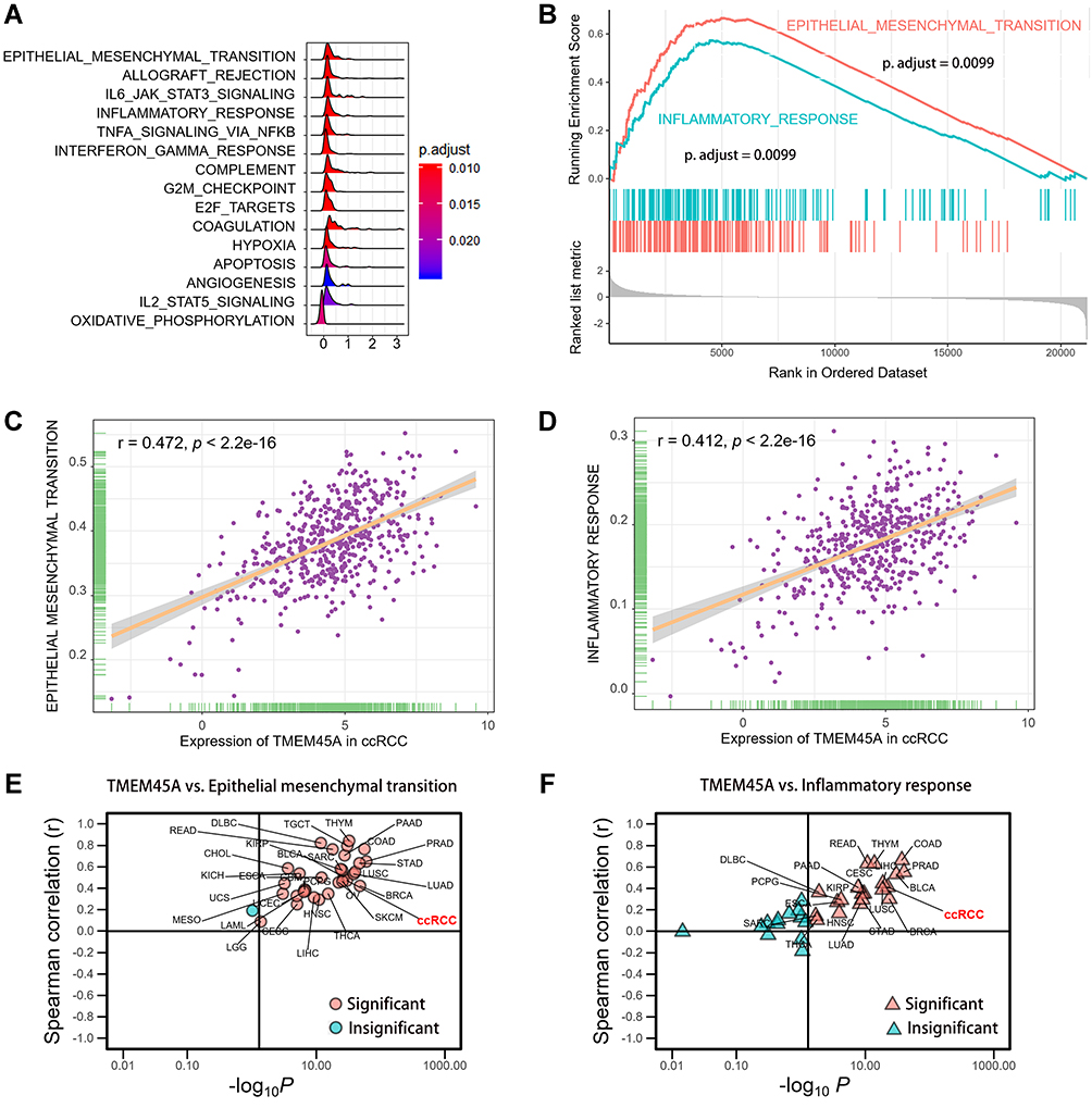 |
Figure 4 Functional enrichment analysis. (A) The results of the gene set enrichment analysis (GSEA). (B) The selected pathways enriched in the GSEA. (C) Significant positive correlation of TMEM45A expression with EMT in patients with ccRCC. (D) Significant positive correlation of TMEM45A expression with the inflammatory response pathway in patients with ccRCC. (E) Correlation analysis of TMEM45A and EMT in the pan-cancer analysis. (F) Correlation analysis of TMEM45A and the inflammatory response pathway in the pan-cancer analysis. Abbreviation: ccRCC, clear cell renal cell carcinoma. |
Discussion
RCC is the most common and aggressive type of primary kidney cancer worldwide. The major histological subtype of the malignancy is ccRCC constituting approximately 75% of the total cases, and is associated with the highest rate of metastasis, mortality and resistance to traditional chemotherapy and radiotherapy. Further understanding of the molecular mechanisms and identification of novel effective prognostic markers is essential for the therapy and prognosis of patients with ccRCC. In the present study, high-throughput RNA sequencing data were analysed from TCGA, and the results were validated using qPCR on real-world samples. In addition, by performing loss-of-function cell assays in vitro, we found that the expression of TMEM45A significantly correlated with various clinicopathological factors, OS, DFS and biological functions (proliferation, migration, and apoptosis) of ccRCC. The findings suggested that TMEM45A is a potential prognostic biomarker and therapeutic target for ccRCC.
TMEM45A, encoding a transmembrane protein, has been reported to be crucial in the progression of several cancers. Several studies have reported high expression of TMEM45A in liver, breast, ovarian cancers and glioma.8,9,22,23 Our study also revealed that the expression of TMEM45A was significantly increased in ccRCC, which is consistent with the results of previous studies.11 Moreover, recent studies have confirmed the prognostic value of TMEM45A in patients with cancer. Overexpression of TMEM45A correlated with higher histological grade and poorer survival in patients with glioma.8 Higher expression of TMEM45A was associated with chemoresistance and an unfavourable prognosis in patients with breast and liver cancer.9 In the current study, we observed a significantly increased expression of TMEM45A with higher histological grade and T stage, distant metastasis and advanced TNM stages. Patients with ccRCC exhibiting high TMEM45A expression had significantly poorer OS and DFS. Consistently, our stratified analysis based on the histological grade and TNM stage confirmed the aforementioned results. However, it should be noted that the survival difference did not exist in patients with TNM stage III/IV disease. The possible reasons may be the relatively lesser number of patients with TNM stage III/IV disease in the low TMEM45A expression group and the highly aggressive nature of the tumour in the aforementioned stages.
It is known that ccRCC is the most common subtype of RCC that arises from the proximal tubular epithelium, and EMT is an important process during the metastasis of the malignancy.24,25 Previous studies have reported that TMEM45A plays an important role in the EMT and migration of cancers. Liu et al disclosed that silencing TMEM45A could inhibit cell migration, invasion and EMT in human papilloma virus (HPV)-positive cervical cancer cells.26 Zhu et al demonstrated that knockdown of TMEM45A suppressed multi-drug resistance-enhanced migration, invasion and EMT in human colorectal cancer cells by inhibiting the transforming growth factor beta (TGF-β) signalling pathway.27 Consistently, the results of the GSEA performed in our study revealed that high expression of TMEM45A was associated with EMT, and the wound-healing assay confirmed that knockdown of TMEM45A inhibited the migration ability of ccRCC cells in vitro. Furthermore, we identified a significant relationship between TMEM45A and EMT in 32 of the 33 cancer types in the pan-cancer analysis. Additionally, IL-6-JAK-STAT3, TNF-α, interferon-γ and inflammatory response signalling were significantly enriched in the GSEA performed in this study. IL-6, TNF-α and interferon-γ are the common and vital inflammatory factors, and inflammation is another crucial parameter associated with cancer progression.28,29 The correlation analysis performed in this study revealed that TMEM45A expression correlated significantly with the inflammatory response pathway in ccRCC. Similarly, a significant relationship existed between TMEM45A and the inflammatory response pathway in 20 of the 33 cancer types in the pan-cancer analysis. Moreover, previous studies have reported that TMEM45A may serve as a potential biomarker and drug target for the diagnosis and treatment of inflammatory bowel disease with arthritis.30 Our study supports the hypothesis that increased expression of TMEM45A can act as a tumour promoter that is involved in EMT and inflammation.
Although the present study indicated the prognostic value and biological function of TMEM45A in ccRCC, several limitations should be acknowledged. First, the survival analysis was chiefly based on the data from TCGA. Another cohort from the real world is necessary to validate the results. Second, the biological function of TMEM45A was only explored in vitro; therefore, in vivo experiments should be conducted in the future. Finally, the exact molecular mechanism by which TMEM45A induced EMT remains unclear and requires further basic research.
Conclusion
Our present study revealed that TMEM45A was significantly upregulated and associated with poorer survival in patients with ccRCC. Acting as a tumour promoter, TMEM45A might be a potential prognostic marker and therapeutic target of ccRCC, which could provide new clues and strategies to treat the malignancy.
Data Sharing Statement
All the figures and tables presented to support the findings of the present study are included in the article.
Acknowledgments
The authors thank TCGA for sharing the sequencing dataset on open access. The authors also thank the associated databases for their contribution to facilitating TCGA analyses. The authors also thank Bullet Edits Limited for the linguistic editing and proofreading of the manuscript. This study was supported by the Peiyu Project of the Meizhou People's Hospital (Grant No. PY-C2020004) and the Medical Science and Technology Research Foundation of Guangdong Province (Grant No. B2021143).
Disclosure
The authors declare that they have no competing interests.
References
1. Linehan WM, Ricketts CJ. The Cancer Genome Atlas of renal cell carcinoma: findings and clinical implications. Nat Rev Urol. 2019;16(9):539–552. doi:10.1038/s41585-019-0211-5
2. Siegel RL, Miller KD, Jemal A. Cancer statistics, 2020. CA Cancer J Clin. 2020;70(1):7–30. doi:10.3322/caac.21590
3. Gray RE, Harris GT. Renal cell carcinoma: diagnosis and management. Am Fam Physician. 2019;99(3):179–184.
4. Padala SA, Barsouk A, Thandra KC, et al. Epidemiology of renal cell carcinoma. World J Oncol. 2020;11(3):79–87. doi:10.14740/wjon1279
5. Pontes O, Oliveira-Pinto S, Baltazar F, Costa M. Renal cell carcinoma therapy: current and new drug candidates. Drug Discov Today. 2021. doi:10.1016/j.drudis.2021.07.009
6. Tacconi EMC, Tuthill M, Protheroe A. Review of adjuvant therapies in renal cell carcinoma: evidence to date. Onco Targets Ther. 2020;13:12301–12316. doi:10.2147/OTT.S174149
7. Schmit K, Michiels C. TMEM proteins in cancer: a review. Front Pharmacol. 2018;9:1345. doi:10.3389/fphar.2018.01345
8. Sun W, Qiu G, Zou Y, et al. Knockdown of TMEM45A inhibits the proliferation, migration and invasion of glioma cells. Int J Clin Exp Pathol. 2015;8(10):12657–12667.
9. Flamant L, Roegiers E, Pierre M, et al. TMEM45A is essential for hypoxia-induced chemoresistance in breast and liver cancer cells. BMC Cancer. 2012;12:391. doi:10.1186/1471-2407-12-391
10. Thibodeau BJ, Fulton M, Fortier LE, et al. Characterization of clear cell renal cell carcinoma by gene expression profiling. Urol Oncol. 2016;34(4):
11. Wrzesiński T, Szelag M, Cieślikowski WA, et al. Expression of pre-selected TMEMs with predicted ER localization as potential classifiers of ccRCC tumors. BMC Cancer. 2015;15:518. doi:10.1186/s12885-015-1530-4
12. Goldman MJ, Craft B, Hastie M, et al. Visualizing and interpreting cancer genomics data via the Xena platform. Nat Biotechnol. 2020;38(6):675–678. doi:10.1038/s41587-020-0546-8
13. Li T, Fan J, Wang B, et al. TIMER: a web server for comprehensive analysis of tumor-infiltrating immune cells. Cancer Res. 2017;77(21):e108–e110. doi:10.1158/0008-5472.CAN-17-0307
14. Tang Z, Kang B, Li C, Chen T, Zhang Z. GEPIA2: an enhanced web server for large-scale expression profiling and interactive analysis. Nucleic Acids Res. 2019;47(W1):W556–w560. doi:10.1093/nar/gkz430
15. Rhodes DR, Yu J, Shanker K, et al. ONCOMINE: a cancer microarray database and integrated data-mining platform. Neoplasia. 2004;6(1):1–6. doi:10.1016/S1476-5586(04)80047-2
16. Edge SB, Compton CC. The American Joint Committee on Cancer: the 7th edition of the AJCC cancer staging manual and the future of TNM. Ann Surg Oncol. 2010;17(6):1471–1474. doi:10.1245/s10434-010-0985-4
17. Subramanian A, Tamayo P, Mootha VK, et al. Gene set enrichment analysis: a knowledge-based approach for interpreting genome-wide expression profiles. Proc Natl Acad Sci U S A. 2005;102(43):15545–15550. doi:10.1073/pnas.0506580102
18. Brabletz T, Kalluri R, Nieto MA, Weinberg RA. EMT in cancer. Nat Rev Cancer. 2018;18(2):128–134. doi:10.1038/nrc.2017.118
19. Lee CH. Epithelial-mesenchymal transition: initiation by cues from chronic inflammatory tumor microenvironment and termination by anti-inflammatory compounds and specialized pro-resolving lipids. Biochem Pharmacol. 2018;158:261–273. doi:10.1016/j.bcp.2018.10.031
20. Sistigu A, Di Modugno F, Manic G, Nisticò P. Deciphering the loop of epithelial-mesenchymal transition, inflammatory cytokines and cancer immunoediting. Cytokine Growth Factor Rev. 2017;36:67–77. doi:10.1016/j.cytogfr.2017.05.008
21. López-Novoa JM, Nieto MA. Inflammation and EMT: an alliance towards organ fibrosis and cancer progression. EMBO Mol Med. 2009;1(6–7):303–314. doi:10.1002/emmm.200900043
22. Lee S, Stewart S, Nagtegaal I, et al. Differentially expressed genes regulating the progression of ductal carcinoma in situ to invasive breast cancer. Cancer Res. 2012;72(17):4574–4586. doi:10.1158/0008-5472.CAN-12-0636
23. Guo J, Chen L, Luo N, Yang W, Qu X, Cheng Z. Inhibition of TMEM45A suppresses proliferation, induces cell cycle arrest and reduces cell invasion in human ovarian cancer cells. Oncol Rep. 2015;33(6):3124–3130. doi:10.3892/or.2015.3902
24. Piva F, Giulietti M, Santoni M, et al. Epithelial to mesenchymal transition in renal cell carcinoma: implications for cancer therapy. Mol Diagn Ther. 2016;20(2):111–117. doi:10.1007/s40291-016-0192-5
25. Wang P, Chen W, Ma T, et al. lncRNA lnc-TSI inhibits metastasis of clear cell renal cell carcinoma by suppressing TGF-β-induced epithelial-mesenchymal transition. Mol Ther Nucleic Acids. 2020;22:1–16. doi:10.1016/j.omtn.2020.08.003
26. Liu Y, Liu L, Mou ZX. TMEM45A affects proliferation, apoptosis, epithelial-mesenchymal transition, migration, invasion and cisplatin resistance of HPV-positive cervical cancer cell lines. Biochem Genet. 2021. doi:10.1007/s10528-021-10094-3
27. Zhu M, Jiang B, Yan D, Wang X, Ge H, Sun Y. Knockdown of TMEM45A overcomes multidrug resistance and epithelial-mesenchymal transition in human colorectal cancer cells through inhibition of TGF-β signalling pathway. Clin Exp Pharmacol Physiol. 2020;47(3):503–516. doi:10.1111/1440-1681.13220
28. Vincent CT, Fuxe J. EMT, inflammation and metastasis. Semin Cancer Biol. 2017;47:168–169. doi:10.1016/j.semcancer.2017.09.003
29. Suarez-Carmona M, Lesage J, Cataldo D, Gilles C. EMT and inflammation: inseparable actors of cancer progression. Mol Oncol. 2017;11(7):805–823. doi:10.1002/1878-0261.12095
30. Verma A, Somvanshi P, Haque S, Rathi B, Sharda S. Association of inflammatory bowel disease with arthritis: evidence from in silico gene expression patterns and network topological analysis. Interdiscip Sci. 2019;11(3):387–396. doi:10.1007/s12539-017-0272-1
 © 2021 The Author(s). This work is published and licensed by Dove Medical Press Limited. The full terms of this license are available at https://www.dovepress.com/terms.php and incorporate the Creative Commons Attribution - Non Commercial (unported, v3.0) License.
By accessing the work you hereby accept the Terms. Non-commercial uses of the work are permitted without any further permission from Dove Medical Press Limited, provided the work is properly attributed. For permission for commercial use of this work, please see paragraphs 4.2 and 5 of our Terms.
© 2021 The Author(s). This work is published and licensed by Dove Medical Press Limited. The full terms of this license are available at https://www.dovepress.com/terms.php and incorporate the Creative Commons Attribution - Non Commercial (unported, v3.0) License.
By accessing the work you hereby accept the Terms. Non-commercial uses of the work are permitted without any further permission from Dove Medical Press Limited, provided the work is properly attributed. For permission for commercial use of this work, please see paragraphs 4.2 and 5 of our Terms.