")
Back to Journals » Infection and Drug Resistance » Volume 17
Unravelling Childhood Tinea Capitis: A Multi-Dimensional Investigation Using Dermoscopy, Scanning Electron Microscopy and Mass Spectrometry
Authors Huang Z, Chen M, Peng Y, Zhang R
Received 22 November 2023
Accepted for publication 21 February 2024
Published 26 February 2024 Volume 2024:17 Pages 727—732
DOI https://doi.org/10.2147/IDR.S451200
Checked for plagiarism Yes
Review by Single anonymous peer review
Peer reviewer comments 2
Editor who approved publication: Prof. Dr. Héctor Mora-Montes
Zeyu Huang,1 Mingyan Chen,1 Yang Peng,1 Ruzhi Zhang2
1Department of Dermatology, The Third Affiliated Hospital of Soochow University, Changzhou, People’s Republic of China; 2Department of Dermatology, The Second Affiliated Hospital of Wannan Medical College, Wuhu, People’s Republic of China
Correspondence: Ruzhi Zhang, Department of Dermatology, The Second Affiliated Hospital of Wannan Medical college, No. 10 Rehabilitation Road, Jinghu District, Wuhu, 241101, People’s Republic of China, Tel +86 18761161826, Email [email protected]
Abstract: Tinea capitis, a common cutaneous fungal infection, shows an increasing prevalence with the increasing number of pets. We present tinea capitis in a 4-year-old girl presenting without typical symptoms such as alopecia or hair breakage. After a comprehensive evaluation including dermoscopy, Wood’s light, direct KOH fluorescent staining, scanning electron microscopy, fungal culture and mass spectrometry analysis, a diagnosis of tinea capitis infected Microsporum canis carried by domestic cats was made. We preliminarily explored the two modes of hair erosion by tinea capitis fungi and analyzed the possibility of the feature in this case. This case highlights the importance of accurate diagnosis and appropriate therapeutic intervention in cases of paediatric tinea capitis, particularly in households with resident pets.
Keywords: dermoscopy, microsporum canis, scanning electron microscopy, mass spectrometry, tinea capitis
Introduction
Tinea capitis, a common cutaneous fungal infection affecting children in midsummer,1 is diagnosed by 10% KOH microscopy and dermoscopy.2 As pets become an integral part of families, outpatient physicians need to focus on fungal diseases of animal origin, particularly those dominated by Microsporum canis.3 Despite challenges in identifying sources of infection, Matrix-Assisted Laser Desorption Ionization-Time Of Flight-Mass Spectrometry (MALDI-TOF MS) is emerges as a prospective tool for efficient identification of animal skin fungi.4 This paper describes the dermoscopic and scanning electron microscopic characterization of a child with tinea capitis, where mass spectrometry analysis confirmed a domestic cat as the source of infection. This highlights the need for a comprehensive diagnostic approach in dermatophytic fungal infections, with MALDI-TOF MS providing a reliable means of rapid and accurate pathogen identification.
Case Report
A 4-year-old girl presented to the dermatology department of our hospital. The patient reported mild pruritus of the scalp. Physical examination revealed an elliptical white lesion on her scalp, surrounded by a prominent white sheath surrounding the hair roots, and several smaller identical skin lesions in its vicinity. Significantly, in contrast to the typical pattern of hair breakage at the scalp, the child’s hair showed no signs of breakage at the time of assessment, and there were no current symptoms of hair loss. No enlarged cervical lymph nodes were found when examined.
Further history revealed that the patient’s family had adopted two English Shorthorn cats as pets two months earlier. One of the cats had ringworm a month earlier, with localized hair loss. The cat was diagnosed, treated and cured at the vet clinic.
Trichoscopy and Wood’s Lamp Examination
On trichoscopy at the first visit, the scalp showed abnormalities with fine scales and the hair roots were encased in a distinctive white sheath, as shown in the accompanying pictures (Figures 1A and B). On close examination, several hairs were twisted and broken, showing the characteristic “comma hairs”. Two weeks later, under the trichoscopy, the child’s hair was broken and thinned, the bacterial sheath had increased in size, and the focus had spread to the periphery, forming a new small focus under the wood light. The patient was then examined in the darkroom under a Wood’s lamb, bright blue-green fluorescence was seen emanating from the hairs, as shown in Figures 1C and D. These findings suggest that tinea capitis is still in progress and that the treatment plan needs to be adjusted.
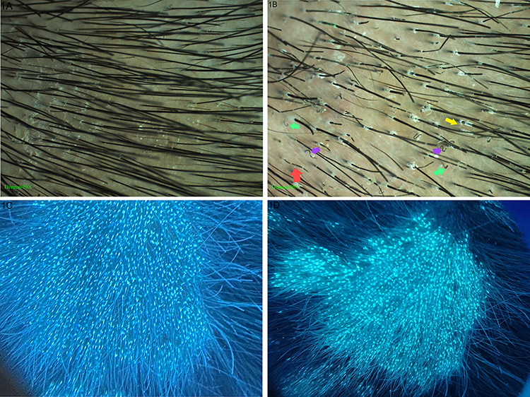 |
Figure 1 Clinical and dermoscopic pictures of skin lesions. Images taken at the patient’s first visit (A and C) and three weeks later (B and D). Trichoscopy of tinea capitis (B) with morse code-like hair (red arrow), corkscrew hair (purple arrows), comma hair (green arrows), and perifollicular scaling (yellow arrow). |
Examination by Scanning Electron Microscope (SEM)
Under dermoscopic guidance, the scalp lesion area was cleaned with 70% alcohol, a hair with a white sheath around the root was extracted with tweezers. The hair was vacuum dried and sprayed with platinum using an ion sprayer, followed by microscopic examination. SEM revealed that the area near the hair root was occupied by a large number of spores, without complete hair cortex or keratin fibre bundles, which were observed in the nearby normal area (Figure 2A). The trichoderms in this region were protruding, splitting and then peeling off (Figures 2B–D). A large number of spores were present along the entire length of the hair shaft, with no empty bands and absence of early breakage (Figure 2C). Dense clusters of spores erode the cuticle and form huge defects with corresponding locations of early hair breakage (Figure 2D).
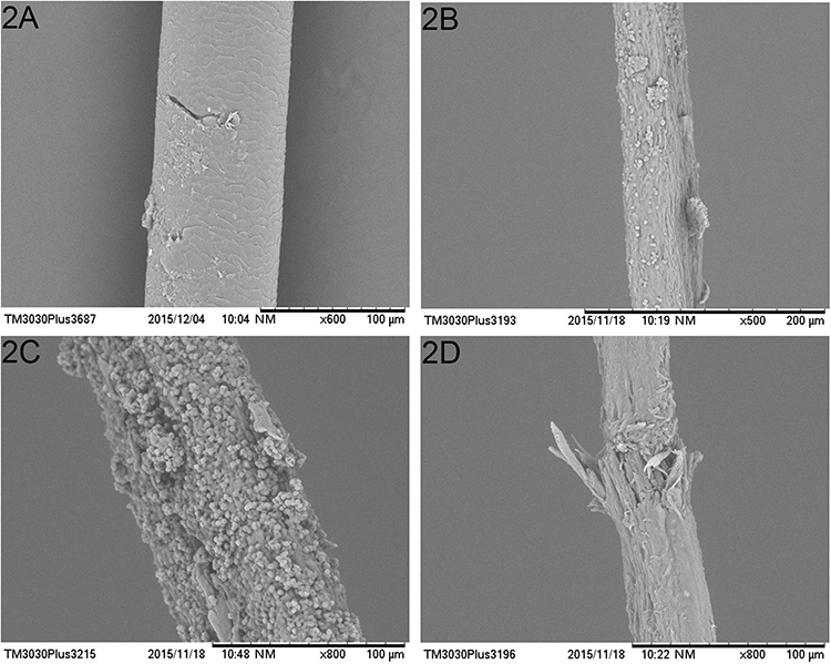 |
Figure 2 Images of fungi eroding hair under the scanning electron microscope. Appearance of normal hair (A) appearance of a hair that has just been eroded by fungus (B); two severely eroded hair features obtained at the centre of the lesion (C and D). |
Under electron microscope, the normal hairs were closely arranged in the shape of scales (2A) before fungal erosion. With the progress of the disease, the stratum corneum of the hair was dissolved and absorbed by fungi and gradually protruded and exfoliated like fragments (2B). In the later stage of erosion, fungi mainly showed two erosion patterns: fungal spores covered the whole hair (2C) or eroded local areas to form defects (2D), while the latter was the main reason, in which the end of the sheath was the most common.
Mycological Examination
Microscopic examination of the hair sample after potassium hydroxide treatment revealed the presence of hyphae and spores on the outer surface of the hair shaft as shown in Figure 3A and B, suggesting a possible ectomycorrhizal infection. The affected hairs were incubated on Sartre dextrose agar slant medium and on YO plate medium, respectively, at 28°C. Dense woolly colonies with golden to brownish yellow surface grew on the medium after 7 days (Figure 3C–E). These colonies showed septate hyphae and a few spores under a Calcofluor White (CFW) fluorescence microscope (Figure 3F), but unfortunately the typical large conidia of Microsporum canis were not observed.
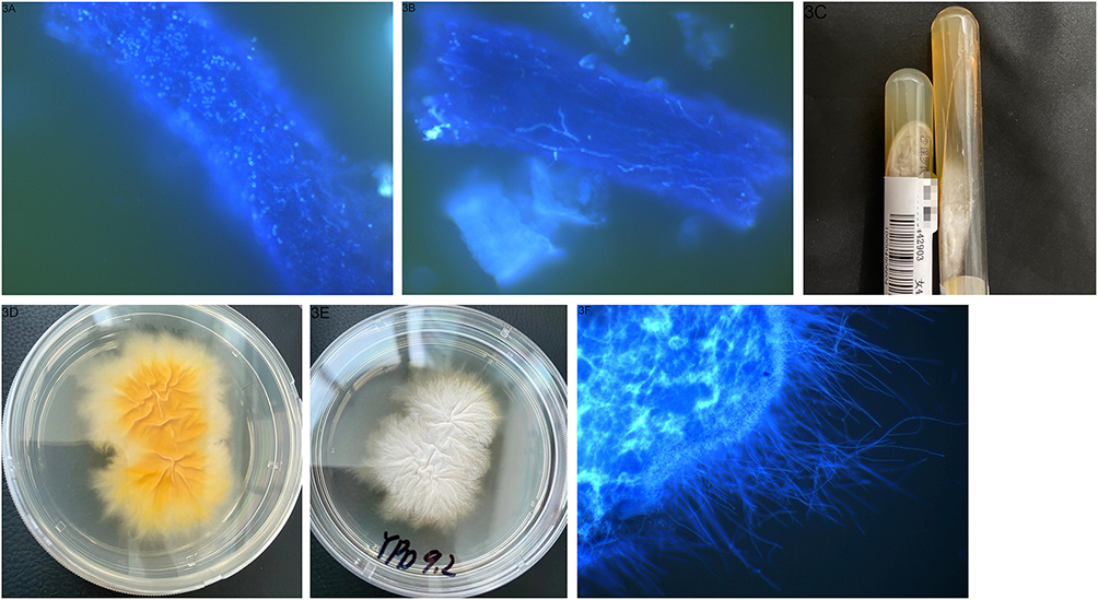 |
Figure 3 Images of diseased hair and fungal culture. Obvious spores (A) and hyphae (B) in the affected hair. The same mycelium as in the previous hair (C). Pathogenic fungi inoculated in Salmonella agar (D) and on YPD medium (E and F) give rise to woolly colonies. |
Mass Spectrometry Analysis
We identified the cultured fungus using matrix assisted laser desorption ionization time-of-flight mass spectrometry (MALDI-TOF MS), which was performed by China-MBT-IVD system in our laboratory under strict biosafety procedures. After ionisation of the fungal sample, the corresponding peak map was formed according to the mass charge ratio and signal value. The identification result was obtained by comparing the mass spectrogram with more than 5900 existing fingerprints in the database, which was confirmed to be Microsporum canis. LogScore values were automatically calculated against the entries in the internal libraries, ranging from 0 (no similarity) to 3 (perfect match). Our identification score for the sample fungi is greater than 1.7, which is an indication of reliability. The results again confirmed the fungus as Microsporum canis. The peak diagram of the mass spectrum is shown below (Figure 4).
 |
Figure 4 Mass spectra obtained from mass spectrometry analysis of fungal samples. By comparison of the mass spectrogram with more than 5900 fingerprints in the database, the result was confirmed to be Microsporum canis. The Abscissa is the value of the ion mass-charge ratio, and the ordinate represents the intensity of each peak. |
On the basis of the comprehensive findings, a definite diagnosis of tinea capitis due to Microsporum canis, carried by the domestic cat, was made. The parents of the girl were given detailed information about the cause of the disease, the course of treatment and what need to be done, in particular to keep their children away from pets. We recommend shaving the affected area and treating the child with oral antifungal medication in combination with topical application of ketoconazole. However, the mother of the patient expressed reluctance to accept oral medication and only agreed to receive topical ketoconazole treatment. Two weeks later, during a follow-up visit, it was observed that the original lesions showed no significant improvement, and new infection areas appeared on other parts of the scalp. Further inquiry revealed that the mother did not diligently apply the medication to the affected areas at home, and the limited efficacy of the topical treatment plan might also contribute to the situation. Considering the lack of griseofulvin, the preferred treatment for tinea capitis in our region, we chose to use locally common antifungal drug terbinafine orally and recommended daily use of 2% ketoconazole shampoo to clean the affected skin areas. The child showed some improvement during the second follow-up visit but was lost to follow-up thereafter. Despite our efforts to contact them, we were unable to ascertain the subsequent treatment progress.
Discussion
In recent years, the growing integration of pets into family life has correlated with an increases incidence of animal-associated ringworm like skin diseases.5 Microsporum canis is the most common extra epidermal fungus in tinea capitis in children and is highly infectious,2,6 invading the epidermal stratum corneum and keratinized tissues such as hair. It reproduces asexually by mitosis during hair/scale invasion and its production of large numbers of conidia can persist on the hair and keratin for months to years. Studies have shown that cats are the most important host.7 Although active infections in animals are rare, many animals can be carriers of cutaneous fungi. Although animals may not show clinical signs of infection, they may still be asymptomatic carriers.
Studies have shown that Microsporum canis can be transmitted to humans through contact with animal hosts, such as contaminants in toys, bowls, and garbage bins.8 For this reason, asymptomatic carriers of Microsporum canis are considered to be a key factor in the epidemiology of the disease. For children and teenagers, the more pets in the home, the greater the likelihood of superficial fungal infection. In adults, although their sebaceous glands are mature enough to secrete sebaceous long-chain fatty acids to protect against fungal colonization and infestation, their prevalence in immunocompromised populations and menopausal women should not be overlooked, with Microsporum canis accounting for 21.1% of the cases.1,9 Studies have also confirmed that 80% of cases indicate the possibility of a fingertip infection.10 Therefore, once the source of infection has been identified, it is recommended dermatologists, veterinarians and mycologists work together to treat patients and animals and to clean contaminated objects and areas.
Tinea capitis, a common fungal infection, predominantly affects children. A retrospective analysis of tinea capitis cases in Xinjiang, China, found that children aged 3–5 years accounted for 54% of patients, with 42.05% infected with Microsporum canis.11 The majority of these cases reported a history of animal contact, and a few reported close contact with individuals harbouring dermatophytes.
In this case report, the girl’s scalp showed symptoms of tinea capitis, with white fungal sheaths visible at the hair roots, but remarkably, her hair showed no signs of alopecia or hair breakage at the sheaths. Fungal microscopy revealed a large number of hyphae and mass spectrometry results quickly confirmed Microsporum canis infection. The results of the SEM revealed two characteristics of fungal hairs: 1) More spores along the hair shaft, no empty strands and premature breakage; 2) Dense spore clusters erode the cuticle, creating large defects with corresponding premature breakage sites, which is consistent with the observation under high-magnification dermoscopy.12 The girl did not show significant hair breakage, probably because the erosion of Microsporum canis had not yet reached the stage of hair breakage. In the early stages of infection, due to the low number of fungi, erosion of the hair cuticle did not spread to the interior of the hair and only raised fissures were formed. As the disease progresses, hair breakage and hair loss may occur.
As the lesions described in this report are scaly and do not show obvious alopecia, it needs to be distinguished from pityriasis capitis, which is characterised by thinner flakes that are less easy to remove. In addition, itching is usually absent in pityriasis capitis. While the fungal microscopic examination confirms tinea capitis.
Identifying the species of pathogens causing superficial fungal infections is crucial for effective treatment. Traditional methods such as fungal culture for dermatophyte identification have limited clinical utility due to their time-consuming nature and low sensitivity. In comparison to the complex and costly procedures involved in ITS sequencing, mass spectrometry analysis offers the advantage of rapid microbial identification at a lower cost, thereby greatly promoting its use as an alternative method for fungal diagnosis. Over the past two decades, as research on mass spectrometry for fungal identification has advanced, significant improvements and supplements have been made to fungal identification rates and reference spectral databases. Despite existing limitations, MALDI-TOF MS undoubtedly represents a significant technological advancement in the field of mycological diagnosis, with the potential for widespread application in clinical diagnostics.
In the treatment of tinea capitis, it’s important to recognise that the fungal infection occurs primarily at the root of the hair follicle. Local treatments are limited in their ability to effectively penetrate the site of infection. This makes them less effective. Oral medication to treat the disease comprehensively is therefore the standard approach.2 Topical medication can be used as a complementary measure to help prevent the growth and spread of the fungal infection. In addition, in cases where patients contract the disease through contact with feline companions, treatment protocols should include strategies aimed at source containment. These strategies may include isolation of infected cats, judicious trimming or shaving of hair in affected areas to reduce bacterial carriage, and the use of selenium disulphide shampoo, among other comprehensive therapeutic interventions. In our report, the girl’s parents refused to give the girl oral medication, and did not strictly isolate the girl from the sick cat. Despite two weeks of topical antifungal ointment, the lesions expanded.
In summary, there is a risk of infection for both in the home and in exposure situations where pets are adopted, and further precautions need to be taken and vigilance exercised regarding the likelihood of infection. Systematic and rapid clinical testing also provides the basis for accurate and effective treatment.
Consent to Participate
Written consent for publication of the case details together with imaging or videos have been obtained from the parents.
Disclosure
The authors report no conflicts of interest in this work.
References
1. Weitzman I, Summerbell RC. The dermatophytes. Clin Microbiol Rev. 1995;8(2):240–259. doi:10.1128/CMR.8.2.240
2. Chen X, Jiang X, Yang M, et al. Systemic antifungal therapy for tinea capitis in children. Cochrane Db Syst Rev. 2016;2016(5). doi:10.1002/14651858.CD004685.pub3
3. Moskaluk AE, VandeWoude S. Current topics in dermatophyte classification and clinical diagnosis. Pathogens. 2022;11(9):957. doi:10.3390/pathogens11090957
4. Baumbach CM, Müller S, Reuschel M, et al. Identification of zoophilic dermatophytes using MALDI-TOF mass spectrometry. Front Cell Infect Mi. 2021;11:631681. doi:10.3389/fcimb.2021.631681
5. Zeng J, Wang S, Guo L, et al. Pediatric tinea capitis in Jilin Province: analyzing previous results from a new perspective. Mycopathologia. 2023;188(5):515–522. doi:10.1007/s11046-023-00718-0
6. Chen XQ, Zheng DY, Xiao YY, et al. Aetiology of tinea capitis in China: a multicentre prospective study. Brit J Dermatol. 2022;186(4):705–712. doi:10.1111/bjd.20875
7. Pasquetti M, Min A, Scacchetti S, Dogliero A, Peano A. Infection by microsporum canis in paediatric patients: a veterinary perspective. Vet Sci. 2017;4(4):46. doi:10.3390/vetsci4030046
8. Moriello KA, Coyner K, Paterson S, Mignon B. Diagnosis and treatment of dermatophytosis in dogs and cats: clinical consensus guidelines of the world association for veterinary dermatology. Vet Dermatol. 2017;28(3):266. doi:10.1111/vde.12440
9. Liang G, Zheng X, Song G, et al. Adult tinea capitis in China: a retrospective analysis from 2000 to 2019. Mycoses. 2020;63(8):876–888. doi:10.1111/myc.13102
10. Grunwald MH, Amichai B, Shemer A. Fingertip contamination after a brief touch of tinea capitis lesions caused by Microsporum canis. Brit J Dermatol. 2015;172(1):291–292. doi:10.1111/bjd.13223
11. Wang X, Abuliezi R, Hasimu H, Zhang L, Abliz P. Retrospective analysis of tinea capitis in Xinjiang, China. Mycopathologia. 2023;188(5):523–529. doi:10.1007/s11046-022-00702-0
12. Lacarrubba F, Verzì AE, Micali G. Newly described features resulting from high-magnification dermoscopy of tinea capitis. JAMA Dermatol. 2015;151(3):308. doi:10.1001/jamadermatol.2014.3313
 © 2024 The Author(s). This work is published and licensed by Dove Medical Press Limited. The full terms of this license are available at https://www.dovepress.com/terms.php and incorporate the Creative Commons Attribution - Non Commercial (unported, v3.0) License.
By accessing the work you hereby accept the Terms. Non-commercial uses of the work are permitted without any further permission from Dove Medical Press Limited, provided the work is properly attributed. For permission for commercial use of this work, please see paragraphs 4.2 and 5 of our Terms.
© 2024 The Author(s). This work is published and licensed by Dove Medical Press Limited. The full terms of this license are available at https://www.dovepress.com/terms.php and incorporate the Creative Commons Attribution - Non Commercial (unported, v3.0) License.
By accessing the work you hereby accept the Terms. Non-commercial uses of the work are permitted without any further permission from Dove Medical Press Limited, provided the work is properly attributed. For permission for commercial use of this work, please see paragraphs 4.2 and 5 of our Terms.