")
Back to Journals » Journal of Experimental Pharmacology » Volume 16
The Effects of Potato (Solanum tuberosum L. vs. Granola; Solanaceae) Peel Extract Gel on Gingival Wound Healing in Wistar Rats
Authors Tiarasanti F, Sufiawati I , Amalia E, Sari KI, Zubaedah C, Takarini V
Received 3 November 2023
Accepted for publication 16 January 2024
Published 26 January 2024 Volume 2024:16 Pages 25—35
DOI https://doi.org/10.2147/JEP.S443355
Checked for plagiarism Yes
Review by Single anonymous peer review
Peer reviewer comments 4
Editor who approved publication: Professor R.D.K. Misra
Faradila Tiarasanti,1,* Irna Sufiawati,1,* Eri Amalia,2 Kartika Indah Sari,3 Cucu Zubaedah,4 Veni Takarini5
1Department of Oral Medicine, Faculty of Dentistry, Padjadjaran University, Bandung, Indonesia; 2Department of Pharmaceutics and Pharmaceutical Technology, Faculty of Pharmacy, Padjadjaran University, Bandung, Indonesia; 3Department of Oral Biology, Faculty of Dentistry, Padjadjaran University, Bandung, Indonesia; 4Department of Dental Public Health, Faculty of Dentistry, Padjadjaran University, Bandung, Indonesia; 5Department of Dental Material Science and Technology, Faculty of Dentistry, Padjadjaran University, Bandung, Indonesia
*These authors contributed equally to this work
Correspondence: Irna Sufiawati, Department of Oral Medicine, Faculty of Dentistry, Padjadjaran University, Jalan Sekeloa No. 1, Bandung, West Java, 40132, Indonesia, Tel +62-22-2504985, Email [email protected]
Purpose: Phenolic compounds with antioxidant, antimicrobial, and anti-inflammatory properties have been identified in potatoes (Solanum tuberosum L.; Solanaceae), which may potentially contribute to wound healing. The study aimed to evaluate the effect of potato peel extract gel Granola variety on oral gingival wound healing in Wistar Rats.
Methods: This research was a true experimental in vivo study, involving 30 male Wistar rats, aged 12– 14 weeks and weighing approximately 150 to 200 grams. Surgical vertical incisions, each 3 mm in length, were made on the mandibular gingiva. The test group consisted of six subgroups, each comprising 5 rats. The negative control group received a base gel, while treatment groups were given 1% povidone-iodine solution, 0.1% triamcinolone acetonide oral paste, and 2%, 4%, and 6% potato peel extract gel. Wound lengths were measured on days 3, 7, and 14 to evaluate the healing process. Statistical analysis used the ANOVA test, a p-value of < 0.05 was considered statistically significant.
Results: All experimental groups showed a reduction in wound length on days 3, 7, and 14. Notably, the application of 4% and 6% potato peel extract gel formulations facilitated to faster wound healing on day 3, surpassing the povidone-iodine and triamcinolone acetonide groups. However, by days 7 and 14, both the povidone-iodine group and the 6% formulation group demonstrated superior outcomes, although the differences in values were not statistically significant (p < 0.05).
Conclusion: Potato peel extract gel formulations at 4% and 6% concentrations were found to expedite the healing of incision wounds, showing no statistically significant difference from the povidone-iodine and triamcinolone acetonide groups. Therefore, potato peel extract gel holds excellent potential for development as an alternative medicine for natural and safe wound healing therapy.
Keywords: potato peel extract, gel, wound healing
Introduction
A wound is characterized as a disruption in the continuous layer of epithelial tissue in the skin or mucosa, often resulting from thermal or physical trauma.1 There are four stages in the process of tissue development and regeneration during wound healing: (i) the hemostasis phase, which immediately follows the injury; (ii) the inflammatory phase, marked by swelling and initiated upon tissue damage; (iii) the proliferation phase, in which new tissue and blood vessels form; and (iv) the maturation phase, during which tissue remodeling takes place.2 Various factors can impede the natural course of wound healing, including factors such as repeated injury, infection, insufficient oxygenation, and the generation of free radicals.3
Various alternatives drugs are used in wound care, such as povidone-iodine and corticosteroids. Povidone-iodine (PVP-I) is widely used, known for its efficacy in penetrating bacterial biofilms, anti-inflammatory properties, and minimal cytotoxicity. It possesses a broad-spectrum antibacterial action and does not impede wound healing.4 While generally safe, concerns about allergies, limited penetration, and host cell toxicity have been raised in some reports.5 Among steroid medications, triamcinolone acetonide is favored for its low potency, making it effective for topical application and tissue penetration. However, long-term use of corticosteroids can lead to local and systemic side effects.6,7 Given these challenges, exploring alternative approaches is imperative. The use of pharmaceuticals or natural ingredients with antibacterial, antioxidant, and anti-inflammatory properties holds promise for enhancing wound healing.8 Additionally, phytoconstituents like flavonoids and polyphenols, known for their antioxidant, anti-inflammatory, and antibacterial effects, contribute to wound healing with minimal side effects, making them safe for consumption.9
Potato (Solanum tuberosum L.), a member of the Solanaceae family, is abundant in phenolic compounds that serve as natural defenses against various pathogens, including bacteria, fungi, and viruses. In Indonesia, potatoes are a readily available natural resource and rank as the third most consumed food crop globally, especially in Asia, Africa, and Latin America.10 Phenolic compounds in potatoes, such as phenolic acids and flavonoids, are recognized for their anti-inflammatory, analgesic, and antioxidant properties.3,11 Additionally, potatoes contain anthocyanins, vitamin B3, pantothenic acid, potassium, manganese, phosphorus, copper, and fiber.12 Potato peel, often discarded in processing, emerges as a potential new source of antioxidants, harboring various intriguing substances like flavonoids, phenolic acids, chlorogenic, cinnamic, and ferulic acids with antioxidant and antibacterial properties.13 The exploration of potato by-products, such as peels, warrants further research.
Previous research has demonstrated the efficacy of topically applying phenolic compounds and anthocyanins in treating oral wounds in rats. Potatoes, recognized for their abundant content of these compounds, emerge as a promising alternative therapy for oral wounds, capable of independently or synergistically promoting wound healing activities.14 Additionally, due to its antibacterial properties, S. tuberosum has been utilized as a treatment for mild acne, harnessing its potential to enhance wound healing.12 Proper wound care not only accelerates wound healing but also fosters the formation of new, healthy tissue. Natural ingredients like potato (Solanum tuberosum L.) can play a vital role in wound treatment.15
Potatoes peel contain a substantial amount of anthocyanins, as much as 0.65g/kg fresh weight, providing them antioxidant, antibacterial, and anti-inflammatory abilities.16 Due to its antibacterial, anti-inflammatory, and antioxidant properties, Solanum tuberosum ointment has been shown in previous research to hasten wound healing on rat skin, contributing to optimal healing outcome.3 Studies have revealed that these anthocyanin antioxidants can effectively eliminate free radicals, reduce inflammation stimulation, decrease the release of inflammatory factors, inhibit the activation of signal pathways associated with inflammation, promote the production of anti-inflammatory factors, and effectively diminish the inflammatory response.17
This study aimed to determine the effect of the gel prepared with ethanolic extract of the potato (Solanum tuberosum L. vs. Granola; Solanaceae) peel on gingival wound healing in Wistar rats.
Materials and Methods
All experimental procedures and protocols conformed to the International Guiding Principles for Biomedical Research Involving Animal. Ethical approval was obtained from the Research Ethics Commission of Universitas Padjadjaran No. 43/UN6.KEP/EC/2023.
Plant Determination and Preparation of Potato Peel Extract
Potatoes (Solanum tuberosum L.) were obtained from Pangalengan District, Bandung Regency, West Java, Indonesia, harvested in June 2023 and determined by Generasi Biologi Indonesia, with the following determination results:
Regnum: Plantae
Division: Magnoliophyta
Class: Magnoliopsida
Order: Solanales
Family: Solanaceae
Genus: Solanum
Species: Solanum tuberosum L.
Potato peel extract was made from 447.03 grams of potato peel, which had been washed and then dried by placing it in the oven at 40°C for 24 hours. Then, the dried potato peel sample was ground using a blender until 62.04 grams of potato peel powder was obtained in small size. The potato peel powder was then extracted using the soxhletation method, which was diluted with 1 liter of 96% ethanol solvent in a flask and then heated until it boiled and evaporated. The 96% ethanol extract of potato peel was then concentrated using a water bath, and the evaporated extract weighed 10.855 grams.18
Phytochemical Screening
A phytochemical screening examination was carried out on the extract, including; (1) Alkaloid test, the test tube was filled with 1 gram of extract and reacted with Dragendorff's reagent. The formation of a precipitate is an indication of the presence of alkaloids; (2) Flavonoid test, namely a test tube filled with 1 gram of extract, reacted with 2–3 drops of 10% NaOH solution, the color changes to yellow, indicating the presence of flavonoids; (3) Terpenoid test, the test tube is filled with 1 gram of extract dissolved in chloroform then one drop of anhydrous acetic acid and one drop of concentrated sulfuric acid are added. A positive reaction is indicated by the formation of a red-green or purple-blue solution; (4) Tannin test, namely by treating 1 gram of extract with 2–3 drops of 1% FeCl3, the sample is positive if a blackish green color form which indicates the presence of condensed tannin. (5) Saponin Test, test tube filled with 1 gram of extract. Aqua destilata (aquadest)/distilled water, was added to the tube and shaken vertically for 1 minute. The sample is positive if foam forms with much intensity.19
Formulation of the Potato Peel Extract Gel and Evaluation
The potato gel formulation involved the combination of carbomer 940 was developed with aquadest in a glass beaker and gradually added to TEA until a gel formed. Methyl paraben and propyl paraben were mixed in a separate beaker while heating until achieving homogeneity. The blend of methyl paraben and propyl paraben was then incorporated into the previously made gel base solution. Potato peel extract was dissolved in glycerin at varying concentrations until homogeneous, then added to the gel base solution and stirred. The remaining aquadest was added until achieving homogeneity. The concentration was determined from previous research on potatoes as an ointment for wound healing that used potato flesh and potato peel together in a 1:1 ratio and the test groups were divided into groups with 1% ointment concentration and groups with 2% ointment concentration. In this study, the concentration was modified by considering using pure potato peel, so the concentration was doubled compared to the previous study. The resulting gel, named formula 1 (F1), contains a 2% concentration of potato peel extract, while formula 2 (F2) contains 4%, and formula 3 (F3) contains 6% (Table 1). The gel preparation of with different concentration was then evaluated.20–22
 |
Table 1 Formulation of Potato Peel Extract Gel |
Animal Preparation and Experimental Design
Thirty (30) male white rats (Rattus norvegicus) of the Wistar strain that were wounded on the gingival are required, which were obtained from Bandung, West Java. Animals were randomly selected with inclusion criteria: weighing 150–200 grams, 12–14 weeks old, and in a healthy condition characterized by active movement. The groups of test animals were divided into six, with each group consisting of 5 Wistar rats. The group divisions consisted of group 1 serving as the negative control, receiving only the gel base. In group 2, wounds were treated with a 1% povidone-iodine solution. Group 3 involved the application of 0.1% triamcinolone acetonide oral paste to the wounds. Wounds in group 4 were treated with a 2% concentration of potato peel extract gel, while those in group 5 received a potato peel extract gel with a concentration of 4%. Lastly, group 6 had wounds treated with 6% potato peel extract gel.
Gingival Wound Healing Evaluation in Wistar Rats
The experimental animals were first acclimatized for seven days in plastic cages. Experimental animals that had been acclimatized were then anesthetized using ketamine at a dose of 20 mg/kg intramuscularly in the upper thigh. The experimental animal was then given a wound as an incision with a length of 3 mm and a depth reaching the alveolar bone in the lower jaw using blade no. 11, which was sterilized using 70% alcohol. The gel was then administered twice daily to each group, as decided by directly applying up to 0.1g of it to the wound using cotton buds.
Observations of incision wound healing were carried out on days 0, 3, 7, and 14. The length of the wound was measured vertically, with the reference point being the farthest point from each side. Measurements were carried out using a measurement instrument with the accuracy of hash marks is 1 mm. Wistar rats were treated by being given food with a soft consistency and drinking water to reduce pain and discomfort during treatment. The data obtained was then analyzed statistically using the One-way analysis of variance (ANOVA) test, then followed by the least significant difference (LSD) test, with a significant value p<0.05.
Results
Based on the results of phytochemical tests conducted at the Central Laboratory of Padjadjaran University, it was determined that potato peel extract contains tannins, flavonoids, saponins, terpenoids, and alkaloids (Table 2). The tannin test demonstrated a color change to blackish green, indicating the presence of condensed tannin. In the flavonoid test, positive results were observed with a color change to brown. The saponin test revealed positive results characterized by the appearance of foam when the solution was heated and shaken vigorously. Positive outcomes in the triterpenoid (terpenoid) test were indicated by a color change to brown. Additionally, the alkaloid test results were declared positive after treating a sample of potato peel extract with Dragendorff’s reagent, resulting in a red precipitate. Table 3 showed the results of the evaluation of the formulation of the potato gel with different concentration.
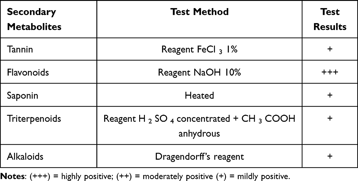 |
Table 2 Phytochemical Screening of Potato Peel Extract |
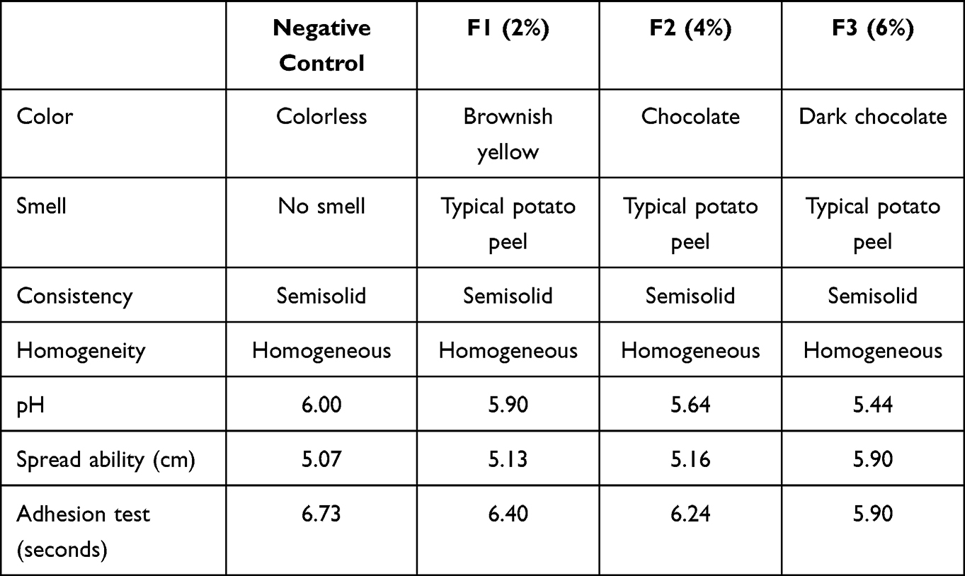 |
Table 3 Evaluation Results of Potato Peel Extract Gel Preparations |
Figure 1 illustrates that, on day 0, the average length of incision wound healing on the mandibular gingiva of Wistar rats was 3 mm. In contrast to normal gingival conditions, during the early stages of treatment, bleeding still occurred, and the wound entered the inflammatory phase.
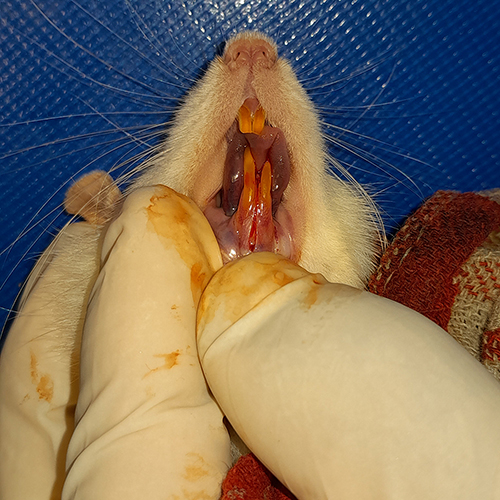 |
Figure 1 The gingival of Wistar rats after incision. |
On the third day after applying the gel to the gingiva of Wistar rats, measurements indicated a change in the length of the wound in each group. The group treated with 6% potato peel extract gel exhibited a more significant reduction in wound length, with an average of 2.892 ± 0.071 mm, compared to the negative control group (average of 3.260 ± 0.397 mm), the povidone-iodine group (average of 3.094 ± 0.243 mm), and the triamcinolone acetonide group (average of 3.14 ± 0.260 mm).
Figure 2 showed that the increase in wound length in the control and treatment groups was caused by swelling in the wound area accompanied by pus with reddish wound edges. The reddish color in the potato peel extract gel treatment group was still within normal limits compared to the negative control group. Compared with measurements on day three, the length of the incision wound appeared to be smaller on day seven after incision. The wounds in the povidone-iodine group and the group that was given potato peel extract gel application had a more significant percentage of wound shrinkage, inversely proportional to the negative control group, which became increasingly swollen (Figure 3). On visual observation, the wound area in each treatment group still had swelling and pus. On the 14th day, long wound closure was marked by the formation of new tissue around the wound area, as seen in the picture. Measurements of the length of the incision observed on days 0, 7, and 14 on each gingiva of Wistar rats are tabulated in Table 4.
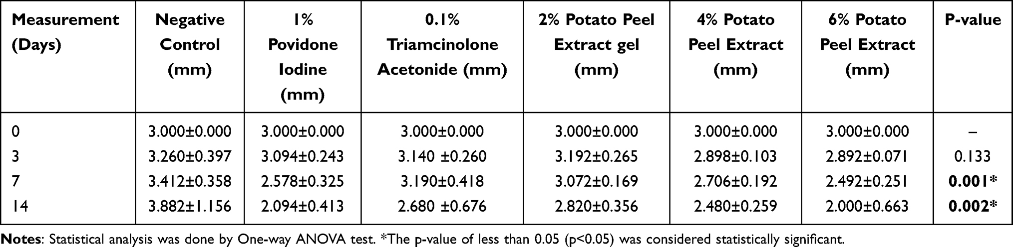 |
Table 4 The Results of Measuring Wound Lengths for All Groups on Days 0, 3, 7, and 14 |
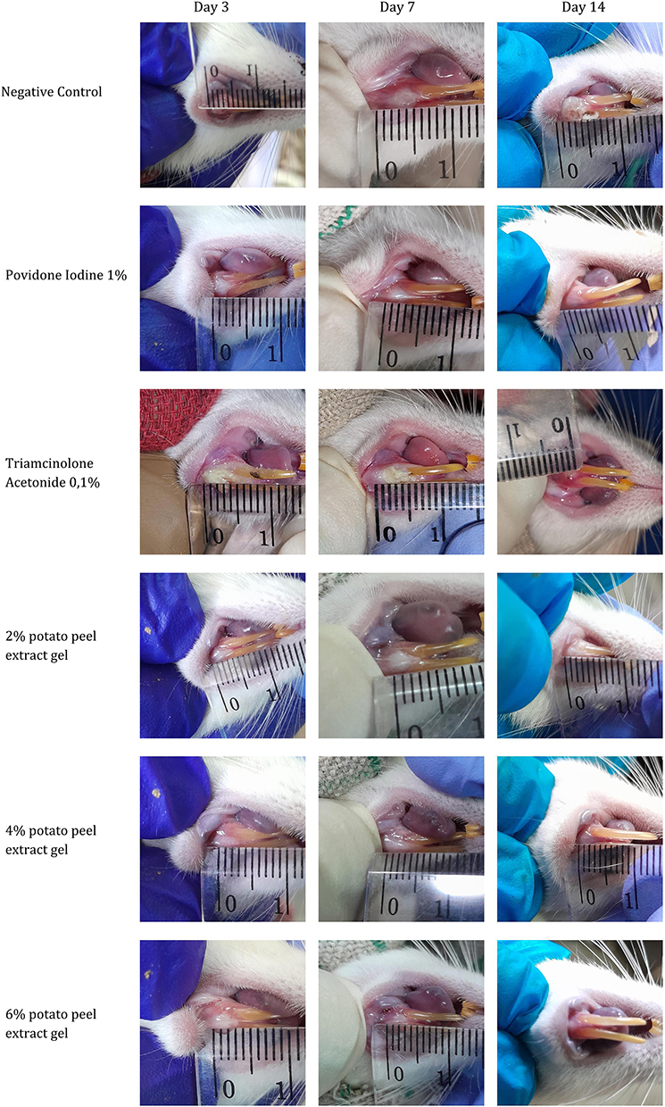 |
Figure 2 Comparison of gingival wound healing between Wistar rats treated with potato peel extract and the control groups. |
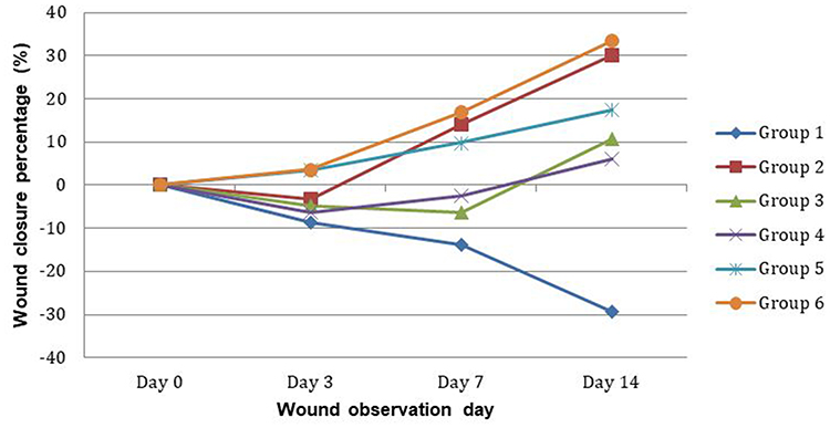 |
Figure 3 Evaluation of wound closure in the experimental groups. Group 1: Negative control; Group 2: 1% povidone-iodine solution; Group 3: 1% triamcinolone acetonide oral paste; Group 4: 2% potato peel extract gel; Group 5: 4% potato peel extract gel; Group 6: 6% potato peel extract gel. |
From the results of statistical tests measuring wound length on day 0, homogeneous data was obtained in all groups, namely wounds with an incision length of 3.00 ± 0.00 mm. There is still bleeding in the wound and it will enter the inflammatory phase because it is still in the early stages of wound healing. Measurements on the third day obtained data with a p value = 0.133 (p value <0.05), which means there was no significant difference between the length of wound healing variables in the treatment groups. In contrast to measurements on the 7th and 14th days, the respective values obtained were p=<0.001 and p=0.002, which means that statistically, there is an average between the wound healing variables on the seventh day with the most significant difference occurring on negative control group with povidone-iodine, negative control with 4% potato peel extract formulation and negative control with 6% potato peel extract formulation (Table 5). Meanwhile, on the 14th day, the most significant value occurred in the negative control group with povidone-iodine and the negative control with 6% potato peel extract formulation, followed by a significant value of p=0.003 in the negative control group with the 4% formulation group, then p=0.008 on comparison of the negative control group with triamcinolone acetonide and p=0.018 in the comparison of the negative control with the 2% formulation group. Therefore, it can be concluded that on the 14th day, there was a significant difference between the negative control group with povidone-iodine, the negative control with triamcinolone acetonide and the negative control with the treatment group. However, there was no significant difference between the povidone-iodine group and the treatment group and the triamcinolone acetonide group with the treatment group.
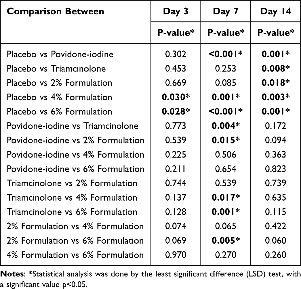 |
Table 5 Comparison of Wound Length Between Treatment Groups |
Discussion
The potato peels from the Solanum genus, known for potent antioxidant activities, due to the concentration of about 50% of phenolic compounds in the peel and surrounding tissues, and diminishing towards the center of the tuber.23 Our study focused on extracting the phenolic compounds of potato peel of Solanum tuberosum L. vs. Granola where the dried peel was grinded to minimize the particle size of powder and was extracted by soxhletation method for obtaining higher yield of extracts.
Observations were conducted on days 0, 3, 7, and 14, focusing on the phases of the wound healing process, namely the hemostasis phase that occurs immediately after the wound is formed, the inflammatory phase, the proliferation phase, and the remodeling/maturation phase. On day 0, there was a homogeneous value, namely 3.00 ± 0.00 mm, and no shrinkage was found in the wound. The severed blood vessels experience constriction and retraction in this phase, followed by the hemostasis phase. The narrowing of blood vessels and formation of fibrin clots mark the beginning of the initial stages of hemostasis.2 Neutrophils, macrophages, mast cells, endothelial cells, and platelets respond chemotactically to cytokines produced and activated by hemostasis components.24 During hemostasis, blood vessels contract to stop bleeding, platelets form a plug strengthened by fibrin polymerization to create a clot and close the wound, and vessels dilate to prevent further bleeding. Fibro-fibronectin clumps act as a temporary ECM matrix and support, allowing fibroblasts and epithelial cells to move to the wound site.25 Collagen, synthesized by fibroblasts, establishes a conducive environment for blood clotting, forming a protective barrier.26 After the initial hemostasis phase, the wound undergoes immediate inflammatory infiltration, triggered by chemokines produced at the incision site. In the early phase of inflammation, there is rapid growth of local fibroblasts in the wound bed.23
On the third day of observation, wounds in the group treated with 1% povidone-iodine exhibited a pale gingival color. A similar study was conducted by Danarti et al, using a fibroblast populated collagen (FPCL) model to test the toxicity of povidone-iodine on human fibroblast cultures. Their study revealed that applying povidone-iodine at concentrations of 0.1, 0.01, 0.001, and 0.0001% significantly reduces fibroblast contraction in humans. Moreover, the application of 1% povidone-iodine can lead to persistent fibroblast damage.26 Fibroblasts have an essential role in the angiogenesis process. Fibroblasts can synthesize various angiogenic factors, including vascular endothelial growth factor (VEGF). These factors promote the migration and proliferation of endothelial cells that are critical for blood vessel development.27 The incision wounds in the group treated with 1% povidone-iodine on the third day of observation showed a wound appearance with the gingiva looking pale, indicating disruption of the angiogenesis process in the wound due to the toxicity of 1% povidone-iodine to fibroblast cells. However, because povidone-iodine is an antiseptic that works as a debridement agent and inhibits microbial contamination of wound tissue, wound closure in the group applied with 1% povidone-iodine was faster compared to the negative control group.
In previous studies involving acute wounds on rat skin, povidone-iodine could accelerate wound healing by increasing the expression of transforming growth factors, neovascularization and re-capitalization.28 This acceleration speeds up the wound’s inflammatory phase, facilitating the prompt start of the subsequent phase.29
The triamcinolone acetonide group exhibited the slowest average healing on days 3 and 7, with significant average healing values compared to the groups using triamcinolone acetonide with povidone-iodine, 4% potato peel extract gel formulation, and 6% potato peel extract gel formulation. However, there was an improvement in wound healing observed on the 14th day. Similar to other corticosteroids, triamcinolone acetonide possesses anti-inflammatory, anti-pruritic, and vasoconstrictive properties. The mechanism through which topical steroids like triamcinolone acetonide reduce inflammation is not entirely clear. It is believed that corticosteroids induce the production of lipocortin, an inhibitory protein for phospholipase A2. Lipocortin prevents the formation of arachidonic acid, a precursor to potent inflammatory mediators like prostaglandins and leukotrienes, which are typically derived from membrane phospholipids released by phospholipase A2.
The application of 2%, 4%, and 6% potato peel extract gel resulted in a reduction in wound length during measurement. This effect is attributed to the inflammatory phase where fibroblasts produce collagen, forming fibers that cover the wound with fibrin fibers. The potato peel extract gel, containing bioactive components like anthocyanins, glycoalkaloids, saponins, phenolic compounds, and flavonoids, contributes to this process.30 Due to its antioxidant and antibacterial properties, the phenolics and flavonoids have been shown to improve the wound healing process by increasing wound contraction and increasing the rate of epithelization. Moreover, these bioactive components have proven effective in promoting wound healing by enhancing oxygen diffusion, reducing free radical production, and increasing collagen synthesis.31,32 Saponins, classified as antibacterial, disrupt bacterial cell membranes, causing damage and releasing essential components such as proteins, nucleic acids, and nucleotides. This destabilization induces cell hemolysis, effectively killing bacteria in the wound and promoting the healing process.33
On the seventh day of observation, the wound size decreased. This reduction is linked to an increase in the extracellular matrix, growth factors, and angiogenesis during the proliferative phase. On day 14, all groups, especially the negative control group, displayed redness, indicating the ongoing activity of angiogenesis in the proliferation phase. The group treated with 6% potato peel extract gel showed the highest closure, followed by the control group receiving 1% povidone-iodine, the 4% potato peel extract gel group, and the triamcinolone-treated group. This highlights the wound-healing capability of potato peel extract gel, attributed to its bioactive content, including alkaloid, saponins, phenolic compounds, and flavonoids as identified in our screening test evaluation.
While indicating a promising effect of potato peel extract on the wound healing process, this study has limitations. Firstly, the study lacks on various wound healing variables, such as histopathological analysis for re-epithelialization, angiogenesis, and inflammatory cells. Molecular evidence for the potato peel extract’s action has also not been investigated yet, highlighting the necessity for further studies to explore its molecular mechanisms comprehensively. Secondly, the study was conducted in an animal model, lacking direct clinical evidence for humans and raising concerns about healing dynamics and skin structure differences. Careful consideration of species variability and metabolic pathway differences is also essential when translating results from rats to humans. Further research is needed to explore the effects of potato peel extract, considering adjustments in dosing frequency due to metabolic rate and pharmacokinetic differences.
Conclusion
Potato peel extract gel at concentrations of 4% and 6% were able to accelerate wound healing, showing no significant difference compared to single treatment of povidone-iodine 1% and triamcinolone acetonide 0.1%. Notably, the application of 6% potato peel extract gel led to a smaller average wound length on the 14th day. Therefore, the 4% and 6% formulations of potato peel extract gel could potentially serve as alternative medicines for the wound healing.
Acknowledgments
The authors thank all members of the staff of the Central Laboratory Padjadjaran University for their technical assistance. This research was supported by the Academic Leadership Grant Universitas Padjadjaran, with grant number 1549/UN6.3.1/PT.00/2023.
Disclosure
The authors report no conflicts of interest in this work.
References
1. Silva-Correa CR, Ortiz-Noriega CM, La Torre VEV, et al. Effect of a Gel Based on Ipomoea batatas (Purple Sweet Potato) on dermal wound healing in mice. Pharmacogn J. 2021;13(6):1720–1726. doi:10.5530/pj.2021.13.222
2. Borbolla-Jiménez FV, Peña-Corona SI, Farah SJ, et al. Films for wound healing fabricated using a solvent casting technique. Pharmaceutics. 2023;15(7):1–27. doi:10.3390/pharmaceutics15071914
3. Rosas-Cruz GP, Silva-Correa CR, Calderón-Peña AA, et al. Wound healing activity of an ointment from Solanum tuberosum L. “Tumbay Yellow Potato” on Mus musculus Balb/c. Pharmacogn J. 2020;12(6):1268–1275. doi:10.5530/pj.2020.12.175
4. Bigliardi PL, Alsagoff SAL, El-Kafrawi HY, Pyon JK, Wa CTC, Villa MA. Povidone iodine in wound healing: a review of current concepts and practices. Int J Surg. 2017;44:260–268. doi:10.1016/j.ijsu.2017.06.073
5. Bigliardi P, Langer S, Cruz JJ, Kim SW, Nair H, Srisawasdi G. An Asian perspective on povidone iodine in wound healing. Dermatology. 2017;233(2–3):223–233. doi:10.1159/000479150
6. Devaraj NK, Rashid AA, Manap AHA, Nasir S. Topical corticosteroids in clinical practice. Med J Malaysia. 2019;74(2):187–189.
7. Abbasi F, Rasoulzadeh Z, Yavari A. The effect of sage (Salvizan gel) compared to triamcinolone acetonide on the treatment of recurrent aphthous stomatitis: a double-blinded randomized clinical trial. BMC Oral Health. 2023;23(1):1–5. doi:10.1186/s12903-023-02861-y
8. Rahman N, Rahman H, Haris M, Mahmood R. Wound healing potentials of Thevetia peruviana: antioxidants and inflammatory markers criteria. J Tradit Complement Med. 2017;7(4):519–525. doi:10.1016/j.jtcme.2017.01.005
9. Hajialyani M, Tewari D, Sobarzo-Sánchez E, Nabavi SM, Farzaei MH, Abdollahi M. Natural product-based nanomedicines for wound healing purposes: therapeutic targets and drug delivery systems. Int J Nanomed. 2018;13:5023–5043. doi:10.2147/IJN.S174072
10. Shin JS, Lee KG, Lee HH, et al. α-Solanine Isolated From Solanum Tuberosum L. cv Jayoung Abrogates LPS-induced inflammatory responses via NF-κB inactivation in RAW 264.7 macrophages and endotoxin-induced shock model in mice. J Cell Biochem. 2016;13(10):2327–2339. doi:10.1002/jcb.25530
11. Akyol H, Riciputi Y, Capanoglu E, Caboni MF, Verardo V. Phenolic compounds in the potato and its byproducts: an overview. Int J Mol Sci. 2016;17(6):835. doi:10.3390/ijms17060835
12. Siti Silfi Ambarwati N, Omar H. Topical herbal therapy with Solanum tuberosum L. to Combat Acne. KnE Soc Sci. 2019;3(12):180.
13. El-Gindy YM, Hafsa SHA, Dosoky WM. Effects of potato peel extract on semen quality, sex hormones and immunity of rabbit bucks under intensive breeding system. Andrologia. 2020;52(11):e13869. doi:10.1111/and.13869
14. Demilew W, Adinew GM, Asrade S. Evaluation of the wound healing activity of the crude extract of leaves of Acanthus polystachyus Delile (Acanthaceae). Evid Based Complement Alternat Med. 2018;2018:2047896. doi:10.1155/2018/2047896
15. Sinno H, Prakash S. Complements and the wound healing cascade: an updated review. Plast Surg Int. 2013;2013:1–7.
16. Raigond P, Singh B, Dutt S, Chakrabarti SK. Potato: Nutrition and Food Security. Springer Nature. 2020:1–287.
17. Ma Z, Du B, Li J, Yang Y, Zhu F. An insight into anti-inflammatory activities and inflammation related diseases of anthocyanins: a review of both in vivo and in vitro investigations. Int J Mol Sci. 2021;22(20):11076. doi:10.3390/ijms222011076
18. Widiyastuti Y, Matoha HK, Fitriana F. Antibacterial activity of melaleuca alternifolia extract from different extraction method. Biosaintifika. 2022;14(2):285–292.
19. Herawati E, Ramadhan R, Ariyani F, et al. Phytochemical screening and antioxidant activity of wild mushrooms growing in tropical regions. Biodiversitas. 2021;22(11):4716–4721. doi:10.13057/biodiv/d221102
20. Bhinge SD, Bhutkar MA, Randive DS, et al. Formulation development and evaluation of antimicrobial polyherbal gel. Ann Pharm Fr. 2017;75(5):349–358. doi:10.1016/j.pharma.2017.04.006
21. Chauhan P. Formulation and evaluation of antibacterial gel containing ethanol extract of thorns of Bombax ceiba. Int J Drug Deliv Technol. 2022;12(3):1015–1019. doi:10.25258/ijddt.12.3.16
22. Safitri FI, Nawangsari D, Febrina D. Overview: application of Carbopol 940 in Gel.
23. DesJardins-Park HE, Mascharak S, Chinta MS, Wan DC, Longaker MT. The spectrum of scarring in craniofacial wound repair. Front Physiol. 2019;10:322. doi:10.3389/fphys.2019.00322
24. Oki AS, Bimarahmanda ME, Rahardjo MB. Increased number of fibroblasts and neovascularization after tooth extraction in Wistar rats with moderate-intensity continuous exercise. J Int Dent Med Res. 2018;11(3):840–845.
25. Toma AI, Fuller JM, Willett NJ, Goudy SL. Oral wound healing models and emerging regenerative therapies. Transl Res. 2021;236(404):17–34. doi:10.1016/j.trsl.2021.06.003
26. Danarti R, Suwardana BA, Wirohadidjojo W. The effect povidone-iodine on the wound healing process: a study on fibroblast populated collagen lattice (FPCL) model. J Thee Med Sci. 2014;46(3):103–107.
27. Frangogiannis NG. Fact and fiction about fibroblast to endothelium conversion: semantics and substance of cellular identity. Circulation. 2020;142(17):1663–1666. doi:10.1161/CIRCULATIONAHA.120.050875
28. Wang L, Qin W, Zhou Y, et al. Transforming growth factor β plays an important role in enhancing wound healing by topical application of Povidone-iodine. Sci Rep. 2017;7(1):991. doi:10.1038/s41598-017-01116-5
29. Alves PJ, Barreto RT, Barrois BM, Gryson LG, Meaume S, Monstrey SJ. Update on the role of antiseptics in the management of chronic wounds with critical colonisation and/or biofilm. Int Wound J. 2021;18(3):342–358. doi:10.1111/iwj.13537
30. Jimenez-Champi D, Romero-Orejon FL, Moran-Reyes A, Muñoz AM, Ramos-Escudero F. Bioactive compounds in potato peels, extraction methods, and their applications in the food industry: a review. CyTA J Food. 2023;21(1):418–432. doi:10.1080/19476337.2023.2213746
31. Paswan SK, Srivastava S, Rao CV. Wound healing activity of ethanolic extract of Selaginella bryopteris on rats. Pharmacogn J. 2020;12(2):335–341. doi:10.5530/pj.2020.12.53
32. Aslam MS, Ahmad MS, Mamat AS, Ahmad MZ, Salam F. Antioxidant and wound healing activity of polyherbal fractions of Clinacanthus nutans and Elephantopus scaber. Evid Based Complement Alternat Med. 2016;2016:4685246. doi:10.1155/2016/4685246
33. Khan MI, Ahhmed A, Shin JH, Baek JS, Kim MY, Kim JD. Green tea seed isolated saponins exerts antibacterial effects against various strains of gram positive and gram negative bacteria, a comprehensive study in vitro and in vivo. Evid Based Complement Alternat Med. 2018;2018:3486106. doi:10.1155/2018/3486106
 © 2024 The Author(s). This work is published and licensed by Dove Medical Press Limited. The full terms of this license are available at https://www.dovepress.com/terms.php and incorporate the Creative Commons Attribution - Non Commercial (unported, v3.0) License.
By accessing the work you hereby accept the Terms. Non-commercial uses of the work are permitted without any further permission from Dove Medical Press Limited, provided the work is properly attributed. For permission for commercial use of this work, please see paragraphs 4.2 and 5 of our Terms.
© 2024 The Author(s). This work is published and licensed by Dove Medical Press Limited. The full terms of this license are available at https://www.dovepress.com/terms.php and incorporate the Creative Commons Attribution - Non Commercial (unported, v3.0) License.
By accessing the work you hereby accept the Terms. Non-commercial uses of the work are permitted without any further permission from Dove Medical Press Limited, provided the work is properly attributed. For permission for commercial use of this work, please see paragraphs 4.2 and 5 of our Terms.