")
Back to Journals » Research and Reports in Neonatology » Volume 13
Neonatal Hydrometrocolpos Secondary to Imperforate Hymen Presented with Acute Urinary Retention: Case Report
Authors Tegene D, Assefa T, Edris A
Received 3 September 2023
Accepted for publication 30 October 2023
Published 2 November 2023 Volume 2023:13 Pages 23—28
DOI https://doi.org/10.2147/RRN.S433727
Checked for plagiarism Yes
Review by Single anonymous peer review
Peer reviewer comments 2
Editor who approved publication: Dr Robert Schelonka
Dereje Tegene, Tesfalem Assefa, Abdurahman Edris
Department of Obstetrics and Gynecology, Adama Hospital Medical College, Adama, Ethiopia
Correspondence: Dereje Tegene, Tel +251913909665, Email [email protected]
Background: Hydrometrocolpos is a rare clinical condition characterized by the accumulation of fluid or mucus secretions in the vagina and uterus due to distal vaginal obstruction. Imperforate hymen is the most common cause of congenital hydrometrocolpos. The diagnosis of congenital hydrometrocolpos can be made prenatally or postnatally. Imperforate hymen is generally managed by hymenotomy or hymenectomy.
Case Presentation: This is a 26-day-old female neonate who was born from a 29-year-old Para − 3 mother after 9 months of amenorrhea. ANC follow-up was uneventful except her obstetric ultrasound scan at 36+3 weeks found a fetal lower intra-abdominal mass. Without proper postnatal evaluation, the neonate was discharged home with the mother after 12 hours of vaginal delivery. Currently, the neonate was referred to our hospital with the diagnosis of Acute Urinary retention, 2o Hydrometrocolpos, after having frequent crying, abdominal distension, and failure to pass urine of 2 days duration. On physical examination, there was a tender, cystic, and mobile mass palpable in the lower abdomen, and up to opening the labia there was a curved membrane covering the vaginal opening. Abdomino-pelvic ultrasound and Magnetic Resonance Imaging confirmed the hydrometrocolpos and imperforate hymen. After obtaining informed consent from the family, a vertical incision and annular hymenotomy were done to drain 200mL of milky fluid. A follow-up ultrasound was done to confirm the resolution of HMC and neonate discharge on the 4th day with improvement.
Conclusion: A high index of suspicion and immediate postnatal evaluation of neonates presented with prenatal U/S showing intra-abdominal mass will result in early diagnosis and treatment of neonatal hydrometrocolpos before the development of complications.
Keywords: Adama, imperforate hymen, neonatal hydrometrocolpos
Introduction
Hydrometrocolpos is a rare clinical condition characterized by accumulation of fluid or mucus secretions in the vagina and uterus due to distal vaginal obstruction.1 The reported incidence of neonatal hydrometrocolpos is around 0.006%.2 Hydrometrocolpos (HMC) is caused by distal vaginal obstruction due to congenital urogenital anomalies,3 or acquired etiologies such as infection, trauma, or sexual abuse.4,5 Imperforate hymen is the most common cause of congenital hydrometrocolpos.2 Female fetuses or neonates with an imperforate hymen leading to hydrocolpos or hydrometrocolpos caused by maternal estrogen may incidentally discover during the antenatal period on obstetrics ultrasound or postnatally present with a variety of clinical manifestations.1,2,6 Early diagnosis of hydrometrocolpos reduces the risk of developing complications,2 such as urinary tract obstruction, renal failure, recurrent UTI, rupture of HMC, peritonitis, and sepsis.1,2,7 Imperforate hymen is generally managed by hymenotomy (surgical incision of the hymen) or hymenectomy (surgical removal of the hymen).5
We reported a 26 -days-old female neonate who was referred to our hospital with the diagnosis of Acute Urinary retention 2o hydrometrocolpos, after having frequent crying, abdominal distension, vomiting of ingested matter, and failure to pass urine of 2 days duration. It emphasizes the need to have a high index of suspicion and immediate postnatal evaluation of neonates with prenatal ultrasound diagnosis of intra-abdominal mass for earlier diagnosis and treatment of neonatal hydrometrocolpos before the occurrence of complications. Relevant literature was also reviewed.
Case Presentation
This is a 26-day-old female neonate who was born from a 29-year-old Para −3 mother after 9 months of amenorrhea. The mother had an ANC follow-up at a share hospital in Batu town, and on her 3rd visit at 36+3 weeks of gestation, the obstetrician informed her that her fetus had a lower abdominal mass measuring 4.53*4.77cm in size (Figure 1).
 |
Figure 1 Obstetric ultrasound at 36+3 weeks, showing fetal intra-abdominal mass. |
At this time, the estimated fetal weight was 3.2kg and the biophysical profile was reassuring with the assessment of 3rd-trimester pregnancy + RBPP + fetal intra-abdominal mass secondary to ?, the plan was to continue ANC follow-up and to have a postnatal evaluation of the neonate. After 1 month of follow-up, she gave birth vaginally and the outcome was an alive female neonate weighing 3.4kg with APGAR of 8 and 9 in the 1st and 5th minutes, respectively. Since the delivery was attended by midwifery nurses, the neonate was discharged without proper postnatal evaluation.
At home, the neonate was on exclusive breastfeeding and passed feces and urine without difficulty. Currently, the neonate was referred to our hospital with the diagnosis of Acute Urinary retention (AUR), 2o hydrometrocolpos + R/O cystic teratoma, after having frequent crying, abdominal distension, vomiting of ingested matter, and failure to pass urine for 2 days duration. Up on physical Examination; vital signs P.R = 149, RR = 53/minute, T = 37.6 ℃; Abdomen is protuberant, and there was a tender, cystic, and mobile mass palpable in the lower abdomen. On the genitourinary system, there was a pediatric nasogastric (NG) tube draining urine, there was no visible abnormality on the labia majora and minora, and on the opening of the labia there was a curved membrane covering the vaginal opening (Figure 2).
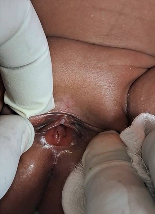 |
Figure 2 Curved membrane covering the vaginal opening which is suggestive of imperforate hymen. |
With the same impression, the neonate was investigated with abdominopelvic ultrasound which showed a 10.2*6.3cm well-defined cystic mass having anechoic fluid with posterior acoustic transmission having internal floating debris posterior to the bladder and anterior to the rectum. The mass displaced the bladder anteriorly. The mass extends down to the vaginal vault. There was also Mild pelvicalyceal separation and ureteral dilation bilaterally. Other abdominal organs appear normal. The conclusion was Hydrometrocolpos with bilateral hydroureteronephrosis. Magnetic Resonance Imaging (MRI) was also done, which shows homogeneous T1 hypointense, and T2 hyperintense collection within the endometrial and vaginal cavity (between the rectum and urinary bladder), causing distention of the uterus above the umbilicus measuring 9.5*4.6*7.6cm. There is a mass effect on the urinary bladder displacing superoanteriorly and stretching the urethra. It also displaces the rectum posteriorly. There is no abnormal communication between the lesions and the rectum. No mass was seen in the vaginal canal. The conclusion was T1 hypointense, T2 hyperintense endometrial, and vaginal cavity collection secondary to Hydrometrocolpos (Figure 3).
 |
Figure 3 Abdomino-pelvic MRI shows homogenous T1 hypointense, T2 hyperintense collection within the endometrial and vaginal cavity causing distension of the uterus above the umbilicus measuring 9.5cm x 4.6cm x 7.6cm. There is a mass effect on the urinary bladder and stretching the urethra. |
With the assessment of Acute Urinary retention 2ᴼ Neonatal Hydrometrocolpos secondary to Imperforate Hymen, a vertical incision and annular hymenotomy were done to drain 200mL of milky fluid. NGT was kept in situ. Follow ultrasound on the 3rd day after the procedure showed normal appearance of the uterus and vagina with no sign of fluid collection. The neonate was discharged with improvement after 4 days of stay and was appointed after 1 month.
Discussions
Hydrometrocolpos is a rare clinical condition characterized by accumulation of fluid or mucus secretions in the vagina and uterus due to distal vaginal obstruction.1 In fetal or neonatal hydrometrocolpos, the collection of cervical mucoid secretions in the obstructed vagina and uterus is caused by the stimulation of the fetal or neonatal cervical mucosal glands by maternal estrogen.8,9 The reported incidence of neonatal hydrometrocolpos is around 0.006%.2
Hydrometrocolpos (HMC) is caused by distal vaginal obstruction due to congenital urogenital anomalies,3 or acquired etiologies such as infection, trauma, or sexual abuse.4,5 Congenital anomalies causing HMC are classified into five types based on the level of obstruction and severity of urogenital or cloacal malformation, which includes type I (imperforate hymen), type II (vaginal septum), type III (distal vaginal atresia), type IV (vaginal atresia with persistent urogenital sinus), and type V (vaginal atresia with cloacal anomaly).7 Inspection of external genitalia may provide clues for initial classification.1 In our case, the cause of HMC was type-1, which is an imperforate hymen.
Imperforate hymen is the most common cause of congenital hydrometrocolpos.2 The hymen is usually perforated at around the eighth week of gestation (embryonic life) to establish a connection between the lumen of the vaginal canal and the vaginal vestibule.2,9 If canalization fails and there are no perforations, the hymen is called imperforate, and it is the most common obstructive congenital lesion of the female genital tract.9 The prevalence of imperforate hymen varies from 1 in 1000 to 1 in 2000 female newborn.10 It is usually an isolated finding with no associated anatomic anomalies;2,9 however, imperforate hymen is sometimes associated with other congenital malformations, such as imperforate anus, bifid clitoris, and polycystic kidney.2 In our index case, imperforate hymen was not associated with other congenital anomalies as evidenced by ultrasound and MRI.
Although an imperforate hymen may be diagnosed at any age by performing a careful inspection of the external genitalia, most of the cases are not detected until puberty.9 Female fetuses or neonates with an imperforate hymen leading with hydrometrocolpos caused by maternal estrogen may incidentally discover during the antenatal period on obstetrics ultrasound or postnatally present with a variety of clinical manifestations.1,2,6 The clinical presentations of HMC depend on the degree of compression of the surrounding structures. Frequently, the urinary tract is compressed causing varying degrees of hydronephrosis.1,11 Rarely, pressure on the bowel cause constipation.1 When the compression is severe, the neonate or infants may present with acute urinary retention or intestinal obstruction.12,13 Acute urinary retention and obstructive uropathy in a patient with HMC is mainly due to compression of the urethra by the distended vagina, which can prevent emptying of the bladder.9 In addition, patients with HMC may manifest as abdominal distension, lower abdominal pain, discomfort in the pelvis, pain in the lower back, and palpable, tender cystic intraabdominal mass or a vulvoperineal mass.5,9 Our index case was presented with abdominal distention, frequent crying, vomiting of ingested matter, and failure to pass urine of 2 days.
The diagnosis of hydrometrocolpos is often missed or delayed owing to its rare incidence and nonspecific symptoms, so that it requires a high index of suspicion for diagnosis.2,5 The diagnosis of congenital hydrometrocolpos can be made prenatally or postnatally.2 Prenatal diagnosis of imperforate hymen and hydrometrocolpos is usually made incidentally with obstetric ultrasound demonstrating an abdominal cystic mass secondary to a distended uterus and vagina and a thin membrane that spreads the labia majora.1,2,9 Antenatal diagnosis of congenital hydrometrocolpos has been reported as early as 25 weeks.2 In our patient, even though intra-abdominal cystic mass was detected on obstetric ultrasound done at 36+3 weeks, HMC was not considered. High index of suspicion and immediate postnatal evaluation of neonates presented with prenatal U/S showing intra-abdominal mass will result in early diagnosis and treatment of neonatal hydrometrocolpos before the development of complications.
Early diagnosis of hydrometrocolpos reduces the risk of developing complications,2 such as urinary tract obstruction, renal failure, recurrent UTI, rupture of HMC, peritonitis, and sepsis.1,2,7 Pelvic ultrasound should be performed in the evaluation of imperforate hymen, particularly prior to surgical intervention.9 Abdominal ultrasonography can reveal a distended uterus and vagina containing echogenic fluid.5 MRI is a valuable imaging tool for assessing the extent of hydrometrocolpos, the thickness of the imperforate hymen, and related complications such as infection, hydronephrosis and endometriosis.10 The diagnosis of HMC was delayed in our case because of lack of communication between healthcare providers and proper postnatal evaluation of the neonate.
The surgical management of imperforate hymen has both long- and short-term goals. Alleviating obstruction of the vagina is the short-term goal, and preserving satisfactory cosmesis, sexual function, and fertility are considered as the long-term goals.9 Imperforate hymen is generally managed by hymenotomy (surgical incision of the hymen) or hymenectomy (surgical removal of the hymen).5 Although no differences in outcomes between the two surgical methods exist, studies show that hymenectomy was associated with more frequent complications than hymenotomy.14 Hymen preserving surgeries such as simple vertical incision and annular hymenotomy are occasionally performed owing to the importance of the first intercourse bleeding.15 Urgent surgical drainage is required for a patient presented acute urinary retention or intestinal obstruction.1 Our index case had an indication for urgent surgical drainage because of acute urinary retention and she was managed with vertical incision and annular hymenotomy.
Timely drainage of the HMC is suggested to avoid complications such as recurrent UTI, pyocolpos, sepsis, failure to thrive and ruptured HMC.16 During the procedure, vagina should be carefully drained with a suction probe, and intrauterine instrumentation to drain the fluid should be avoided to prevent perforation of the thin, overstretched uterine wall.9 In addition, the uterus should not be squeezed for drainage as it can facilitate dissemination of endometrial cells through the fallopian tube which leads to peritoneal endometriosis, tubal adhesions, chronic pelvic pain, and infertility later life.17 Patients should be followed for 2 to 3 weeks to ensure adequate resolution of the HMC.9 The neonate in our case was appointed after 1 month of the procedure and was evaluated clinically and with ultrasound showing resolution of the HMC with no complications.
Conclusion
This is a neonatal hydrometrocolpos secondary to an imperforate hymen diagnosed in a 26 days old female neonate who had intra-abdominal cystic mass on obstetrics ultrasound done during the antenatal period. This is a rare clinical condition characterized by accumulation of fluid within the vagina and uterus. High index of suspicion and immediate postnatal evaluation of neonates presented with prenatal U/S showing intra-abdominal mass will result in early diagnosis and treatment of neonatal hydrometrocolpos before the development of complications.
Data Sharing Statement
All data generated in the preparation of this document are included in the submitted manuscript, and the data sets used in this case report can be obtained from the corresponding author.
Ethical Approval and Consent to Participate
Informed written consent to participate in this case report was obtained from the parents of the neonate. Ethical clearance was obtained from the Institutional Ethics Review Board of Adama Hospital Medical College.
Consent for Publication
Informed written consent was obtained from the parents of the neonate for publication of this case and accompanying images.
Author Contributions
All authors made a significant contribution to the work reported, whether that is in the conception, study design, execution, acquisition of data, analysis and interpretation, or in all these areas; took part in drafting, revising or critically reviewing the article; gave final approval of the version to be published; have agreed on the journal to which the article has been submitted; and agree to be accountable for all aspects of the work.
Funding
There is no funding to report.
Disclosure
All authors declare that they have no competing interests.
References
1. Chen M-C, Chang Y-L, Chao H-C. Hydrometrocolpos in infants: etiologies and clinical presentations. Children. 2022;9(2):219. doi:10.3390/children9020219
2. Vitale V, Cigliano B, Vallone G. Imperforate hymen causing congenital hydrometrocolpos. J Ultrasound. 2013;16:37–39. doi:10.1007/s40477-013-0009-x
3. Celayir AC, Kurt G, Sahin C, Cici I. Spectrum of etiologies causing hydrometrocolpos. J Neonatal Surg. 2013;2(1):5. doi:10.47338/jns.v2.17
4. Hwang HJ, Lim HW, Han YS, Choi JI, Kim MJ. Hematocolpos as a result of delayed treatment of acute straddle injury in an adolescent girl. Case Rep Obstet Gynecol. 2016;2016:1–3. doi:10.1155/2016/1987690
5. Tanitame K, Tanitame N, Urayama S, Ohtsu K. Congenital anomalies causing hemato/hydrocolpos: imaging findings, treatments, and outcomes. Jpn J Radiol. 2021;39:733–740. doi:10.1007/s11604-021-01115-7
6. Hamouda B, Ghanmi S, Soua H, Sfar M. Spontaneous rupture of the imperforate hymen in two newborns. Arch de Pediat. 2016;23(3):275–278. doi:10.1016/j.arcped.2015.11.022
7. Khanna K, Sharma S, Gupta D. Hydrometrocolpos etiology and management: past beckons the present. Pediatr Surg Int. 2018;34:249–261. doi:10.1007/s00383-017-4218-9
8. Bajaj SK, Misra R, Thukral BB, Gupta R. OHVIRA: uterus didelphys, blind hemivagina and ipsilateral renal agenesis: advantage MRI. J Hum Reprod Sci. 2012;5(1):67. doi:10.4103/0974-1208.97811
9. Jones HW, Rock JA. Te Linde’s Operative Gynecology.
10. Fischer JW, Kwan CW. Emergency point-of-care ultrasound diagnosis of hematocolpometra and imperforate hymen in the pediatric emergency department. Pediatr Emerg Care. 2014;30(2):128–130. doi:10.1097/PEC.0000000000000080
11. Mallmann MR, Reutter H, Mack-Detlefsen B, et al. Prenatal diagnosis of hydro (metro) colpos: a series of 20 cases. Fetal Diagn Ther. 2019;45(1):62–68. doi:10.1159/000486781
12. Arriola-Montenegro L, Arriola-Montenegro J, Pia-Balmaceda M, et al. Hydrometrocolpos and post-axial polydactyly complicated with acute intestinal obstruction and hydroureteronephrosis. Cureus. 2021;13(8). doi:10.7759/cureus.17612
13. Okoro PE, Obiorah C, Enyindah C. Experience with neonatal hydrometrocolpos in the Niger Delta area of Nigeria: upsurge or increased recognition? Af J Paediat Surg. 2016;13(4):161. doi:10.4103/0189-6725.194666
14. Lee KH, Hong JS, Jung HJ, et al. Imperforate hymen: a comprehensive systematic review. J Clinl Med. 2019;8:1.
15. Cetin C, Soysal C, Khatib G, Urunsak IF, Cetin T. Annular hymenotomy for imperforate hymen. J Obstet Gynaecol Res. 2016;42(8):1013–1015. doi:10.1111/jog.13010
16. Bischoff A, Levitt MA, Breech L, Louden E, Peña A. Hydrocolpos in cloacal malformations. J Pediatr Surg. 2010;45(6):1241–1245. doi:10.1016/j.jpedsurg.2010.02.097
17. Anselm OO, Ezegwui UH. Imperforate hymen presenting as acute urinary retention in a 14-year-old Nigerian girl. J Surg Tech Case Rep. 2010;2(2):84. doi:10.4103/2006-8808.73623
 © 2023 The Author(s). This work is published and licensed by Dove Medical Press Limited. The full terms of this license are available at https://www.dovepress.com/terms.php and incorporate the Creative Commons Attribution - Non Commercial (unported, v3.0) License.
By accessing the work you hereby accept the Terms. Non-commercial uses of the work are permitted without any further permission from Dove Medical Press Limited, provided the work is properly attributed. For permission for commercial use of this work, please see paragraphs 4.2 and 5 of our Terms.
© 2023 The Author(s). This work is published and licensed by Dove Medical Press Limited. The full terms of this license are available at https://www.dovepress.com/terms.php and incorporate the Creative Commons Attribution - Non Commercial (unported, v3.0) License.
By accessing the work you hereby accept the Terms. Non-commercial uses of the work are permitted without any further permission from Dove Medical Press Limited, provided the work is properly attributed. For permission for commercial use of this work, please see paragraphs 4.2 and 5 of our Terms.