")
Back to Journals » Clinical, Cosmetic and Investigational Dentistry » Volume 15
Morphological Interface Between Restorative and Pulp-Capping Materials: A Systematic Review
Authors Desta NT
Received 27 March 2023
Accepted for publication 1 June 2023
Published 5 June 2023 Volume 2023:15 Pages 99—108
DOI https://doi.org/10.2147/CCIDE.S414418
Checked for plagiarism Yes
Review by Single anonymous peer review
Peer reviewer comments 2
Editor who approved publication: Professor Christopher E. Okunseri
Natnael Teshome Desta
Department of Stomatology, Universitat de València, Valencia, Spain
Correspondence: Natnael Teshome Desta, Department of Stomatology, Universitat de València, C/ Gasco Oliag 1, Valencia, 46010, Spain, Email [email protected]
Background: Bioceramic materials (BCMs) are widely used in vital pulp therapy (VPT) for their biocompatibility and bioactivity; however, their mechanical properties are equally important in the clinical outcome of the pulp-capped teeth.
Objective: To carry out an analysis of the research produced on the morphology of the interface between BM and restorative materials (RM) through a systematic review.
Methodology: An electronic search was performed in Scopus, PubMed, and Web of Science until December 9, 2022. The keywords used in combination with truncation and Boolean operators were: (morphology OR filtration OR porosity) AND (silicate OR composite) AND (cement) AND (“pulp capping” OR “vital pulp therapy” OR “vital pulp treatment”).
Results: Of the 387 articles initially retrieved from the electronic search databases, 5 articles met the criteria for qualitative data collection. MTA and Biodentine were the most studied BCMs. All the articles used a scanning electron microscope as a method of evaluating the samples. Sample sizes and setting times of RM and BCMs differed between studies. Three out of 5 studies used similar conditions of recorded temperature and humidity of 37°C and 100%, respectively.
Conclusion: The different biomaterials used, the application of adhesive systems, humidity and restoration time affect the bonding performance and the ultrastructural interface between BCMs and RMs. The scarcity of research on this point makes it necessary to delve into it and study new materials to obtain more scientific evidence.
Keywords: silicate, cement, vital pulp therapy, morphology, interface
Introduction
Background
In recent years, bioceramic materials (BCM) have gained considerable attention in dentistry due to their excellent biocompatibility properties, their tissue regeneration capacity, their intrinsic osteoconductive capacity and their ability to establish excellent hermetic seal, among other advantages they present.1,2 BCMs are a biocompatible version of ceramics, which are inorganic, non-metallic materials that are produced by heating raw minerals at high temperatures.3,4 Research and development of BCMs date back to the 1960s and today, they include hydroxyapatite, zirconia, alumina, bioactive glass, glass ceramics, resorbable calcium phosphates and radiotherapy glasses.4,5
Due to their enhanced sealing ability, antibacterial and antifungal activity, BCMs are currently used in various dental specialities for treatments including, but not limited to, filling up bony defects, to repair roots, to seal perforations, as root canal sealers, as apical filling materials and vital pulp therapy (VPT).4–7 VPT aims to preserve and maintain pulp tissue that has been compromised but not destroyed by caries, trauma, or restorative procedures in a healthy state.8 Evidence suggests that VPT is particularly important in the young adult tooth with incomplete apical root development due to the high healing of pulp tissue in younger patients.9 Furthermore, VPT lends the benefit of protective resistance to masticatory forces compared with root canal treated teeth.10
Objective
BCMs have grown popular for their use in VPT owing to their excellent biocompatibility properties, however their mechanical properties are just as important in the long-term clinical outcome of the pulp-capped tooth. In today’s market, there are over a dozen commercially available pulp capping materials in the form of BCMs, each with their own specific composition, setting mechanism and consistency.1–5,7,11 Similarly, there is a wide variety of adhesives and direct restorative materials (RM).12 The bonding performance and the ultrastructural interface between BCMs and RMs are fundamental to the clinical outcome of pulp-capped teeth, and clinicians need to be aware of which combinations of BCMs & RMs provide a sound biomechanical interaction for long-term success in treatments such as VPT. Therefore, this review aims to evaluate the morphology in the interface between BCMs and RMs.
Materials and Methods
Method
The report of this systematic review was adapted from the latest PRISMA (Preferred Reporting Items for Systematic Reviews and Meta-Analysis) statement published in 2021.13 The research protocol was registered prior to the extraction of data in Open Science Framework (OSF) Registries (https://osf.io/e872w/).14
Employing the PICO (Population, Intervention, Comparison, Outcome) framework; due to the vast variety of BCMs and RMs available, this review aims to evaluate the morphology (O) in the interface (P) between BCMs (I) and RMs (C), to potentially elucidate the best combination for clinical use in VPT.
To identify relevant studies to be included in this review, an electronic search was conducted on Scopus, PubMed and Web of Science utilizing the Faculty of Medicine and Dentistry, University of Valencia, Valencia, Spain, system from March 22 until December 9, 2022. The keywords used in combination with truncation and Boolean operators on each individual database were as such: (morphology OR filtration OR porosity) AND (silicate OR composite) AND (cement) AND (“pulp capping” OR “vital pulp therapy” OR “vital pulp treatment”).
Eligibility Criteria
All in-vitro studies that evaluated the morphology or mechanical interaction in the interface between BCMs and RMs were included, without language restrictions. Articles that principally studied a different topic, so long as they also evaluated the morphology in the interface between a BCM and a RM, were also included. However, case series, case reports, conference abstracts, editor’s notes and reviews were excluded. Articles that were published before the year 1999 were also excluded from data collection due to the technological advancement of BCMs over the past two decades.
Selection of Studies and Data Collection
All references gathered from each electronic database were introduced to a reference manager software (Mendeley; Elsevier, AMS), and duplicate articles were manually removed. The screening and selection of the studies to be included in the review were conducted by the lead author according to the eligibility criteria, employing a two-phase screening process. In Phase-one, all the titles and abstracts gathered from the electronic databases were screened, from which only the relevant articles were selected to proceed to Phase-two. In Phase-two, the articles considered eligible from Phase-one were read in full and identified for qualitative data collection, with the author citing reasons for any exclusion.
Due to the absence of quantitative data on the objective of the study; the morphology of the interface between BCMs and RMs, only qualitative data were extracted from the studies. A customized table that had been previously prepared with variables of interest for this study was used for data collection.
Risk of Bias – Methodology Quality Assessment
The Consolidated Standards of Reporting Trials (CONSORT) statement is a tool to improve transparency of reporting in Randomized Controlled Trials (RCTs). However, since the studies included are not RCTs, the “Modified CONSORT checklist of items for reporting in vitro studies of dental materials” (Faggion, 2012)15 was used to assess the quality of their methodologies. Each study was individually evaluated for fulfilment or nonfulfillment of the 14 parameters considered in the quality assessment tool. The percentage was subsequently calculated as the number of complied items divided by the total number of items multiplied by 100 and reported rounded to the nearest tens.
Results
Search Result Findings
The search strategy in the electronic databases retrieved a total of 387 articles, of which 110 duplicates appeared and hence were discarded. During Phase-one of screening, the titles and abstracts of the individual articles were read, and 249 articles were excluded because they were irrelevant to the subject of this review. A total of 10 studies remained for Phase-two of screening, where each article was read in full. From the studies perused, 5 articles were subsequently excluded because they did not meet the inclusion criteria for data collection. The publications by Voicu et al,13 Farrugia et al16 and Gandolfi et al17 were discarded for data collection because they studied the surface microstructure and/or characteristics between dental BCMs, but not between BCMs & RMs. de Souza et al18 investigated the physical-mechanical properties and micromorphology of calcium cements exposed to polyacrylic and phosphoric acids. Similarly, a study by Kitasako et al19 was also not included as it investigated the interface between resin and pulp tissues under a Scanning Electron Microscope (SEM). Therefore, as Figure 1 search flowchart demonstrates, 5 studies were included for qualitative data synthesis.20–24 In Table 1, the literature search findings are outlined.
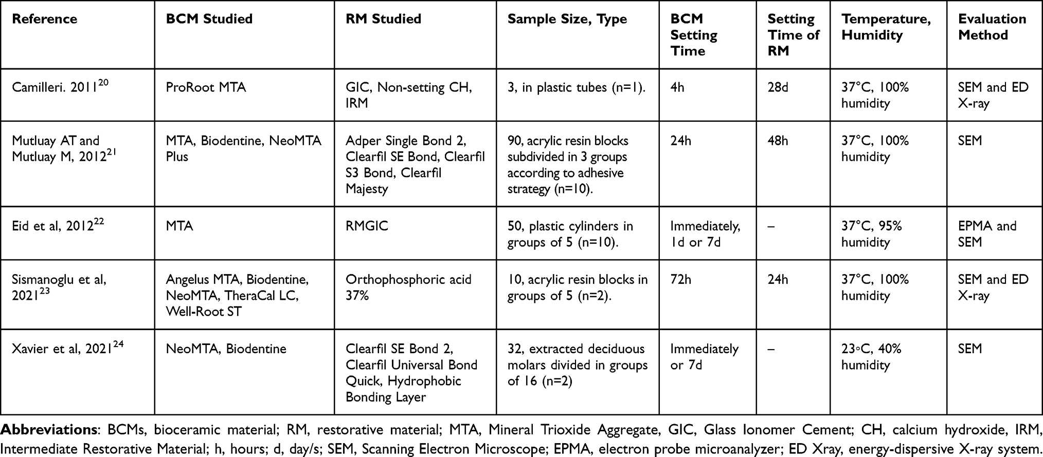 |
Table 1 Findings of the Literature |
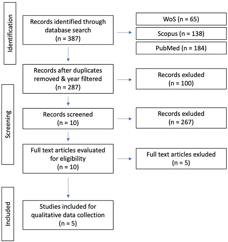 |
Figure 1 Search strategy flowchart. |
Quality Assessment
The quality assessment results using the “Modified CONSORT checklist of items for reporting in vitro studies of dental materials” (Faggion, 2012)15 are presented in Table 2. From a total of 5 studies assessed, 4 studies had results between 50% and 60% in terms of the quality of the data reported in their respective papers,20–23 while the study by Xavier et al24 had the highest quality assessment report at 70%.
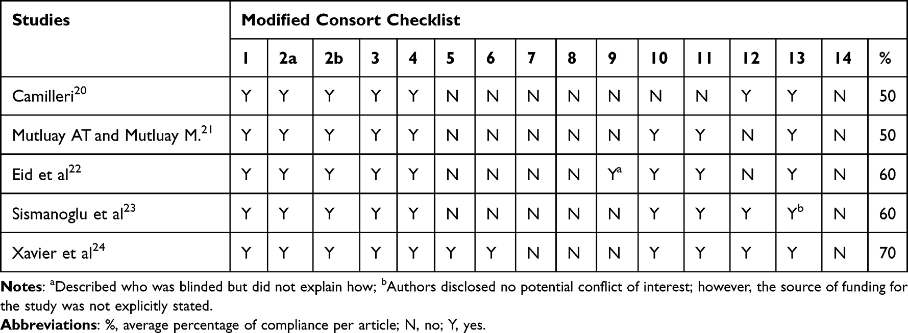 |
Table 2 Modified Consort Checklist |
All the articles presented a well-summarized abstract, specific background, and explanation of rationale, as well as clear objectives and/or hypotheses (items 1, 2a and 2b). In the methodologies, the interventions were presented with sufficient detail and the outcomes along with their measures were clearly defined (items 3 and 4). However, regarding items 5–8, which concerned the sample size determination, the randomization sequence generation, the allocation concealment mechanism, and the implementation of the randomization sequence, only the manuscript by Xavier et al fulfilled those assessments. In this study, the researchers calculated the sample size using a software G*Power 3.1.9.2 (University of Düsseldorf), specimens were randomly selected from a pooled biobank of extracted teeth and were randomly allocated into 16 groups (n = 2) after being mounted in self-cure acrylic resin blocks. Item 9 assessed blinding in the studies and how the researchers were blinded, and only Eid et al partially fulfilled that as they included two blinded observers to investigate the microstructural analysis of the interface although they did not describe how. While the other two randomized controlled trials reported to have included a blinded researcher, they did not fulfill those parameters as they were used to carry out fracture type analysis on a stereomicroscope21 and shear bond strength tests,24 investigations out of the scope of this review. Statistical analyses and outcomes along with their significance levels (items 10 and 11) were performed and reported in all studies except for the manuscript by Camilleri20 which investigated only three samples and therefore not suitable to carry out. Three out of the 5 studies addressed the potential limitations and the sources of potential bias of their respective methodologies (item 12). All included studies reported sources of funding (item 13), however the study by Sismanoglu et al23 does not explicitly report the source of funding but was registered as having fulfilled the requirement as indicated by their conflict-of-interest disclosure statement. No reference to their respective full trial protocol was made by any of the studies (item 14). The percentage of complied items varied from 50% to 70%, indicating that the studies rated at a moderate risk of bias.
Discussion
As demonstrated in Table 1, the selected studies used different materials, setting times, in-vitro setting conditions, sample sizes and means of data acquisition. Biodentine and MTA were the most frequently studied BCM, with Biodentine used in 3 out of 5 studies and the latter in all studies. Three out of 5 studies used similar registered temperature conditions of 37°C and 100% humidity, while Eid et al22 had clinical settings of 37°C and 95% humidity, and Xavier et al24 23°C and 40% humidity. In all the studies, a SEM was utilized to evaluate the interface morphology between the BCM and RM.
Due to the discrepancy of the methodologies between the studies, the first phase of the discussion will explore the procedure of each article, the second phase will examine their findings and the final phase; the strengths, limitations, and future perspectives on this subject.
Methodology of the Studies: Considerations on Material Choice, Sample Preparation and Evaluation Settings
In the study by Camilleri,20 three samples were prepared according to the manufacturer’s mixing instructions where the MTA (ProRoot MTA; Dentsply, Tulsa Dental Products, Tulsa, OK, USA) was left to set undisturbed for 4 hours: MTA with GIC (Fuji IX GP, GC America, Alsip IL, USA), MTA with non-setting calcium hydroxide (CH) (Calasept, Nordiska Dental, Angelholm, Sweden) and MTA with Immediate Restorative Material (IRM) (IRM, Dentsply DeTrey, Konstanz, Germany). A plastic tube 48 mm long and with a 12 mm internal diameter was prepared to place 1.5 grams of MTA using a glass spatula, together with a similar quantity of one of the other three cements, so that the two materials in each plastic tube were in 3–4mm thick layers with an approximately horizontal interface. The tubes were sealed at both ends and placed vertically in an incubator to cure for 28 days at 37◦C and 100% humidity. The author noted that a drop of water was placed on the MTA surface after 24h to avoid desiccation.
Once the mixes were cured, they were separated from the plastic tubes and dried in a vacuum desiccator for eight hours. The mixes were embedded in epoxy resin to prepare polished sections using diamond polishing compound to examine the cross-section of the interfaces. After applying a thin conductive coating of evaporated carbon to the polished sections, the samples were examined under a SEM in black scatter mode and by elemental analysis of the different hydration products by X-ray energy dispersive analysis. To draw atomic ratio plots, lines parallel to the interface at widening incremental distances of 50 μm up to 600 μm were scanned and spectra collected.
Mutluay AT and Mutluay M.21 investigated three BCMs, namely MTA (Pro-Root MTA, Dentsply Tulsa Dental, Tulsa, OK), Biodentine (Septodont, Saint-Maur-des-Fosses, France) and NeoMTA Plus (Avalon Biomed Inc, Bradenton, FL). Ninety cylindrical acrylic blocks with 2mm depth and 4mm diameter were prepared and the samples were randomly divided into the three main groups based on BCM utilized (n = 30, each). The BCMs were prepared according to manufacturer’s instructions and placed in the cavities to be kept at 37°C for 24 h at 100% humidity. Each sample surface was wet sanded for ten seconds until surface standardization was attained, then subsequently subdivided into three groups as per the different adhesive strategies: two-stage self-etch system (SE Bond, Kuraray, Japan), two-stage total-etch system (acid etching with 35% phosphoric acid + Single Bond 2 3M ESPE, USA) and a one-stage self-etch system (S3 Bond, Kuraray, Japan). Once the bonding agent was polymerized using an LED device with a light intensity of 1200 mW cm2, a composite resin layer was also placed using a cylindrical metal mould of 3.5 mm internal diameter and 5 mm height and then light cured.
A single researcher carried out all the restorative procedures and the samples were kept at 37°C and 100% humidity for 48 hours. Using a water-cooled cutting device (Mikracut 152, Metkon, Turkey) and a diamond saw (19–125 Diamond Cutting Disc, Metkon, Turkey), the specimens were cut vertically, sputter-coated with gold–palladium, and examined under a SEM at 91,000 magnification.
Unlike the previous two studies which examined various BCMs and RMs, in the research by Eid et al,22 they examined the interface between a single BCM which was MTA (ProRoot MTA; Dentsply, Tulsa, OK), and one RM, resin-modified GIC (RMGIC) (Fuji II LC; GC Corporation, Tokyo, Japan), and instead had the independent variables as time and wet condition of RMGIC placement. Fifty plastic cylinders of 4 mm diameter and 7 mm length were half filled with MTA which was mixed with sterile water according to the manufacturer’s instructions. While the base of the cylinders was in contact with wet gauze, the top surface of the MTA was smoothed with a plastic plunger, after which the specimens were randomly divided into 5 equal groups (n = 10) according to time and wetness (covered with a wet cotton pellet) before being light cured, which were; immediately (IMM), after 24 hours in a wet condition (1Dw) and an IRM temporary filling (Dentsply DeTrey, Konstanz, Germany) was added, after 24 hours in a dry condition (1Dd), after 7 days in a wet condition (7Dw) and after 7 days in a dry condition (7Dd). In the wet condition groups, a sharp excavator was used to remove the IRM and cotton pellets without touching the MTA surface, after which a gentle stream of air was used to remove excess moisture before finally placing the GIC. On the other hand, in the dry condition groups, IRM was placed on the most external part of the plastic cylinders without having contact with the MTA surface, after which it was removed with a sharp excavator and any debris was carefully blown away. All the specimens were stored at 37◦C and 100% humidity.
The MTA-GIC specimens were prepared by embedding them in transparent epoxy resin and thereafter sectioned perpendicularly to the interface using a low-speed saw, polished and covered with a thin layer of carbon before being analyzed with an SEM. Furthermore, an electron probe microanalyzer was used to detect the elemental distribution of calcium, fluorine, silicon and bismuth along the MTA-GIC interface in an area of 512×512 mm.
The study by Sismanoglu et al23 was focused not only on the morphology interface, but also the bond strength and the elemental composition of Calcium-Silicate Cements (CSCs) to composite resin. For the part of SEM-EDX analysis, the study observed morphological differences between the intact BCMs and the surface of BCMs exposed to 37% orthophosphoric acid. The BCMs under investigation were Angelus MTA, Biodentine, NeoMTA, TheraCal LC and Well-Root ST.
A total of 160 acrylic resin blocks with a central hole of 2 mm depth and 4-mm diameter were prepared; however, for the SEM analysis, two specimens from each group of 5 BCMs (n = 10) were selected. The Biodentine and MTA-based cements were mixed according to the manufacturers’ instructions and were filled inside the resin holes. The material surfaces were covered with wet cotton pellets and the specimens were stored at 37°C and 100% relative humidity for 72 hours to ensure that it was completely set.
On the contrary, TheraCal LC was loaded into the holes in two successive increments with a thickness of 1 mm before being polymerized with an LED light curing device for 20 seconds. Due to its on-demand setting characteristics, the TheraCal LC specimens were readied without an additional waiting time. Like the study by Mutluay AT and Mutluay M,21 before applying the adhesive layer, the specimen surfaces were polished to provide flat surfaces using silicon carbide papers under water cooling.
Once the CSC specimens were prepared, to examine the elemental mapping and chemical composition of the CSCs, a SEM-EDX analysis was conducted. Two specimens from each group (n = 10) were selected and substrate surfaces were either acid etched for 15 seconds with 37% orthophosphoric acid or not. The substrate surfaces were investigated under a SEM (Apreo S; Thermo Fisher Scientific, USA), fitted with an EDX system (UltraDry EDX Detector; Thermo Fisher Scientific, USA) for microstructural surface analyses in low vacuum mode using an accelerating voltage of 10 kV.
Like the previous research discussed, the study by Xavier et al24 was focused not only on the morphology interface, but also on the shear bond strength and the elemental composition of CSCs to adhesive composite restorations. The four independent variables were BCMs, different adhesive systems, the use of additional Hydrophobic Bonding Layer (HBL) and immediate or 7 day delayed restoration time. The BCMs under investigation were NeoMTA & Biodentine, while the adhesive composite restorations included were Clearfil SE Bond 2 (CSEB2) and Clearfil Universal Bond Quick (CUBQ).
Having gained consent from patients and parents, as well as approval from the Ethics Committee of Faculty of Medicine of the University of Coimbra, 32 deciduous molars with at least one-third of the root and without furcation involvement were randomly selected from a pooled biobank of extracted teeth. The extracted teeth were stored in an aqueous chloramine solution 0.5% at 4◦C for up to 6 months, which was renewed every month following the norm ISO/TS 11405:2015.
Occlusal cavities exposing the pulp chamber were prepared in each tooth and the remaining pulp tissue was removed with a spoon excavator, rinsed with sterile saline solution, and air-dried. The teeth were then mounted in self-cure acrylic resin blocks at the level of the cement-enamel junction and randomly allocated into 16 groups (n = 2), according to the independent variables previously described.
Each BCM was prepared following the manufacturer’s instructions before they were digitally compressed with a humid cotton pellet inside the cavity and allowed to set; the NuSmile® NeoMTA had a 3-minute setting time while the Biodentine had a 12-minute setting time. In the immediate restoration group, once the respective BCM was set, the adhesive systems were applied over the BCM surface for 20 seconds, dried with air and light-cured for 10 seconds at “High Power” mode (Bluephase® Style M, Ivoclar, Vivadent, Schaan, Liechtenstein). In groups with application of an additional HBL, an extra layer of CSEB2 Bond was applied over the adhesive, dried with a mild airflow and light-cured.
In the delayed restoration group, the materials were covered by glass ionomer cement (GIC) (Ionostar® Molar—VOCO® GmbH, Cuxhaven, Germany) and stored in an incubator at 37 ◦C with 100% humidity for 7 days. After the storage duration, the GIC was separated with black coarse aluminum oxide abrasive discs, 3MTM Sof-LexTM (3M ESPE, St. Paul, MN, USA), until a flat surface of the BCM was uncovered and polished using water sandpaper. The same adhesive and restorative procedures were applied as outlined for the immediate groups. A single researcher carried out all the adhesive and restorative procedures. During all specimen preparations, the registered room temperature was about 23 ◦C, with 40% humidity.
After 1 week of storage, the teeth were multi-sectioned in a buccolingual direction along their longitudinal axis using a high-precision diamond cut-off wheel (Accutrom 50 machine, Struers, Denmark) with approximately 1000 μm thickness. The specimens were polished and treated by 35% phosphoric acid gel for 15 seconds, washed, dried and sequentially dehydrated in increasing concentration of ethanol (50%, 75%, 95%, 100%) and were mounted on aluminum stubs, sputter-coated with gold–palladium and observed under a SEM (Hitachi S-4100, Tokyo, Japan) at various magnifications.
Findings of the Studies
Besides the studies by Mutluay AT and Mutluay M, 201221 and Xavier et al, 2021,24 each study had an individualized methodology which consequentially led to varying results that made it difficult to compare among one another. In short, the studies evaluated the interface between one or multiple BCMs and: CH, GIC and IRM (Camilleri 201120), RMGIC (Eid et al22), exposure to 37% orthophosphoric acid (Sismanoglu et al, 202123) and different adhesive systems (Mutluay AT and Mutluay M, 201221 and Xavier et al, 202124).
In the study by Camilleri20 where the author investigated the interface of MTA with CH, GIC and IRM, it was found that MTA interacts with other BCMs with varying elemental migration. While the MTA-CH specimen did not show any evidence of changes in microstructure, the MTA-GIC and MTA-IRM specimens exhibited a high degree and some degree of micro-cracking, respectively, and some porosity in the interfacial region. Furthermore, with regard to the elemental analysis of the different hydration products by X-ray energy dispersive analysis, lines parallel to the interface at increasing incremental distances of 50 μm up to 600 μm were scanned and spectra collected to draw atomic ratio plots. From this aspect of the research, the author concluded that zinc oxide eugenol (ZOE)-based cements should be avoided in conjunction to MTA as zinc causes retardation of cement hydration with greater porosity. Similarly, GIC absorbs the water of hydration from MTA thereby resulting in greater porosity and incomplete hydration of MTA.
In the research by Mutluay AT and Mutluay M.21 the different adhesive types used over the three BCMs they investigated, namely MTA, NeoMTA Plus and Biodentine, did not influence the interfacial gap in the specimens. The samples demonstrated no gaps, cracks or delamination within the adhesive layer, which reveals a good adhesion between the CSC–composite interface in all subgroups. Moreover, different CSCs used with different adhesive families exhibited similar interface morphology.
In the study by Eid et al22 where they examined the interface between MTA and RMGIC at different times and wetness conditions, the SEM indicated that all the groups suffered adhesive separation and gap formation at the interface. They found that although cohesive separation in MTA was found in all groups, it was detected with greater frequency in the dry condition groups (1Dd and 7Dd) in comparison to the wet condition groups (1Dw and 7Dw). All groups demonstrated horizontal and vertical cracks in GIC that interconnected with one another in the internal voids within the GIC. However, these observed changes were limited to the outermost layer of the MTA, and neither the deeper layers of MTA nor the GIC itself seemed affected.
With regard to the elemental migration, the growth of calcium crystals was evident at the interface in the wet condition groups and showed to enlarge with time. Silicon was observed on both the MTA and GIC side, whereas fluorine was detected exclusively on the GIC side as densely packed small particles.
In the study by Sismanoglu et al23 where they observed morphological differences between the intact BCMs and the surface of BCMs exposed to 37% orthophosphoric acid, the results of the SEM showed distinct morphological alterations after acid etching. Surface irregularity, capillary voids and microcracks due to superficial dissolution caused by acid etching were common to all the CSC specimens under investigation; Angelus MTA, Biodentine, NeoMTA, TheraCal LC and Well-Root ST. After acid etching, Biodentine and TheraCal LC exhibited much more distinct woodpecker holes, whereas the MTA-based cements displayed “needle-shaped and coral-like crystalline structures”.
With regard to the quantitative results of the chemical composition under the EDX microanalysis, high levels of calcium (Ca) in the Well Root (26.5% by weight), NeoMTA (31.7% by weight), Biodentine (33.1% by weight) and Angelus MTA (38.6% by weight) groups were found rather than silicon (Si), as compared to TheraCal LC which consisted similar amounts of Ca (9.2% by weight) and Si (11.1% by weight) and higher amount of carbon (45% by weight). However, after acid etching, the Ca/Si ratio of the cement surface decreased by approximately 4-fold in all the samples.
Xavier et al, 202124 evaluated the morphological interface between two CSCs and various adhesive systems, and reported that all the specimens observed under the SEM formed a hybrid layer presented as interpenetrations between the CSCs and the adhesive systems. Moreover, its depth and thickness vary according to the adhesive type and the timing of the restoration, where it was evident that the interpenetration was less deep in the DR groups than the IMM groups. The authors hypothesize that this may be due to the hydrophilic nature of the adhesive systems and the presence of water from the unset CSC.
With regard to the morphological patterns, there was greater superficial dissolution of CSC and incorporation of particles into the adhesive layer in the IMM and the CSEB2 groups, in addition to the adhesive filling of spaces between the inorganic content of the CSC. The authors note that some of those spaces seemed empty under the SEM due to a possible wash-out effect of the adhesive as well as the CSC particles during the preparation of the cuts for examination. It was also reported that the groups with an additional HBL exhibited greater thickness in the adhesive layer.
Strengths, Limitations, and Future Perspectives
The use of different-novel materials, setting times, in-vitro clinical settings, sample sizes and means of data acquisition posed a challenge to extrapolate the qualitative data in a comprehensive manner. Moreover, the absence of real-life external factors does not reflect its reproducibility in clinical scenarios. Nonetheless, in-vitro studies are an important step in the ladder of assembling rigorous, high-level evidence-based research for the future, and as such, this systematic review sought to evaluate the morphology in the interface between BCMs and RMs, with the aim of elucidating how these materials interact with one another.
A Modified CONSORT checklist of items for reporting in-vitro studies of dental materials15 was utilized to assess the quality of the evidence reported in this review. The 14 parameters considered by the quality evaluation tool were individually assessed for each study to see whether they were met or not and presented as a percentage. The percentage of complied items varied from 50% to 70%, indicating that the studies rated at a moderate risk of bias.
Future studies on the topic of the morphological interaction between BCMs and RMs should aim to replicate the methodology and conditions of one of the discussed studies (preferably one with the least risk of bias), while evaluating similar and/or novel RMs or BCMs, to compare the reproducibility of the qualitative results.
Conclusion
Understanding the morphology in the interphase between BCMs and RMs enables the practitioner to determine the most effective combination of materials in treatments such as VPT to potentially achieve excellent long-term clinical outcome of a pulp-capped tooth.
The different biomaterials used, the application of adhesive systems, humidity and restoration time affect the bonding performance and the ultrastructural interface between MR and MB. The scarcity of research on this point makes it necessary to delve into it and study new materials to obtain more scientific evidence. Within the limitation of the revised in-vitro studies’ conclusions, these are the recommendations that were reached:
- Place adhesive immediately after the BCM has set.
- Adequate adhesive performance can be achieved without acid etching.
- When pulp-capping with MTA, avoid placing GIC or IRM and instead opt for calcium hydroxide.
Ethical Statement
The articles and data were obtained lawfully, as the articles were retrieved in academic databases by the aforementioned search engines.
Funding
The studies that required payment for access were collected through the University of Valencia Online Library Portal access.
Disclosure
The author declares no conflicts of interest in this work.
References
1. Piva E, Da Rosa WLO, Coco AR, et al. Systematic review of dental pulp capping materials. Dent Mater. 2017;32:e89. doi:10.1016/j.dental.2016.08.185
2. Surya Raghavendra S, Jadhav GR, Gathani KM, Kotadia P. Bioceramics in endodontics – a review. J Istanbul Univ Fac Dent. 2017;51. doi:10.17096/jiufd.63659
3. Shenoy A, Shenoy N. Dental ceramics: an update. J Conserv Dent. 2010;13(4):195. doi:10.4103/0972-0707.73379
4. Jain P, Ranjan M. The rise of bioceramics in endodontics: a review. Int J Pharma Bio Sci. 2015;6:416–P422.
5. Best SM, Porter AE, Thian ES, Huang J. Bioceramics: past, present and for the future. J Eur Ceram Soc. 2008;28(7):1319–1327. doi:10.1016/j.jeurceramsoc.2007.12.001
6. Leong DJX, Yap AU. Vital pulp therapy in carious pulp–exposed permanent teeth: an umbrella review. Clin Oral Investig. 2021;25:6743–6756. doi:10.1007/s00784-021-03960-2
7. Jitaru S, Hodisan I, Timis L, Lucian A, Bud M. The use of bioceramics in endodontics – literature review. Med Pharm Rep. 2016;89(4):470–473. doi:10.15386/cjmed-612
8. Ghoddusi J, Forghani M, Parisay I. New approaches in vital pulp therapy in permanent teeth. Iran Endod J. 2014;9(1):15–22.
9. Kharchi AS, Tagiyeva-Milne N, Kanagasingam S. Regenerative endodontic procedures, disinfectants and outcomes: a systematic review. Prim Dent J. 2020;9(4):65–84. doi:10.1177/2050168420963302
10. Caplan DJ, Cai J, Yin G, White BA. Root canal filled versus non-root canal filled teeth: a retrospective comparison of survival times. J Public Health Dent. 2005;65(2):90–96. doi:10.1111/j.1752-7325.2005.tb02792.x
11. Iftikhar S, Jahanzeb N, Saleem M, Ur Rehman S, Matinlinna JP, Khan AS. The trends of dental biomaterials research and future directions: a mapping review. Saudi Dent J. 2021;33(5):229–238. doi:10.1016/j.sdentj.2021.01.002
12. Hickel R, Dasch W, Janda R, Tyas M, Anusavice K. New direct restorative materials*. Int Dent J. 1998;48(1):3–16. doi:10.1111/j.1875-595X.1998.tb00688.x
13. Voicu G, Didilescu AC, Stoian AB, Dumitriu C, Greabu M, Andrei M. Mineralogical and microstructural characteristics of two dental pulp capping materials. Materials. 2019;12(11):1772. doi:10.3390/ma12111772
14. Desta NT. Morphology of the interface between restorative and pulp-capping materials: a systematic review. OSF. 2022. doi:10.17605/OSF.IO/E872W
15. Faggion CM. Guidelines for reporting pre-clinical in vitro studies on dental materials. J Evid Based Dent Pract. 2012;12(4):182–189. doi:10.1016/j.jebdp.2012.10.001
16. Farrugia C, Lung CYK, Schembri Wismayer P, Arias-Moliz MT, Camilleri J. The relationship of surface characteristics and antimicrobial performance of pulp capping materials. J Endod. 2018;44(7):1115–1120. doi:10.1016/j.joen.2018.04.002
17. Gandolfi MG, Siboni F, Botero T, Bossù M, Riccitiello F, Prati C. Calcium silicate and calcium hydroxide materials for pulp capping: biointeractivity, porosity, solubility and bioactivity of current formulations. J Appl Biomater Funct Mater. 2014;13(1):1.
18. De Souza GF, Arrais AB, Aragão CFS, Ferreira IA, Borges BCD. Physical-mechanical properties and micromorphology of calcium cements exposed to polyacrylic and phosphoric acids. Scanning. 2018;2018:3197510. doi:10.1155/2018/3197510
19. Kitasako Y, Arakawa M, Sonoda H, Tagami J. Light and scanning electron microscopy of the inner surfaces of resins used in direct pulp capping. Am J Dent. 1999;12(5):217–221.
20. Camilleri J. Scanning electron microscopic evaluation of the material interface of adjacent layers of dental materials. Dent Mater. 2011;27(9):870–878. doi:10.1016/j.dental.2011.04.013
21. Mutluay AT, Mutluay M. Characterisation of the calcium silicate-based cement-composite interface and the bonding strength with total-etch or single/two-stage self-etch adhesive systems. Aust Endod J. 2021;48(3):501–509. doi:10.1111/aej.12600
22. Eid AA, Komabayashi T, Watanabe E, Shiraishi T, Watanabe I. Characterization of the mineral trioxide aggregate-resin modified glass ionomer cement interface in different setting conditions. J Endod. 2012;38(8):1126–1129. doi:10.1016/j.joen.2012.04.013
23. Sismanoglu S, Yildirim-Bilmez Z, Gurcan AT, Gumustas B. Influence of application mode of universal adhesive on the surface morphology, elemental composition and bond strength of calcium silicate-based cements to composite resin: a SEM-EDX microanalysis study. J Adhes Sci Technol. 2021;36(17):1833–1846. doi:10.1080/01694243.2021.1992979
24. Xavier MT, Costa AL, Caramelo FJ, Palma PJ, Ramos JC. Evaluation of the interfaces between restorative and regenerative biomaterials used in vital pulp therapy. Materials. 2021;14(17):5055. doi:10.3390/ma14175055
 © 2023 The Author(s). This work is published and licensed by Dove Medical Press Limited. The full terms of this license are available at https://www.dovepress.com/terms.php and incorporate the Creative Commons Attribution - Non Commercial (unported, v3.0) License.
By accessing the work you hereby accept the Terms. Non-commercial uses of the work are permitted without any further permission from Dove Medical Press Limited, provided the work is properly attributed. For permission for commercial use of this work, please see paragraphs 4.2 and 5 of our Terms.
© 2023 The Author(s). This work is published and licensed by Dove Medical Press Limited. The full terms of this license are available at https://www.dovepress.com/terms.php and incorporate the Creative Commons Attribution - Non Commercial (unported, v3.0) License.
By accessing the work you hereby accept the Terms. Non-commercial uses of the work are permitted without any further permission from Dove Medical Press Limited, provided the work is properly attributed. For permission for commercial use of this work, please see paragraphs 4.2 and 5 of our Terms.