")
Back to Journals » Infection and Drug Resistance » Volume 12
Lateral flow biosensor combined with loop-mediated isothermal amplification for simple, rapid, sensitive, and reliable detection of Brucella spp
Authors Li S, Liu Y, Wang Y, Chen H, Liu C, Wang Y
Received 9 April 2019
Accepted for publication 8 July 2019
Published 30 July 2019 Volume 2019:12 Pages 2343—2353
DOI https://doi.org/10.2147/IDR.S211644
Checked for plagiarism Yes
Review by Single anonymous peer review
Peer reviewer comments 3
Editor who approved publication: Professor Suresh Antony
Shijun Li,1 Ying Liu,1 Yue Wang,1 Hong Chen,2 Chunting Liu,1 Yi Wang3,4
1Laboratory of Bacterial Infectious Disease of Experimental Center, Guizhou Provincial Center for Disease Control and Prevention, Guiyang 550004, People’s Republic of China; 2Laboratory of Guiyang Center for Animal Disease Control and Prevention, Guiyang 550081, People’s Republic of China; 3Key Laboratory of Major Diseases in Children, Beijing Pediatric Research Institute, Beijing Children’s Hospital, Capital Medical University, National Center for Children’s Health, Beijing 10045, People’s Republic of China; 4Ministry of Education, National Key Discipline of Pediatrics (Capital Medial University), Beijing Pediatric Research Institute, Beijing Children’s Hospital, Capital Medical University, National Center for Children’s Health, Beijing 10045, People’s Republic of China
Abstract: Brucella species is responsible for brucellosis in human and animals, which is still of public health, veterinarian, and economic concern in many regions of the world. Here, a novel molecular diagnosis assay, termed loop-mediated isothermal amplification coupled with nanoparticles-based lateral flow biosensor (LAMP-LFB), was developed and validated for simply, rapidly, and reliably detecting all Brucella spp. strains. A set of six primers was designed based on the Brucella-specific gene Bscp31. The Brucella-LAMP results were visually reported by biosensor within 2 mins. A variety of bacterial strains representing several Brucella species, as well as several Gram-negative and Gram-positive bacterial species were used to determine the analytical sensitivity and specificity of the assay. Optimal LAMP conditions were 63°C for 40 mins, and the assay’s sensitivity was found to be 100 fg of genomic DNA in the pure cultures. No cross-reactions to non-Brucella strains were obtained; thus, analytical specificity of LAMP-LFB assay is of 100%. Using the protocol, 20 mins for rapid DNA preparation followed by isothermal amplification (40 mins) combined with biosensor detection (2 mins) resulted in a total assay time of approximately 65 mins. In the case of 117 whole blood samples, 13 (11.11%) samples were Brucella-positive by LAMP-LFB, and the diagnostic accuracy was 100% when compared to the culture-biotechnical method. In conclusion, Brucella-LAMP-LFB technique developed in this study is a sensitive and specific method to rapidly identify all Brucella spp. strains, and can be applied as a potential diagnostic tool for brucellosis in basic, clinical, and field laboratories.
Keywords: Brucella spp., brucellosis, loop-mediated isothermal amplification, lateral flow biosensor, limit of detection
Introduction
Brucellosis, which is one of the worldwide zoonoses, is still of public health, veterinarian, and economic concern in many regions of the world.1 Brucellosis is caused by a number of host adapted species of facultative intracellular bacteria of the genus Brucella.2 Brucella spp. comprises six classical species (B. melitensis, B. ovis, B. abortus, B. suis, B. neotomae, B. canis), two marine species (B. pennipedilis and B. ceti), one human origin species (B. innoponita), and several newly described species (eg, B. vulpis and B. microti).3,4 Brucella spp. is responsible for disease in both humans and animals.5 Human brucellosis is considered as a life-threatening illness, and is the result of direct exposure to infected animals and their carcass, or animal products consumption.6 Animal brucellosis is characterized by fetal death and spontaneous abortion in animals, and causes severe economic losses and restrictions on international animal movement and trade. In order to effectively prevent and control brucellosis, accurate early identification of Brucella spp. is very important.
Serology has been used in control and surveillance programs for brucellosis, while serological methods usually cause false-negative and false-positive results.7 Isolation of the aetiological agent is the most specific diagnostic examination for the detection of Brucella spp. strains, but suffers from the disadvantages of low sensitivity, needing a long incubation period, especially in the chronic stage of the illness. Most importantly, the culture material must be carefully performed in bio-safety level laboratories, because several species (such as B. melitensis and B. abortus) of Brucella spp. are a class III organism and infects livestock and humans at a very low infectious dose.8 Molecular techniques, such as traditional PCR and real-time PCR, have been proved to be efficient and rapid at different stages of the brucellosis, more sensitive than culture-based methods, more specific than serological diagnostics.9 However, PCR-based techniques require the use of complex apparatus and skilled personnel, thus these assays are not readily available in all diagnostic laboratories in resource-poor settings and limits their use for wide-scale application at field level.10
To overcome the drawbacks posed by PCR-based assays, various other techniques, on the basis of isothermal amplification of nucleic acid, have been designed, which are able to rapidly synthesize DNA in large amounts without the use of any sophisticated apparatus.11 Among these isothermal amplification methods, loop-mediated isothermal amplification (LAMP) is one of the most widely used techniques, which has been employed for rapid, sensitive, and specific detection of many bacterial pathogens including Brucella spp.1,12–16 However, the results of Brucella-LAMP assays were displayed using agarose gel electrophoresis, color indicator (such as hydroxynaphthol blue, SYBR green I, calcein dye et al.), and real-time turbidity equipment. The use of gel electrophoresis to analyze Brucella-LAMP products is a complex procedure, and increases the risk of carryover contamination and degradation. Analysis of Brucella-LAMP products by color with the naked eyes is potentially subjective; thus, a practical sample may be somewhat ambiguous to the unaided eye when the concentration of Brucella genomic templates is very low. The analysis of Brucella-LAMP results by real-time turbidity requires a special optical instrument, and easily suffers from background interference.
The increasing application of LAMP assay has emphasized simplicity and speed as crucial criteria for adoption in “on-site” diagnosis, point-of-care (POC) testing, field detection and more. To well-suit for these applications, nanoparticles-based lateral flow biosensors (LFBs) have been designed, and increasingly employed as alternative tools for reporting LAMP results due to their simplicity, rapidness, and low cost.17–19 Herein, we reported on the establishment of a visual, sensitive, and specific LAMP-based method, which successfully incorporated conventional LAMP assay with LFB (LAMP-LFB) for rapid and reliable detection of Brucella strains. The optimal conditions, analytical sensitivity, specificity, and feasibility of Brucella-LAMP-LFB method were validated using pure cultures and clinical samples.
Methods and materials
Reagents and instruments
Colorimetric indicator (Malachite Green, MG), universal isothermal amplification kits and biotin-14-dCTP were purchased from Bei-Jing HaiTaiZhengYuan. Co., Ltd. (Beijing, China). The LFB materials, including sample pad, nitrocellulose membrane (NC), conjugate pad, absorbent pad, and backing card were purchased from the Jie-Yi Biotechnology. Co., Ltd. (Shanghai, China). Dye (Crimson red) streptavidin-coated polymer nanoparticles (129 nm, 10 mg mL−1, 100 mM borate, pH 8.5 with 0.1% BSA, 0.05% Tween 20 and 10 mM EDTA) were purchased from Bangs Laboratories, Inc.. (Indiana, USA). Anti-FITC (rabbit anti-fluorescein antibody) and biotin-BSA (biotinylated bovine serum albumin) were purchased from Abcam. Co., Ltd. (Shanghai, China). Genomic template extraction kits (QIAamp DNA minikits; Qiagen, Hilden, Germany) were purchased from Qiagen (Beijing, China).
Design of LAMP primer
A set of six primers, including two outer primers (F3 and B3), two inner primers (FIP and BIP), and two loop primers (LF and LB), was designed according to the Bscp31 gene of the Brucella spp. published in GenBank in NCBI (Genbank accession no. M20404). Bscp31 gene encodes a 31-kDa surface protein in all Brucella species and biovars.20 According to the design principle of LAMP primer, Primers Explorer V4 (http://primerexplorer.jp/e/; Eiken Chemical Co., Ltd., Tokyo, Japan) online primer design software was employed for screening out good LAMP primer sets. In addition, FITC (fluorescein isothiocyanate) was labeled at 5ʹ end of the FIP primer, and the new primer was named as FIP* and used for LAMP-LFB assay. The details, including primer sequences, modifications, and locations in the expression site of the Bscp31 gene, are shown in Table 1 and Figure 1.
 |
Table 1 The primers used in the current report |
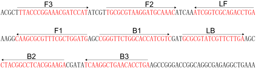 |
Figure 1 Sequence and location of Bscp31 gene used to design loop-mediated isothermal amplification primers. The nucleotide sequences of the sense strand of Bscp31 are listed. Right arrows and left arrows indicate sense and complementary sequences that are used. |
Bacterial strains and template preparation
A total of 42 Brucella strains (including B. abortus, B. melitensis, B. ovis, B. canis, B. suis, B. neotomae) and 19 non-Brucella isolates were employed at the current study (Table 2). Vaccine strain B. suis (GZ-CDC-S2) was used as a reference strain for optimizing the LAMP-LFB assay. According to the manufacture’s instructions, the templates were prepared using DNA extraction Kits, and the extracted DNA were quantified using ultraviolet spectrophotometer (Nano drop ND-1000, Calibre, Beijing, China) at A260/280. The templates extracted from GZCDC-S2 were serially diluted ranging from 10 ng/μL to 100 aq/μL (10 ng/μL, 10 pg/μL, 1 pg/μL, 100 fg/μL, 10 fg/μL, 1 fg/μL, and 100 aq/μL), which was used for optimizing reaction temperature and testing assay’s sensitivity. A volume of 1 μL of each dilution was used as a template for LAMP reactions.
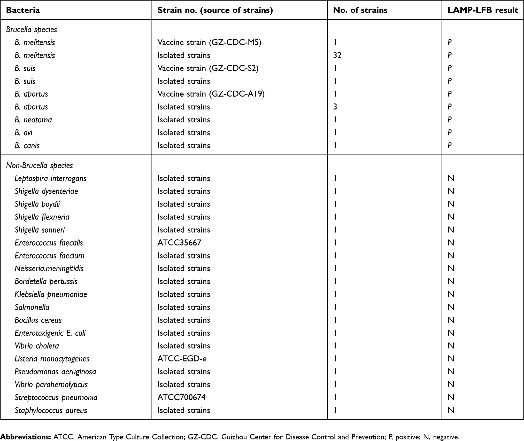 |
Table 2 Bacterial strains used in the current study |
Preparation of lateral flow biosensor
In this report, LFB was used for reporting LAMP results, and was constructed as previous publications.17–19 In brief, LFB includes an absorbent pad, NC membrane, an immersion pad, a conjugate pad, and a backing pad. SA-PNPs (Dye streptavidin-coated polymer nanoparticles) were gathered in the conjugate pad. Then, anti-FITC and biotin-BSA were immobilized at test line (TL) and control line (CL), respectively.
Brucella-LAMP assay
Brucella-LAMP amplification was carried out in a one-step reaction in a 25-μL mixture containing 12.5 μL of supplied buffer (2 X), 0.4 μM each of outer primer F3 and B3, 0.8 μM each of loop primers LF and LB, 1.6 μM each of inner primers FIP and BIP, 0.4 mM biotin-14-dCTP, 1 μL (8 U) of Bst 2.0 DNA polymerase and 1 μL of template. Double distilled water (DW) was used as the template in the blank control (BC) sample, and non-Brucella genomic DNAs, including Staphylococcus aureus and Salmonella DNAs, were used as the templates in the negative control (NC) sample.
LAMP-LFB assay
A 1 μL aliquot of LAMP products, which were FITC and biotin-labeled LAMP products, was loaded into the sample zone. Then, a volume of 60 μL of running buffer was also loaded into the same region. As a result, the capillary flow can simultaneously transfer LAMP products and SA-PNPs from the conjugate region to TL and CL. Biotin-labeled LAMP products form complex with SA-PNPs via biotin-streptavidin-biotin interactions at the conjugated zone, and biotin/LAMP complexes were immobilized at the test band by interaction between hapten (FITC) and anti-FITC. SA-PNPs that did not construct complexes were captured at CL by interaction between streptavidin and biotin. Thus, SA-PNPs/LAMP/FITC complexes and non-complexed SA-PNPs were reported by visible line at TL and CL, respectively. Moreover, additional monitoring techniques, including real-time turbidity (LA-320C) and colorimetric indicator (Malachite Green, MG), were also used for reporting the Brucella-LAMP products.
Optimizing the reaction temperature of LAMP-LFB assay
The effect of different temperatures (from 60ºC to 67ºC, with 1ºC intervals) on LAMP amplification was determined. Amplification mixtures with 1 μL of template of S. aureus (isolated strain) and Salmonella (isolated strain) were used as NCs. Amplification mixtures with 1 μL of double DW were used as a BC.
Sensitivity of LAMP-LFB assay
Analytical sensitivity of LAMP-LFB assay was tested using serially diluted B. suis (GZ-CDC-S2) genomic DNA, and assay’s sensitivity was verified as the last dilution of each positive test. For comparison, sensitivity of Brucella-LAMP assay using real-time turbidity and colorimetric indicator (MG) was also examined.
Optimizing the reaction time of LAMP-LFB assay
The effect of different times (from 30 mins to 60 mins, with 10 mins intervals) on LAMP amplification was evaluated, and the results were reported using LFB.
Specificity of LAMP-LFB assay
The specificity of LAMP-LFB was demonstrated using genomic DNA (at least 10 ng per microliters) from 42-Brucella strains and 19 non-Brucella strains (Table 2), and a 1 μL aliquot of genomic DNA was used as a template for LAMP reactions. All LAMP results were indicated using biosensor. All samples were repeated two times.
Evaluation of the feasibility of LAMP-LFB assay
A total number of 117 whole blood samples, which were suspected from human (21) and goat (96) brucellosis, were collected from different regions of Guizhou province, China (Ethic consideration: The National Health and Family Planning Commission of China determined that the collection of data from human cases of brucellosis was part of continuing public health surveillance of a notifiable infectious disease and was exempt from institutional review board assessment. All data were supplied and analyzed in an anonymous format, without access to personal identifying information).21 These samples were used for Brucella spp. diagnosis using culture-based technique, PCR detection, and LAMP-LFB test. Traditional blood cultures were conducted with BACTEC FX system (Becton-Dickinson, Sparks, MD), incubated for six weeks and sub-cultured weekly.22 In brief, about 3 mL fresh venous blood samples was aseptically inoculated into a two-phase culture flask (BIOVD, Zhengzhou, Henan, China) to cultivate and isolate Brucella strains. Post incubation at the conditions of 37ºC with 5% CO2 for 3–5 days (or more 3–5 days cultivation for blind passage), the bacteria strain was streaked on blood agar plate and Brucella agar plate for pure cultivation. Subsequent methods including Gram stain, serum agglutination test, phage lysis test, were applied for the identification of the Brucella suspicious isolates. In addition, DNA templates from these practical samples (500 μL) were directly extracted using protocol of QIAamp for PCR and LAMP-LFB assays. PCR diagnosis was carried out using Brucella spp. specific primers (B4 and B5 primers) targeting bscp31 gene having an amplicon size of 224 bp.23 The examination results produced from LAMP-LFB method were compared with culture-based assay and PCR detection.
Results
Confirmation and demonstration of LAMP products
An appreciable LAMP reaction was observed when the assay was conducted at a fixed temperature of 63ºC for a duration time of 1 hr. The Brucella-LAMP amplified products exhibited light green under visible light, while the negative samples remained colorlessness (Figure 2A). Further validation of Brucella-LAMP amplification was visualized through the appearance of two crimson red bands (TL and CL) in biosensor, while the negatives samples only appeared a crimson red line (CL) in biosensor (Figure 2B). Our results confirmed that the LAMP primer set targeting Bscp31 gene was appropriate candidate for establishment of LAMP-LFB assay for Brucella spp. detection.
 |
Figure 2 Validation of Brucella-LAMP products. (A), Color change of Brucella-LAMP tubes; (B), biosensor applied for visual detection of Brucella-LAMP products. Tube A1 (biosensor B1), positive amplification; tube A2 (biosensor B2), negative amplification (Salmonella), tube A3 (biosensor B3), negative amplification (S. aureus), tube A4 (biosensor B4), negative control (DW). |
Optimal temperature of LAMP primer set
Brucella-LAMP reaction condition was standardized to find out the optimum temperature. Eight amplification temperatures (ranging 60–67ºC, with 1ºC interval) were examined and compared under Brucella-LAMP protocol presented above. By real-time turbidity, all tested temperatures produced the kinetics graphs, and faster reactions were obtained for assay temperature of 62–64ºC (Figure 3). Assay temperature of 63ºC was employed for conducting the rest of LAMP reactions in the current study.
 |
Figure 3 Optimal temperature for Brucella-LAMP primer set. The LAMP amplifications for detection of Bscp31 sequence were examined by real-time measurement of turbidity. The corresponding curves of concentrations of templates were marked in the figures. Eight kinetic graphs (1–8) were generated at various temperatures (60–67°C, 1°C intervals) with target pathogens DNA at the level of 1 pg per tube (The threshold value was 0.1 and the turbidity of >0.1 was considered to be positive). The graphs from 3 (62°C) to 6 (64°C) showed robust reaction. |
Sensitivity of Brucella-LAMP method
Analytical sensitivity of Brucella-LAMP assay was evaluated by limiting dilution of Brucella genomic DNA. The lowest limit of detection by Brucella-LAMP assay was found to be 100 fg per reaction. TL and CL were observed on the biosensor, displaying the positive LAMP results for Bscp31 gene (Figure 4B). Moreover, the analytical sensitivity of Brucella-LAMP using biosensor was consistent with real-time turbidity detection (Figure 4C) and colorimetric indicator analysis (Figure 4A).
 |
Figure 4 Sensitivity of Brucella-LAMP-LFB method using serially diluted genomic DNA with B. suis strain GZ-CDC-S2. Signals (A)/Tubes (B)/Biosensors (C) 1–8 represented the DNA levels of 10 ng, 10 pg, 1 pg, 100 fg, 10 fg, 1 fg, 100 atto gram per reaction and blank control (DW). The genomic DNA levels of 10 ng to 100 fg per reaction yielded the positive reactions. |
Optimal duration time of Brucella-LAMP assay
We evaluated the effect of different duration time (ranging from 20 mins to 50 mins with 10 mins interval) at optimal amplification temperature (63ºC), and the target DNA at the LoD level (100 fg) was detected when Brucella-LAMP reaction lasted only for 40-mins (Figure 5). Hence, an amplification time of 40-mins was used as the optimal LAMP time for conducting the rest LAMP amplifications performed at the current report. As a result, the whole procedure, including target DNA preparation (20 mins), Brucella-LAMP reaction (40 mins) and result indicating (2 mins), was completed within 65 mins.
 |
Figure 5 Optimal duration of time required for Brucella-LAMP-LFB assay. Four reaction times (A), 20 mins; (B), 30 mins; (C), 40 mins; and (D), 50 mins were tested and compared at optimal temperature (63°C). Biosensors 1, 2, 3, and 4 represent DNA levels of 10 ng μL−1, 10 pg, 1 pg μL−1, and 100 fg. The best sensitivity was obtained when the amplification lasted for 40 mins (C). |
Specificity of Brucella-LAMP method
The analytical specificity of Brucella-LAMP method was determined using Brucella vaccine strains, Brucella isolated strains, and non-Brucella bacterial pathogens. As shown in Figure 6 and Table 2, Brucella-LAMP assay specifically detected all Brucella species/strains, while non-Brucella bacterial pathogens were not detected. By biosensor, TL and CL simultaneously appeared at detection regions of LFB, suggesting the positive results for Brucella pathogens (Figure 6, biosensor 1–10). Only one crimson band (CL) appeared at the detection zone of LFB, reporting the negative results for non-Brucella strains and BC (DW) (Figure 6, biosensor 11–30).
 |
Figure 6 Specificity of Brucella-LAMP-LFB assay using different bacterial strains. The Brucella-LAMP-LFB was tested using different genomic DNAs. Biosensor 1, B. melitensis (GZ-CDC-M5); biosensor 2, B. suis (GZ-CDC-S2); biosensor 3, B. abortus (GZ-CDC-A19); biosensor 4, B. canis (isolated strain); biosensor 5, B. ovi (isolated strain); biosensor 6, B. neotoma (isolated strain); biosensor 7–8, B. melitensis (isolated strains); biosensor 9, B. suis (isolated strain); biosensor 10, B. abortus (isolated strain); Biosensor 11–29, Leptospira interrogans; Shigella dysenteriae; Shigella boydii; Shigella flexneria; Shigella sonneri; Enterococcus faecalis; Enterococcus faecium; Neisseria meningitidis; Bordetella pertussis; Klebsiella pneumoniae; Salmonella; Bacillus cereus; Enterotoxigenic E. coli; Vibrio cholera; Listeria monocytogenes; Pseudomonas aeruginosa; Vibrio parahemolyticus; Streptococcus pneumonia; Staphylococcus aureus; Biosensor 30, negative control (DW). |
Demonstrating the feasibility of Brucella-LAMP-LFB assay by whole blood sample
We further determined the feasibility of Brucella-LAMP-LFB as a valuable tool for target pathogen detection. A total of 117 whole blood samples suspected from human and goat brucellosis were tested by LAMP-LFB assay, conventional PCR detection, and culture-biotechnical method. The results are summarized in Table 3. In the case of 117 biological samples, 13 (11.11%) and 13 (11.11%) samples were Brucella-positive by LAMP-LFB and traditional culture-biotechnical method, respectively. Thus, diagnostic accuracy obtained from LAMP-LFB assay was 100% when compared to the culture-biotechnical method. However, only 11 (9.40%) were Brucella-positive by conventional PCR technique. These results indicated that the Brucella-LAMP-LFB assay devised here was a valuable tool for target pathogen detection, and exhibited higher diagnostic ability when compared to the PCR method.
 |
Table 3 Comparison of conventional LAMP-LFB, culture-biotechnical and PCR methods for the detection of Brucella in whole blood samples of human and goats |
Discussion
Brucella spp. is responsible for brucellosis in both human and animals, and human brucellosis remains the world’s most common bacterial zoonosis with more than 500,000 new cases annually.24 Particularly, human brucellosis is related to substantial residual disability, and is a vital cause of travel-associated morbidity.25 Diagnostic techniques, including microbiological isolation and identification of pathogens and nucleic acid amplification-based techniques, have been established for the detection of the target pathogens. However, traditional diagnostic assays for Brucella detection, such as culture-biotechnical methods and PCR-based techniques, are time-consuming and laborious.2 Hence, a newer diagnostic technique is required for rapid, simple, reliable detection of Brucella spp. strains in clinical, basic, and field laboratories.
To actualize more such effective detection tool, a LAMP-LFB assay (loop-mediated isothermal amplification coupled with nanoparticles-based LFB) has been developed for detection of Brucellaspp. in this report. A set of LAMP primers (F3, B3, FIP, BIP, LF, and LB), which specifically recognized eight regions of the Brucella-specific gene (Bscp31 gene), was designed on the basis of LAMP rules, thus providing a high degree of selectivity for target pathogen diagnostic (Figure 1).26,27 To practically demonstrate the specificity of LAMP-LFB method, genomic DNAs extracted from Brucella spp. strains and non-Brucella bacterial strains were examined (Figure 6 and Table 2). Brucella-LAMP-LFB assay was able to detect all Brucella strains. All negative results were observed from the assay of non-Brucella spp. strains, and thus no cross-reactions to non-Brucella strains were obtained according to the specificity test. Herein, LAMP-LFB assay devised in the report detected these target pathogens with 100% specificity.
As an isothermal amplification technique, LAMP assay like multiple cross displacement amplification and cross-priming amplification only utilizes Bst polymerase enzyme which displays high resistance to known Taq polymerase inhibitors such as NaCl, hemoglobin, EDTA, N-acetyl cysteine, and bile salts.28,29 So far, LAMP assay has been used to detect a variety of pathogens, including Brucella spp.14,30,31 Unfortunately, these Brucella-LAMP assays required the use of agarose gel electrophoresis, color indicator (such as hydroxynaphthol blue, SYBR green I, calcein dye et al) and real-time turbidity equipment for indicating the amplification results. As a result, these Brucella-LAMP methods relied on a complex procedure (gel electrophoresis), a special optical instrument (real-time turbidity instrument), or special indicator (hydroxynaphthol blue or SYBR green I), which hampered their wider application in field, “on-site” or POC laboratories.
Here, the first report, which employed LFB for analyzing LAMP products, was developed for rapid, visual, simple, and reliable detection of Brucella spp. strains (Figures 2–6). Comparing with the other monitoring techniques (ie, colorimetric indicator, gel electrophoresis, and turbidity) employed in previous publications, LFB showed its superiority on simple operation, rapid results, and ease of use in basic, clinical, and field laboratories. In particular, reporting Brucella-LAMP results by biosensor was able to avoid the use of special apparatus, reagent, and additional procedure, thus, Brucella-LAMP-LFB assay was more suitable than other Brucella-LAMP methods developed by previous studies for simple, visual, and rapid diagnostic of target pathogens.
The analytical sensitivity results showed that the Brucella-LAMP-LFB assay devised here was able to detect the target gene (Bscp31 gene) even from 100 fg of the strain. The assay’s sensitivity obtained from LFB detection was consistent with real-time turbidity detection and colorimetric indicator (MG) analysis (Figure 4). The whole procedure of LAMP-LFB detection, including genomic template preparation (20 mins), LAMP reaction (40 mins), and LFB analysis (2 mins), was competed within 65 mins. For further evaluating the practical availability of LAMP-LFB assay to target pathogens, this report evaluated 117 whole blood samples of goats and human using culture bio-technique method, PCR diagnostic and LAMP-LFB detection. The LAMP-LFB technique exhibited high detection analysis for Brucella spp. Particularly, two biological samples were determined to be positive by culture-biotechnical assay and LAMP-LFB, but negative by traditional PCR detection. Lower diagnostic rate of PCR method may be due to the reasons that the copy numbers of target genomic templates were lower than the limit of detection or the presence of inhibitors specific to the conventional PCR technique affected the detection sensitivity. Moreover, comparing with culture-based assays and PCR method, Brucella-LAMP-LFB technique was conducted with only a simple instrument that provides a constant temperature of 63ºC, avoiding the long turnaround times and removing the use of expensive apparatus. But the LAMP-LFB assay has its own limitation as the LAMP results are indicated qualitatively as red colored bands.
In conclusion, a LAMP-LFB assay, which targeted the Brucella-specific Bscp31 gene, was successfully developed and validated for the detection of Brucella spp. The assay showed high analytical specificity for Brucella detection, and had the LoD of 100 fg per vessel with pure culture. The LAMP results were visually, rapidly, and indirectly reported using biosensor, which were objective, easy-to-use, and disposable. Brucella-LAMP-LFB assay established here was a simple, rapid, sensitive, and reliable method, which could be used as a potential diagnosis tool for target pathogens in basic, clinical, and field laboratory.
Acknowledgments
This study was supported by the grant of the research team for experimental diagnostic techniques and molecular epidemiological studies of major infectious diseases in Guizhou Province (Program of Scientific and Technological Innovation Team of Guizhou Province. Grant No. Qian Ke He Platform talent [2018]5606), Special Funds for High-Level Creative Talents Cultivation in Guizhou Province (Qian Ke He (2016)4021), and Special Funds for the Cultivation of Outstanding Youth Talents of Science and Technology in Guizhou Province (No. Qian Ke He Ren Word [2015] 09).
Disclosure
The authors declare that the research was conducted in the absence of any commercial or financial relationships that could be construed as a potential conflict of interest.
References
1. Zadon S, Sharma NS, Arora AK, Chandra M. Development of the novel loop mediated isothermal amplification (LAMP) of IS711 sequence for rapid detection of Brucella species. Proc Natl Acad Sci India Sect B. 2015;85(2):685–691. doi:10.1007/s40011-014-0377-9
2. Kaden R, Ferrari S, Alm E, Wahab T. A novel real-time PCR assay for specific detection of Brucella melitensis. BMC Infect Dis. 2017;17(1):230. doi:10.1186/s12879-017-2757-2
3. De BK, Stauffer L, Koylass MS, et al. Novel Brucella strain (BO1) associated with a prosthetic breast implant infection. J Clin Microbiol. 2008;46(1):43–49. doi:10.1128/JCM.01494-07
4. Scholz HC, Revilla-Fernández S, Dahouk SA, et al. Brucella vulpis sp. nov., isolated from mandibular lymph nodes of red foxes (Vulpes vulpes). Int J Syst Evol Microbiol. 2016;66(5):2090–2098. doi:10.1099/ijsem.0.000998
5. Wasl Al-Adsani AA, Al-Mousa M. A case of Brucella melitensis endocarditis in a patient with cardiovascular implantable electronic device. Infect Drug Resist. 2018;11:387. doi:10.2147/IDR.S152771
6. Traxler RM, Guerra MA, Morrow MG, et al. Review of brucellosis cases from laboratory exposures in the United States in 2008 to 2011 and improved strategies for disease prevention. J Clin Microbiol. 2013;51(9):3132–3136. doi:10.1128/JCM.00813-13
7. Muñoz PM, Marín CM, Monreal D, et al. Efficacy of several serological tests and antigens for diagnosis of bovine brucellosis in the presence of false-positive serological results due to yersinia enterocolitica O:9. Clin Diagn Lab Immunol. 2005;12(1):141–151. doi:10.1128/CDLI.12.1.141-151.2005
8. Schwarz NG, Loderstaedt U, Hahn A, et al. Microbiological laboratory diagnostics of neglected zoonotic diseases (NZDs). Acta Trop. 2017;165:40–65. doi:10.1016/j.actatropica.2015.09.003
9. Gupta VK, Verma DK, Rout PK, Singh SV, Vihan VS. Polymerase chain reaction (PCR) for detection of Brucella melitensis in goat milk. Small Ruminant Res. 2006;65(1):79–84. doi:10.1016/j.smallrumres.2005.05.024
10. Law JW, Ab Mutalib NS, Chan KG, Lee LH. Rapid methods for the detection of foodborne bacterial pathogens: principles, applications, advantages and limitations. Front Microbiol. 2014;5:770. doi:10.3389/fmicb.2014.00547
11. Moosavian M, Seyed-Mohammadi S, Saki M, et al. Loop-mediated isothermal amplification for detection of Legionella pneumophila in respiratory specimens of hospitalized patients in Ahvaz, southwest Iran. Infect Drug Resist. 2019;12:529. doi:10.2147/IDR.S198099
12. Bhat IA, Mashooq M, Kumar D, Varshney R, Rathore R. Development of probe based real time loop mediated isothermal amplification for detection of Brucella. J Appl Microbiol. 2018;126(5):1332–1339.
13. Prusty BR, Chaudhuri P, Chaturvedi VK, Saini M, Mishra BP, Gupta PK. Visual detection of Brucella spp. in spiked bovine semen using loop-mediated isothermal amplification (LAMP) assay. Indian J Microbiol. 2016;56(2):142–147. doi:10.1007/s12088-015-0563-3
14. Soleimani M, Shams S, Majidzadeh AK. Developing a real-time quantitative loop-mediated isothermal amplification assay as a rapid and accurate method for detection of Brucellosis. J Appl Microbiol. 2013;115(3):828–834. doi:10.1111/jam.12290
15. Pérez-Sancho M, García-Seco T, Arrogante L, et al. Development and evaluation of an IS711-based loop mediated isothermal amplification method (LAMP) for detection of Brucella spp. on clinical samples. Res Vet Sci. 2013;95(2):489–494. doi:10.1016/j.rvsc.2013.05.002
16. Kang S-I, Her M, Kim J-Y, et al. Rapid and specific identification of Brucella abortus using the loop-mediated isothermal amplification (LAMP) assay. Comp Immunol Microbiol Infect Dis. 2015;40:1–6. doi:10.1016/j.cimid.2015.03.001
17. Wang Y, Wang Y, Li D, Xu J, Ye C. Detection of nucleic acids and elimination of carryover contamination by using loop-mediated isothermal amplification and antarctic thermal sensitive uracil-DNA-glycosylase in a lateral flow biosensor: application to the detection of Streptococcus pneumoniae. Microchimica Acta. 2018;185(4):212.
18. Wang Y, Liu D, Deng J, Wang Y, Xu J, Ye C. Loop-mediated isothermal amplification using self-avoiding molecular recognition systems and antarctic thermal sensitive uracil-DNA-glycosylase for detection of nucleic acid with prevention of carryover contamination. Anal Chim Acta. 2017;996:74–87. doi:10.1016/j.aca.2017.10.022
19. Wang Y, Li H, Wang Y, Zhang L, Xu J, Ye C. Loop-mediated isothermal amplification label-based gold nanoparticles lateral flow biosensor for detection of enterococcus faecalis and staphylococcus aureus. Front Microbiol. 2017;8:192. doi:10.3389/fcimb.2018.00192
20. Ohtsuki R, Kawamoto K, Kato Y, Shah MM, Ezaki T, Makino SI. Rapid detection of Brucella spp. by the loop-mediated isothermal amplification method. J Appl Microbiol. 2008;104(6):1815–1823. doi:10.1111/j.1365-2672.2008.03732.x
21. Lai S, Zhou H, Xiong W, et al. Changing epidemiology of human brucellosis, China, 1955–2014. Emerg Infect Dis. 2017;23(2):184. doi:10.3201/eid2311.170833
22. Sagi M, Nesher L, Yagupsky P. The BACTEC FX blood culture system detects Brucella melitensis bacteremia in adult patients within the routine one-week incubation period. J Clin Microbiol. 2017. doi:10.1128/JCM.02320-16
23. Baily GG, Krahn JB, Drasar BS, Stoker NG. Detection of Brucella melitensis and Brucella abortus by DNA amplification. J Trop Med Hyg. 1992;95(4):271–275.
24. Pappas G, Papadimitriou P, Akritidis N, Christou L, Tsianos EV. The new global map of human brucellosis. Lancet Infect Dis. 2006;6(2):91–99. doi:10.1016/S1473-3099(06)70382-6
25. Memish ZA, Balkhy HH. Brucellosis and international travel. J Travel Med. 2004;11(1):49–55. doi:10.2310/7060.2004.13551
26. Notomi T, Okayama H, Masubuchi H, et al. Loop-mediated isothermal amplification of DNA. Nucleic Acids Res. 2000;28(12):e63. doi:10.1093/nar/28.12.e63
27. Nagamine K, Hase T, Notomi T. Accelerated reaction by loop-mediated isothermal amplification using loop primers. Mol Cell Probes. 2002;16(3):223–229.
28. Francois P, Tangomo M, Hibbs J, et al. Robustness of a loop-mediated isothermal amplification reaction for diagnostic applications. FEMS Immunol Med Microbiol. 2011;62(1):41–48. doi:10.1111/j.1574-695X.2011.00785.x
29. Kaneko H, Kawana T, Fukushima E, Suzutani T. Tolerance of loop-mediated isothermal amplification to a culture medium and biological substances. J Biochem Biophys Methods. 2007;70(3):499–501. doi:10.1016/j.jbbm.2006.08.008
30. Song L, Li J, Hou S, Li X, Chen S. Establishment of loop-mediated isothermal amplification (LAMP) for rapid detection of Brucella spp. and application to milk and blood samples. J Microbiol Methods. 2012;90(3):292–297. doi:10.1016/j.mimet.2012.05.024
31. Karthik K, Rathore R, Thomas P, et al. Rapid and visual loop mediated isothermal amplification (LAMP) test for the detection of Brucella spp. and its applicability in epidemiology of bovine brucellosis. Veterinarski Arhiv. 2016;86(1):35–47.
 © 2019 The Author(s). This work is published and licensed by Dove Medical Press Limited. The full terms of this license are available at https://www.dovepress.com/terms.php and incorporate the Creative Commons Attribution - Non Commercial (unported, v3.0) License.
By accessing the work you hereby accept the Terms. Non-commercial uses of the work are permitted without any further permission from Dove Medical Press Limited, provided the work is properly attributed. For permission for commercial use of this work, please see paragraphs 4.2 and 5 of our Terms.
© 2019 The Author(s). This work is published and licensed by Dove Medical Press Limited. The full terms of this license are available at https://www.dovepress.com/terms.php and incorporate the Creative Commons Attribution - Non Commercial (unported, v3.0) License.
By accessing the work you hereby accept the Terms. Non-commercial uses of the work are permitted without any further permission from Dove Medical Press Limited, provided the work is properly attributed. For permission for commercial use of this work, please see paragraphs 4.2 and 5 of our Terms.