")
Back to Journals » Clinical, Cosmetic and Investigational Dentistry » Volume 15
Correlation Between Maxillary Anterior Teeth and Common Facial Measurements
Authors Alshamri HA, Al Moaleem MM , Al-Huthaifi BH, Al-labani MA, Naseeb WR, Daghriri SM, Suhail IM, Hamzi WH, Abu Illah MJ, Thubab AY, Aljabali SA, AlNehmi MM
Received 26 September 2023
Accepted for publication 31 October 2023
Published 7 November 2023 Volume 2023:15 Pages 289—300
DOI https://doi.org/10.2147/CCIDE.S438302
Checked for plagiarism Yes
Review by Single anonymous peer review
Peer reviewer comments 2
Editor who approved publication: Professor Christopher E. Okunseri
Hameed A Alshamri,1 Mohammed M Al Moaleem,2 Basem H Al-Huthaifi,3 Mohammed A Al-labani,4 Weaam RB Naseeb,5 Shahad Mohammed Daghriri,5 Ibtihaj Mohammed Suhail,5 Wasan H Hamzi,5 Mohammed J Abu Illah,5 Abdulaziz Yahya Thubab,5 Shuaib A Aljabali,3 Mohammed M AlNehmi1
1Department of Restorative and Prosthodontics, College of Dentistry, University of Science and Technology, Sana’a City, Yemen; 2Department of Prosthetic Dental Science, College of Dentistry, Jazan University, Jazan, Saudi Arabia; 3Department of Preventive and Biomedical Science, College of Dentistry, University of Science and Technology, Sana’a City, Yemen; 4Department of Orthodontics, College of Dentistry, University of Sana’a, Sana’a City, Yemen; 5College of Dentistry, Jazan University, Jazan, Saudi Arabia
Correspondence: Mohammed M Al Moaleem, Department of Prosthetic Dental Science, College of Dentistry, Jazan University, Jazan, 45142, Saudi Arabia, Email [email protected]
Background: The symmetry between maxillary anterior teeth and the face holds significant importance. This study assessed and analyzed the relationship between facial parameters and anterior teeth in the maxillary arch of male and female subjects. Specifically, individual width and combined width (CW) measurements of the maxillary anterior teeth were investigated.
Methods: This study involved a total of 150 dentate Yemeni subjects (74 men and 76 women), whose ages ranged from 18 years old to 30 years old. A maxillary cast was created, and two digital photographs of the face of each subject were taken and analyzed. Digital calipers and AutoCAD were used to gather measurement data of the dental parameters (intercanthal distance [ICD], interpupillary distance [IPD], interalar width [IAW], intercommissural width [ICW], and bizygomatic width [BZW]) and facial parameters (profile distance).
Results: Significant correlations were found for the following: IPD and width of six maxillary anterior teeth of each of the study subjects; ICD and their central incisors; and BZW and their canine width measurements. In contrast, IAW and ICW were not correlated with all tooth measurements. Linear regression findings showed that the CW measurement of the four incisors was significantly correlated with all facial parameter measurements, excluding the ICW and IAW in females and the IAW, ICW, and profile distance in males.
Conclusion: The IPD and ICD of males and females may be used to determine their CW measurements. The BZW and IPD of males can be used to take precise anthropological measurements of the width of the central canines and incisors. Meanwhile, the IPD distance of females can be used to assess the central and lateral incisor widths.
Keywords: dental measurement, facial measurement, anterior teeth, Yemen
Introduction
The symmetry between maxillary anterior teeth and face is intricately connected to notion of attractiveness and beauty, and dental and facial measurements are strongly associated with aesthetic smiles. Clinicians may provide proper treatment plans that take into account the various potential factors influencing the perception of an aesthetic smile by using self-satisfaction measurements.1,2 Dental aesthetics is a main influence affecting people’s psychosocial wellbeing. Among the most critical components of an aesthetic smile is proportion within the maxillary anterior dentition and the surrounding oral structures.3,4 A recent review supported that the existence of evidence of the golden proportion in natural smiles is lacking and the existence of this proportion in dentistry is a myth and not a fact.5
Furthermore, the general appearance of the maxillary anterior teeth considerably influences dental and facial aesthetics. Several anatomical facial measurements, including the bizygomatic width (BZW), interpupillary distance (IPD), interalar width (IAW), intercanthal distance (ICD), and intercommissural width (ICW), can help in determining the individual and combined dimensions of the maxillary incisor teeth.6–8
When pre-extraction records are unavailable, evaluation of the combined width (CW) measurements of the six anterior maxillary teeth can be challenging. Meanwhile, the correlation between the width of the maxillary anterior teeth and the width of the face can be used to generate samples of denture teeth and determine the proper size of teeth for fixed restoration.9 The measurements of the individual width and CW of the six maxillary anterior teeth can be predicted on the basis of the distance between the intercanthus, between the inter ala of the nose, or between corners of the mouth width (ie, ICD, IAD, or ICW, respectively).8,10,11 Multiple facial measurements are crucial in making informed decision regarding the width of maxillary anterior teeth.9,12
Facial and dental measurements exhibit variations across geographic locations, climate conditions, and the historical backgrounds of different subjects; these measurements are closely associated with human DNA, such as facial shape and features.13 When restoring teeth for facial aesthetics, dental professionals should take into account the patient’s heritage, cultural background, and societal norms. However, pre-extraction records of the Yemeni population are typically unavailable. The selection of the proper maxillary anterior tooth size can be complex and may result in unsatisfactory aesthetic outcomes. On the other hand, even when dealing with individuals who have preextraction records, different measurement points on the face still need to be assessed because older dental records of a patient might not be readily accessible.14 These gaps can account for the numerous anatomical measurement methods that have been proposed by scholars.15
Hasanreisoglu et al examined the relationship between maxillary anterior tooth crown dimensions and facial measurements and found that the dimensions of central incisors and canines vary between gender groups.16 Significant differences were observed in the perceived widths in females, and proportional relationships were found between BZW and central incisor width. The ICD to central incisor width and IAD to anterior tooth straight-line width were used as dependent parameters for Kurdish male subjects. A L-Kaisy and Garib analyzed the frontal facial measurements and found correlations between the mesiodistal width of the maxillary teeth and the ICD, IPD, and ICM.17 Gomes et al18 confirmed the strong correlation between the mesiodistal width of the maxillary teeth and other facial measurements. However, no significant difference was established between the crown width and the face type of Bangladeshi subjects, of whom approximately 55.7% had narrow faces, with mean crown width–length ratios of central and lateral incisors and canines of 0.92±0.078, 0.88±0.172, and 0.89±0.097 mm.19 Parciak et al9 did not find correlations between the facial and mesiodistal dimensions of the six maxillary anterior teeth of subjects from three ethnicities, except for the central incisor width-to-BZW ratio; however, the ICWs of female subjects were higher than those of the general group across ethnicities. Flavie et al used the width of the central incisor and the distance between the two maxillary canine pointers to determine the bizygomatic distance.20
Regarding mesiodistal tooth width and tooth size discrepancies of Yemenis subjects, Al-Gunaid et al did not find any significant differences in the tooth size and width between the right and left sides of the jaw.21 Men have larger teeth than women, with clinically significant differences between their anterior and overall tooth size ratios. Alaghbari et al1 found that men have larger mean dental arch dimensions and facial measurements than women, with the greatest difference between their maxillary arch widths. Significant correlations were found between ICW and maxillary canine width. Nevertheless, dental arch width was not significantly correlated with BZW, IAW, or MW.
Numerous studies1,21–23 have investigated the relation of teeth with certain anatomical features of the faces of Yemeni subjects. However, none of these studies have evaluated the association between dental and facial aesthetic measurements of Yemeni adults. This work is the first correlation study to evaluate and compare dental and facial measurements between men and women using maxillary casts and digital images. In formulating the null hypothesis, a nonsignificant relationship between gender and dental or facial aesthetic measurement was assumed.
Subjects and Methods
Study Design and Subject Size Calculation
This analytical cross-sectional study was approved by the Medical Ethics Committee of the Dental Faculty of the University of Science and Technology (UST), Sana’a, Yemen (MECA No.: EAC/UST168). The research was conducted between October 2022 and April 2023. The patients attended by the Prosthodontics Department of UST and Sana’a University College of Dentistry in Sana’a City, Yemen, were selected as research subjects. All recruited subjects were informed about the research purpose, and those who agreed to participate signed consent forms. The subject size was calculated using G*Power version 3.1.9.4 at an 85% confidence level and 0.05 precision, and the actual proportion was 71.4%. The selection of 150 subjects for the study was based on the number of clinically managed patients by the departments 3 years prior to the study. A consent form signed by each participant was sought again before performing impression and facial measurements.
Inclusion and Exclusion Criteria
One hundred and fifty male and female Yemeni participants were recruited. The age was between 18 and 30 years old were enrolled in this cross-sectional study. The criteria included the following: the patient must have a symmetrical face, complete permanent maxillary and mandibular dentition, class I molar and canine occlusion, normal hard and soft tissues with no abnormalities, no crossbite, crowding of ≤2 mm, and spacing of ≤2 mm in either the maxillary or mandibular arches, overjet of ≤3 mm, no attrition on teeth, no retained deciduous teeth, and no peg lateral incisors. Patients who were using dental appliances, had undergone orthodontic treatment, had maxillofacial surgery, had dental implants, or had periodontal surgery in the maxillary arch were excluded from the study.
Data Collection, Study Tools, and Study Cast Measurements
The data were produced using a study cast, and then the research subjects were photographed. The castes have analyzed the dental arch dimensions on the dental stone casts (Dental Yellow Buff Stone, Type IV, USA) of a maxillary impression constructed using a silicone putty-type impression material (Vinyl Polysiloxane, Hamburg, Germany). Teeth were measured using a sliding caliper following the methods in the literature,1,9,20 and the width of each tooth in the anterior of the arch was measured using a Digital Vernier Caliper (Mitutoyo, São Paulo, Brazil) with a sharp tip and an accuracy of 0.01 mm. Dental parameters, such as individual mesiodistal width of maxillary central incisors, lateral incisors, and canines, were measured on the dental cast (Figure 1A). The clinical crowns of the maxillary anterior teeth were measured mesiodistally at the greatest dimension for each tooth width from the right to the left canine. CW measurements of the six maxillary anterior teeth were measured in a straight line at the distal surface of the canines as shown in Figure 1B.
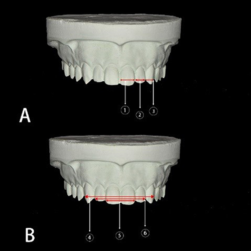 |
Figure 1 Study cast of the participant during width measurements of central, lateral, canines (A) and both incisors, both laterals, and maxillary 6 anterior teeth width (B). |
Facial Measurements
Each research subject was instructed to sit upright and face forward while sitting on a dental chair, and then their facial features were measured. Data on the frontal parameters (BZW, IPD, ICD, IAW, and ICW) were acquired using frontal view images (Figure 2A), while data on the width of the profile distance were obtained using lateral view images (Figure 2B). Then, all images were inputted into AutoCAD, and digital calipers were used to measure the facial features (in mm). The distance between the medial canthi of the eyes represents the ICD. Participants in Figure 2 have provided a written informed consent for the image to be published. Table 1 presents the measurements for all parameters related to the maxillary dental arch or facial dimensions measurements. A single investigator (BM) performed all dental and facial measurements.
 |
Table 1 Parameters, Abbreviations and Their Definitions, Views of the Study Casts and Photographs |
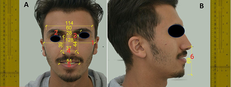 |
Figure 2 Parameters in facial measurements for frontal view (A) and lateral view (B). |
Data Analysis
The statistical analysis was performed in SPSS version 24.0 (SPSS Inc., Chicago, IL, USA). The p value was set to a significant level of 0.05. The Shapiro‒Wilk test was used to verify a normal distribution. Descriptive statistics (mean and standard deviation [SD]) and inferential statistics were used to compare the dental and facial measurements of the male and female subjects. The mean differences in dental and facial measurements were assessed through a T-test. The relationship between the face and maxillary anterior teeth was assessed by Pearson’s correlation coefficient test (r), with values set between −1 and +1 (ie, −1 ≥ r ≥ 1, where r > 0 means a positive correlation, r < 0 denotes a negative correlation, and r = 0 indicates the absence of correlation).
Results
Among the 150 participants, 74 (49.3%) were males and 76 (50.7%) were females, with a mean age and SD of the study sample was 23.431±1.3. As shown in Table 2, the males and females differed significantly in terms of mesiodistal width of the left lateral incisor, right central and canine, and CW measurements. The maxillary anterior teeth of males have a wider mesiodistal dimension, but they are not significantly different from those of females. The widest tooth width (in mm) was the left central incisor (8.05 mm) in the male subjects, whereas the narrowest tooth width was for the lateral incisors on the left and right sides (5.83 mm) among the female subjects. The facial measurements on both sides were near to each other’s or similar for the male and female subjects, presenting only slight variations (ie, nonsignificant difference; Table 3).
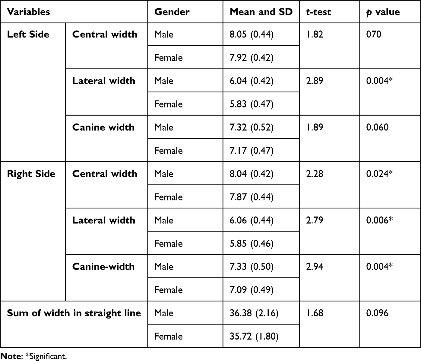 |
Table 2 Comparison of Individual Tooth Measurements by Gender (n = 150) |
 |
Table 3 Comparison of the Mean Differences in Facial Measurements by Gender |
A weak correlation is established between the summation widths of the six maxillary anterior teeth measured in a straight line (Table 4). By contrast, the IPD and CW measurements of the two central incisors, four incisors, and six maxillary anterior teeth were strongly correlated. The CW measurements of the two central incisors and the summation widths measured in a straight line were significantly correlated with ICD. In contrast to CW measurements of six maxillary anterior teeth and four incisors, ICD presents a moderate correlation. These findings indicate that the ICD of Yemeni adults can be estimated through CW measurements of the two central incisors and the summation width measured in a straight line.
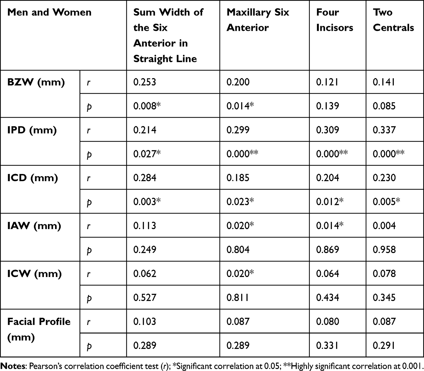 |
Table 4 Correlation Between Gender and Facial and CW Dental Measurements (n = 150) |
The summation width is strongly correlated with BZW but not with the six maxillary anterior teeth measured in a straight line (Table 5). The IPD and CW measurements of the two central incisors, four incisors, and six maxillary anterior teeth were strongly correlated. The CW data of two central incisors and the summation width measured in a straight line were significantly correlated with ICD. ICD only had a moderate correlation compared with CW measurement of maxillary six anterior teeth and four incisors. Our findings indicate that the ICD of Yemeni adults can be estimated using CW measurement of the two central incisors and summation width measured in a straight line. Meanwhile, the IPD is strongly correlated with the width of the right and left central incisors but only weakly correlated with the left canine. The ICD was also poorly correlated with the widths of the left central incisor, left lateral incisor, and right lateral incisor.
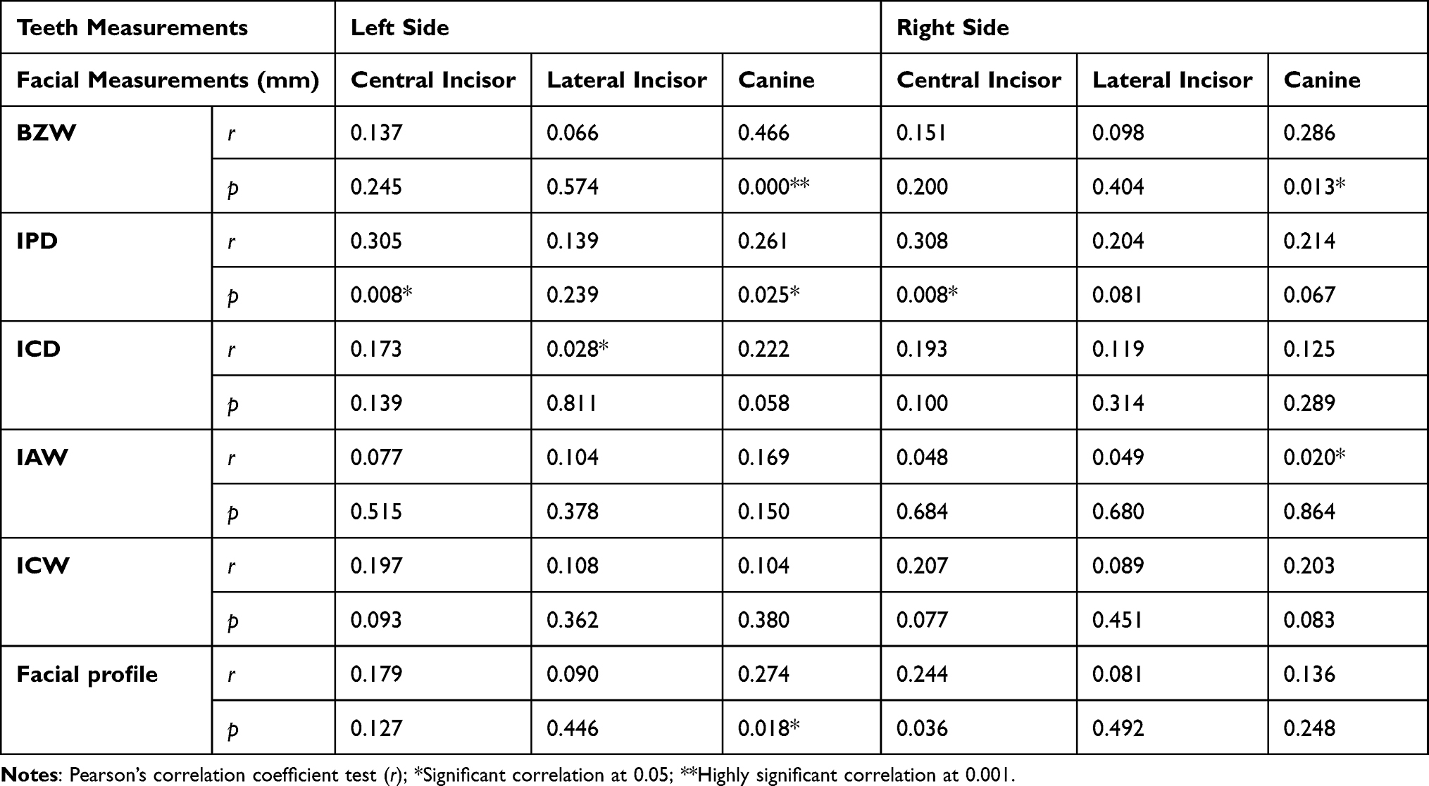 |
Table 5 Correlation Between Facial Measurements and Individual Width of Six Maxillary Anterior Teeth Measurements for Males (n = 74) |
In the female subjects (Table 6), statistically significant correlations were found between BZW and the width of canines on the left and right sides and the width of the right central incisor (p=0.001). However, no correlation was found between the width of the profiles and the width of the six maxillary teeth. The IPD was also not correlated with the width of the canines. Nonetheless, the IPD was significantly correlated with the central and lateral incisors on the left and right sides.
 |
Table 6 Correlation Between Facial Measurements and Individual Width of Six Maxillary Anterior Teeth Measurements for Females (n = 76) |
Discussion
Given the importance of correlation in facial measurement studies, numerous scholars worldwide have used face measurement parameters to determine tooth size for edentulous individuals or for fixed or removable prostheses.9–24
The main goal of this study was to establish accurate relationships between face measurements and maxillary anterior tooth width. The mean widths of six maxillary anterior teeth are wider for men than women, as supported by previous studies.21,24,25 However, few studies on the African population have shown contrasting results.20,26 The average distance between the distal surfaces of maxillary canines is 36.013 mm, which is similar to the values reported for Croatian and Kashmiri populations.27,28 The minor variations may be attributed to differences in measurement methods.
In the current study, the results of facial parametric analyses for BZW, IPD, IAW, and ICW revealed greater mean values in the Yemeni male than female subjects, which is consistent with earlier research.1 In relation to the mean facial measurement distance, the mean value of BZW was higher in the Yemeni male than female subjects, which aligns with the findings about the Kashmiri population28 but not with the Iraqi population29 or among other races.9 The mean IPD obtained in this study was 60.4 mm, which is consistent and within the range of values in earlier studies conducted in Turkey, the United States, and Malaysia;14,30,31 however, the values were less than those in prior studies that reported larger means of 73 and 69 mm.9,18
Meanwhile, no differences in the mean ICD were found between the Yemeni male and female subjects, which is comparable to the 28–35 mm range reported in the literature.32,33 In contrast with the research of other scholars, Abdullah et al,34 Al Wazzan et al35 and Dwivedi et al36 documented wider mean values of 28 and 34–36 mm, which can be explained by their research subjects having much wider eyes, and their ethnicity varied. Additionally, the IAW in this work showed a mean of 34 mm, with statistically nonsignificant differences between males and females, which is similar to the findings about Iraqi subjects29 but contrasts with some published investigations.18,36 In terms of the ICW, the mean of males was 46.87 mm higher than that of females (45.69 mm), with a statistically significant difference. This finding is consistent with the research results for Pakistan (45.24 mm)37 and Yemen by Alaghbari et al1 who reported males with a larger ICW mean than females (30.94 and 27.69 mm, respectively). However, the ICW mean obtained in this work contrasts with those of other studies.9,38 Overall, the male subjects had a greater mean for facial characteristics compared with female subjects. The human faces of males and females vary, especially during puberty, which may explain the derived results.39 Females recorded a higher psychological impact than males in relation to maxillary anterior teeth symmetry and this reflected on their quality of life in Polish subpopulation.40
BZW was significantly correlated with summation width. This finding aligns with a previous finding12 that showed BZ distance and age acting as predictors of central incisor and maxillary anterior teeth widths. Mishra et al24 and Ellakwa et al41 found weak correlations between IAW and the six maxillary anterior teeth. Ariani et al42 who studied the Indonesian population did not find any correlation between IAW and ICW and canine distance. Hunter and Priest43 used soft tissue measurements to demonstrate correlations between IAW and the width of six maxillary anterior teeth; nonetheless, this finding contrasts with the findings on subjects from Mongolia.44 Meanwhile, despite the method of recruiting live participants, this study could not confirm the conclusions provided by Hunter and Priest,43 which found that IAW is not equal to the CW measurement of six maxillary anterior teeth when multiplied by a factor of 1.31. The finding of this study also differs from that documented by Hoffman et al45 but it aligns with Parciak et al.9
A strong correlation was found between the BZW and the width of the left and right canines of males and the general population. This scenario differed for females. Meanwhile, the finding regarding the 1:14 ratio for the BZW to central incisor width was similar to the range of 1:13–1:19 reported by Flavie et al20 but it differed from the 1:16 ratio reported by Ellakwa et al41 and Bozkir et al.46 To the best of our knowledge, no research has been conducted on the use of BZW to measure anterior teeth as suggested earlier.47 Among Chinese population, wider maxillary incisors and canines were recorded, which is not in parallel with this study findings.48
The width of the left canine and the right and left central incisors of the male subjects recruited in this study were significantly correlated with the IPD. Meanwhile, the IPD of the female subjects was significantly correlated with the width of the right central incisor, the right and left lateral incisors, and the left central incisor but poorly correlated with the central incisor. This finding implies that the width of six maxillary anterior teeth and IPD have a strong statistical correlation across the population. The aforementioned finding is consistent with that found by Al-Kaisy and Garib17 but contradicts the results obtained by Parciak et al,9 which did not find a correlation between IPD and the width of six maxillary teeth. The ratio of IPD to the width of the maxillary central incisor, which was equal to 6.6,30 was not found in this study; rather, the ratios of 7.55 mm for males and 7.56 mm for females were established, which accords with the ratios of 7.7 and 7.5 mm for males and females, respectively.16
Furthermore, this study did not find significant differences in ICD with respect to sex orientation, which is consistent with the findings reported in the literature.1,20,38 The width of six maxillary anterior teeth and ICD were not significantly correlated among the male subjects, which is similar to that for the Iraqi population.29 The ICD was significantly correlated with the right and left central incisors of the female subjects, which is consistent with the findings with Köseoğlu et al.14 In terms of the total population, the ICD was correlated with the left and right central incisors but weakly correlated with the right lateral incisors and left canine width. The minor variations across the different scholarly findings may be related to genetic variability attributable to geographical origins and historical backgrounds. Nonetheless, ICD may be regarded as a reliable measurement of facial features for determining the width of maxillary anterior teeth among Yemeni adults.
This study did not find any correlation between IAW, ICW, and six maxillary anterior teeth, which is consistent with previous results.20,38 The central incisor width was statistically greater among the female subjects and lower than the IAW/4 ratio, confirming the Turkish and Saudi population studies.14,49 Furthermore, ICW was not significantly correlated with all intraoral measurements, confirming the findings of Hoffman et al and QAMAR et al.45,50 No correlations were established between profile measurements and the six maxillary anterior teeth among males, females, and the general population; this finding differs from those of other races/ethnicities, as reported in the literature.9,14,15,20,34
A commonly accepted view is that genetic causes and environmental adaptation both affect craniofacial features.51 Another explanation is that people of the same ethnicity, even those from the same villages, may have different facial physical characteristics, as illustrated in the literature.42,52,53 In this study, the male subjects presented longer facial measurements and lower face heights than the female subjects. This finding is supported by similar studies conducted in other countries or regions, such as Turkey, Cameroon, Iraq, and the central part of India.14,20,54–56 Furthermore, this study found significantly positive aesthetic correlations in the facial dimensions and dental characteristics of the Yemeni population. The aesthetic measurements of Yemenis people frequently match the acknowledged aesthetic norms established by dentists. Alone orthodontic treatment in participants with teeth and face asymmetry appears to significantly impact a range of psychological and aesthetic measurements.57 Orthodontics and prosthodontics have a moderate quality of evidence on influence on the self-esteem of both genders.58,59
One of the limitations of this study is that the research subjects were recruited from only one city and did not include different cities in the south, east, and west. Second, it does not include some other facial parameters, and categorizes the participants into different age groups. For research recommendation, future research may use digital calipers to evaluate the left and sides of the teeth. Also, to use some digital x-rays measurements or check the possibility of included artificial inelegancy in such topic.
Conclusions
IPD and ICD can be used to determine CW measurement of four incisors of male and female research subjects. Furthermore, BZW and IPD can be used to gather specific anthropological measurements for the width of central incisors and canines in males. Meanwhile, IPD can be used to measure the width of central and lateral incisors in females.
Ethical Approval
The procedures were conducted according to the principles outlined in the Declaration of Helsinki and Ethical Conduct for Research with Human Beings and the Good Clinical Practice Guidelines. Ethical approval was granted by the Ethics Board of the Medical Ethics Committee of the Dental Faculty of the University of Science and Technology (UST), Sana’a, Yemen in January/5/2018, No.: EAC/UST168.
Consent to Participate
Informed consent was taken from all participants included in the study.
Acknowledgment
We wish to express our gratitude to our colleagues at UST Yemen for their assistance in gathering and processing the data. We would like to extend our special thanks to our supervisor, Prof. Mohsen Alhamzi, for his support and for all of the opportunities we were given to advance this research.
Author Contributions
All authors made a significant contribution to the work reported, whether that is in the conception, study design, execution, acquisition of data, analysis and interpretation, funding or in all these areas; took part in drafting, revising, or critically reviewing the article; gave final approval of the version to be published; have agreed on the journal to which the article has been submitted; and agree to be accountable for all aspects of the work.
Funding
This study received no fund from any institution or company.
Disclosure
The authors declare that they have no conflicts of interest in this work.
References
1. Alaghbari SS, Mohammed BSA, Alalwani NN, et al. Analysis of the facial measurements and dental arch dimensions for the construction of dental prostheses among adult Yemenis. J Contemp Dent Pract. 2023;24(8):595–604. doi:10.5005/jp-journals-10024-3511
2. Shaabi FI, Al-Makramani BM, Al-Sanabani FA, Alraawi MA, Al Ahmari NM, Al Moaleem MM. The potential factors affecting the perception of aesthetic smile among adult patients attending dental clinics of Jazan University. Acta Stomatol Naissi. 2020;36(81):2022–2035. doi:10.5937/asn1980956S
3. Chrapla P, Paradowska-Stolarz A, Skoskiewicz-Malinowska K. Subjective and objective evaluation of the symmetry of maxillary incisors among residents of Southwest Poland. Symmetry. 2022;14(6):1257. doi:10.3390/sym14061257
4. Natanek Ł, Adamiecki MK, Kłosek S. Motivational interviewing in promoting oral health: a literature review. Dent Med Probl. 2023;60(2):355–362. doi:10.17219/dmp/140221
5. Londono J, Ghasemi S, Lawand G, Dashti M. Evaluation of the golden proportion in the natural dentition: a systematic review and meta-analysis. J Prosthet Dent. 2023;129(5):696–702. doi:10.1016/j.prosdent.2021.07.020
6. Latta GH, Weaver JR, Conkin JE. The relationship between the width of the mouth, interalar width, bizygomatic width, and interpupillary distance in edentulous patients. J Prosthet Dent. 1991;65(2):250–254. doi:10.1016/0022-3913(91)90170-2.
7. Hossain S, Islam KZ, Islam KM. Correlation between maxillary canines and facial anatomical landmarks in a group of Bangladeshi people. City Dent College J. 2012;9(2):12–14. doi:10.3329/cdcj.v9i2.12315
8. Shetty K, Kumar M, Palagiri K, Amanna S, Shetty S. Facial measurements as predictors of the length of the maxillary central incisor in a cross section of the Indian population - A clinical study. J Oral Hyg Health. 2013;1(01):106. doi:10.4172/2332-0702.1000106
9. Parciak EC, Dahiya AT, AlRumaih HS, Kattadiyil MT, Baba NZ, Goodacre CJ. Comparison of maxillary anterior tooth width and facial dimensions of 3 ethnicities. J Prosthet Dent. 2017;118(4):504–510. doi:10.1016/j.prosdent.2016.10.035.
10. Sinavarat P, Anunmana C, Hossain S. The relationship of maxillary canines to the facial anatomical landmarks in a group of Thai people. J Adv Prosthodont. 2013;5(4):369–373. doi:10.4047/jap.2013.5.4.369
11. Hussain MW, Qamar K, Naeem S. The role of interpupillary distance in the selection of anterior teeth. Pakistan Oral Dent J. 2012;32(1):165–169.
12. Scandrett FR, Kerber PE, Umrigar ZR. A clinical evaluation of techniques to determine the combined width of the maxillary anterior teeth and the maxillary central incisor. J Prosthetic Dent. 1982;48(1):15–22. doi:10.1016/0022-3913(82)90041-5
13. Richmond S, Howe LJ, Lewis S, Stergiakouli E, Zhurov A. Facial genetics: a brief overview. Front Genet. 2018;9:462. doi:10.3389/fgene.2018.00462.
14. Köseoğlu M, Özdemir H, Bayındır F. Evaluation of different smile parameters in the Turkish population. Inter Dent Res. 2018;8(1):1–6. doi:10.5577/intdentres.2018.vol8.no1.1
15. Kazanji M, Qadir AQM, Habeeb SH, Hasan AS. Relation of maxillary central incisors width to some facial measurements. J Oral Dent Res. 2017;4(2):93–101. doi:10.12816/0038704
16. Hasanreisoglu U, Berksun S, Aras K, Arslan I. An analysis of maxillary anterior teeth: facial and dental proportions. J Prosthet Dent. 2005;94(6):530–538. doi:10.1016/j.prosdent.2005.10.007.
17. Neda AK, Garib BT. Selecting maxillary anterior tooth width by measuring certain facial dimensions in the Kurdish population. J Prosthet Dent. 2016;115(3):329–334. doi:10.1016/j.prosdent.2015.08.012.
18. Gomes VL, Gonçalves LC, Do Prado CJ, Junior IL, de Lima Lucas B. Correlation between facial measurements and the mesiodistal width of the maxillary anterior teeth. J Esthet Restor Dent. 2006;18(4):196–205. doi:10.1111/j.1708-8240.2006.00019_1.x.
19. Jamayet N, Viwattanatipa N, Amornvit P, Alam MK. Comparison of crown width/length ratio of six maxillary anterior teeth between different facial groups in Bangladeshi. Inter Medi J. 2014;21(1):49–54.
20. Flavie AME, Pierrot KN, Omer N, Michael A, Paul SIBJ, Fidele NB. Correlation between the bizygomatic distance and the width of the upper central incisor in the Cameroonian melanoderma adult. Open J Stomatol. 2022;12:294–304. doi:10.4236/ojst.2022.1210026
21. Al-Gunaid T, Yamaki M, Saito I. Mesiodistal tooth width and tooth size discrepancies of Yemeni Arabians: a pilot study. J Orthod Sci. 2012;1(2):40–45. doi:10.4103/2278-0203.99760
22. Al-Zubair NM. The relationship between mandibular arch length and widths in a sample of Yemeni subjects with normal Dento-Skeletal relationship. J Orthod Sci. 2013;2(4):120–123. doi:10.4103/2278-0203.123198
23. Alhadad A, Aldhorae K, Al Moaleem MM, et al. Epidemiology of facial profiles, occlusal features, and orthodontic treatment need among adolescence: a cross-sectional study. J Contemp Dent Pract. 2022;23(3):313–319. doi:10.5005/jp-journals-10024-3258
24. Mishra MK, Singh RK, Suwal P, Parajuli PK, Shrestha P, Baral D. A comparative study to find out the relationship between the inner inter-canthal distance, interpupillary distance, inter-commissural width, inter-alar width, and the width of maxillary anterior teeth in Aryans and Mongoloids. Clin Cosmet Investig Dent. 2016;8:29–34. doi:10.2147/CCIDE.S87837.
25. Hashim H, Al-Najoomi H. Tooth width among Qatari with different Malocclusion. Int J Dent Oral Health. 2018;4(2):1–7. doi:10.16966/2378-7090.252
26. Fernandes TM, Sathler R, Natalício GL, Henriques JF, Pinzan A. Comparison of mesiodistal tooth widths in Caucasian, African and Japanese individuals with Brazilian ancestry and normal occlusion. Dental Press J Orthod. 2013;18(3):130–135. doi:10.1590/s2176-94512013000300021.
27. Ibrahimagić-šeper L, Čelebić A, Petričević N, Selimović E. Anthropometric differences between males and females in face dimensions and dimensions of central maxillary incisors. Med Glas. 2006;3(2):58–62.
28. Nazir S, Zargar NM, Khurshaid SZ, Shah AF, Mir S, Rashid R. The selection of maxillary anterior teeth width in Kashmiri population. J Orofac Res. 2015;5(2):40–42. doi:10.5005/jp-journals-10026-1175
29. Ahmed HM, Al-Labban YR, Nahidh M. Facial measurements and maxillary anterior teeth mesio-distal dimensions, is there a relationship? Iraqi Dent J. 2013;35(2):41–45.
30. Cesario VA, Latta GH. Relationship between the mesiodistal width of the maxillary central incisor and interpupillary distance. J Prosthet Dent. 1984;52(5):641–643. doi:10.1016/0022-3913(84)90133-1.
31. Isa ZM, Tawfiq OF, Noor NM, Shamsudheen MI, Rijal OM. Regression methods to investigate the relationship between facial measurements and widths of the maxillary anterior teeth. J Prosthet Dent. 2010;103(3):182–188. doi:10.1016/S0022-3913(10)60028-5.
32. Al Wazzan KA. The relationship between intercanthal dimension and the widths of maxillary anterior teeth. J Prosthet Dent. 2001;86(6):608–612. doi:10.1067/mpr.2001.119682.
33. Kini AY, Angadi GS. Biometric ratio in estimating widths of maxillary anterior teeth derived after correlating anthropometric measurements with dental measurements. Gerodontology. 2013;30(2):105–111. doi:10.1111/j.1741-2358.2012.00648.x.
34. Abdullah MA, Stipho HD, Talic YF, Khan N. The significance of inner canthal distance in prosthodontics. Saudi Dent J. 1997;9(1):36–39.
35. Al Wazzan K, Al Haidan A, Al Madi E, Al Murfarj A. The relationship between facial references and mesiodistal width of maxillary anterior teeth among Saudi patients. Alexandria Dent J. 1991;20(4):39–45.
36. Dwivedi A, Yadav NS, Mishra SK. Inter-canthal and inter alar distance as a predictor of width of maxillary central and lateral incisor â an in vivo study. Anna Medi Health Sci Res. 2017;7:276–279.
37. Baleegh S, Choudhry Z, Malik S, Baleegh H. The relationship between width of the upper anterior teeth and facial width. Paki Oral Dent J. 2015;35(4):742–747.
38. Deogade SC, Mantri SS, Sumathi K, Rajoriya S. The relationship between innercanthal dimension and interalar width to the intercanine width of maxillary anterior teeth in central Indian population. J Indian Prosthodont Soc. 2015;15(2):91–97. doi:10.4103/0972-4052.155028.
39. Kurkcuoglu A, Bahadıroglu S, Buyukberber SG, Guclu S, Gurbuz S, Karslıoglu A. Evaluation of lower face heights and ratios according to sex. Rev Arg Anat Clin. 2013;5(3):213–221. doi:10.31051/1852.8023.v5.n3.14078
40. Paradowska-Stolarz A, Kawala B. Dental anomalies in maxillary incisors and canines among patients with total cleft lip and palate. Appl Sci. 2023;13(11):6635. doi:10.3390/app13116635
41. Ellakwa A, McNamara K, Sandhu J, et al. Quantifying the selection of maxillary anterior teeth using intraoral and extraoral anatomical landmarks. J Contemp Dent Pract. 2011;12(6):414–421. doi:10.5005/jp-journals-10024-1069.
42. Ariani N, Hendrik H, Masulili C, Kusdhany L. Correlation between the width of maxillary anterior teeth and several facial landmarks of Indonesian female and male. J Inte Dent Med Res. 2017;10:648–651.
43. Hunter WS, Priest WR. Errors and discrepancies in measurement of tooth size. J Dent Res. 1960;39(2):405–414. doi:10.1177/00220345600390022301.
44. Ul Ayoub W, Malik UM. New technique to determine maxillary anterior teeth dimensions in Kashmiri population. Int J Appl Dent Sci. 2017;3(4):499–501.
45. Hoffman W, Bomberg TJ, Hatch RA. Interalar width as a guide in denture tooth selection. J Prosthet Dent. 1986;55(2):219–221. doi:10.1016/0022-3913(86)90348-3.
46. Bozkir MG, Karakaş P, Oĝuz O. Measurements of soft tissue orbits in Turkish young adults. Surg Radiol Anat. 2003;25(1):54–57. doi:10.1007/s00276-002-0092-8.
47. Liu CYJ, Wilkinson C. Image conditions for machine-based face recognition of juvenile faces. Sci Justice. 2020;60(1):43–52. doi:10.1016/j.scijus.2019.10.001.
48. Su ED, Chen YH, Zhang CY, Yu H. Effect of smile esthetics on the quality of life in a Han Chinese population. J Esthet Restor Dent. 2023;35(2):303–308. doi:10.1111/jerd.12999
49. Algarni AM, Alazmi KF, AlGhamdi AM, Eskandrani RM. A comparative study to find out the aesthetic relationship between facial and dental parameters in Saudi population. Int J Dent Sci Res. 2019;7(2):38–43. doi:10.12691/IJDSR-7-2-3
50. Qamar K, Das G, Naeem S. Effects of gender and facial profiles of the size of maxillary central incisors. Pakistan Oral Dent J. 2017;371.
51. Djordjevic J, Zhurov AI, Richmond S, et al. Genetic and environmental contributions to facial morphological variation: a 3D population-based twin study. PLoS One. 2016;11(9):e0162250. doi:10.1371/journal.pone.0162250.
52. Barbosa M, Vieira EP, Quintão CC, Normando D. Facial biometry of Amazon indigenous people of the Xingu River - Perspectives on genetic and environmental contributions to variation in human facial morphology. Orthod Craniofac Res. 2016;19(3):169–179. doi:10.1111/ocr.12125.
53. Ngeow WC, Aljunid ST. Craniofacial anthropometric norms of Malaysian Indians. Indian J Dent Res. 2009;20(3):313–319. doi:10.4103/0970-9290.57372.
54. Kadhim HA, Al Azzawi AM, Uraibi AH, Hasan HS. Iraqi adult cephalometric standards: an analytical approach. Asian J Dent Sci. 2020;3(1):9–20.
55. Al-Janabi MF, Kadhom Z. Soft-tissue cephalometric norms for a sample of Iraqi adults with class I normal occlusion in natural head position. J Baghdad Coll Dent. 2011;23(3):160–166.
56. Chhajed S, Kodumuru S, Singh G, Arun A, Cholleti SK, Kothari S. Facial soft tissue cephalometric norms in a central Indian ethnic population. J Indian Ortho Soc. 2014;48(1):7–13. doi:10.5005/jp-journals-10021-1211
57. Johal A, Amin M, Dean R. The impact of orthodontic treatment on a young person’s quality of life, esthetics, and self-esteem in hypodontia: a longitudinal study. Am J Orthod Dentofacial Orthop. 2023;S0889–S5406(23):423. doi:10.1016/j.ajodo.2023.05.030
58. Bekes K, Kuhr K, Ohm C, Frenzel Baudisch N, Jordan AR. Does orthodontic treatment need have an impact on oral health-related quality of life? J Orofac Orthop. 2023;84(Suppl 1):19–25. doi:10.1007/s00056-022-00438-y
59. Göranson E, Sonesson M, Naimi-Akbar A, Dimberg L. Malocclusions and quality of life among adolescents: a systematic review and meta-analysis. Eur J Orthod. 2023;45(3):295–307. doi:10.1093/ejo/cjad009
 © 2023 The Author(s). This work is published and licensed by Dove Medical Press Limited. The full terms of this license are available at https://www.dovepress.com/terms.php and incorporate the Creative Commons Attribution - Non Commercial (unported, v3.0) License.
By accessing the work you hereby accept the Terms. Non-commercial uses of the work are permitted without any further permission from Dove Medical Press Limited, provided the work is properly attributed. For permission for commercial use of this work, please see paragraphs 4.2 and 5 of our Terms.
© 2023 The Author(s). This work is published and licensed by Dove Medical Press Limited. The full terms of this license are available at https://www.dovepress.com/terms.php and incorporate the Creative Commons Attribution - Non Commercial (unported, v3.0) License.
By accessing the work you hereby accept the Terms. Non-commercial uses of the work are permitted without any further permission from Dove Medical Press Limited, provided the work is properly attributed. For permission for commercial use of this work, please see paragraphs 4.2 and 5 of our Terms.