")
Back to Journals » Clinical, Cosmetic and Investigational Dentistry » Volume 16
Comparative Analysis of Three Nickel–Titanium Rotary Files in Severely Curved L-Shaped Root Canals: Preparation Time, Aberrations, and Fracture Rates
Authors Almnea RA, Albeaji SMAA, Alelyani AA, AlHarith D , Alshahrani AS, Al Malwi AA , Alobaid MA , Al Moaleem MM
Received 13 December 2023
Accepted for publication 13 February 2024
Published 16 February 2024 Volume 2024:16 Pages 1—9
DOI https://doi.org/10.2147/CCIDE.S452742
Checked for plagiarism Yes
Review by Single anonymous peer review
Peer reviewer comments 2
Editor who approved publication: Professor Christopher E. Okunseri
Raid Abdullah Almnea,1 Sadun Mohammad Al Ageel Albeaji,2 Ahmed Ali Alelyani,3 Dalia AlHarith,4 Abdulmajeed Saeed Alshahrani,1 Ahmed Abdullah Al Malwi,5 Mohammed A Alobaid,6 Mohammed M Al Moaleem7
1Department of Restorative Dentistry, Division of Endodontics, College of Dentistry, Najran University, Najran, Saudi Arabia; 2Consultant Endodontics, Dental Center, Hafar al Batin, Saudi Arabia; 3Restorative Department, Endodontic Division, College of Dentistry, Najran University, Najran, Saudi Arabia; 4Department of Restorative Dentistry, Riyadh Elm University, Riyadh, Saudi Arabia; 5Department of Restorative Dentistry, Division of Endodontics, College of Dentistry, King Khalid University, Abha, Saudi Arabia; 6Restorative Dental Science Department & Department of Dental Education, College of Dentistry, King Khalid University, Abha, Saudi Arabia; 7Department of Prosthetic Dental Science, College of Dentistry, Jazan University, Jazan, 45142, Saudi Arabia
Correspondence: Mohammed M Al Moaleem, Department of Prosthetic Dental Science, College of Dentistry, Jazan University, Jazan, 45142, Saudi Arabia, Tel +966-550599553, Email [email protected]
Background: This simulated study of 30 severely curved L-shaped root canals aimed to compare preparation time, aberrations, width measurements, and fractured files of three nickel–titanium (Ni–Ti) files, namely, ProTaper, ProTaper Next (PTN), and WaveOne (WO).
Methods: Thirty simulated L-curved root canals of resin blocks were randomly divided into three groups. The canals were prepared to a tip size of 25 using ProTaper, PTN, and WO rotary file systems. Pre- and post-operative views for each sample were captured by a professional camera at a standardized distance and position. Blue India ink was injected into the pre-operative canals, and red India ink was injected into the post-operative canals to give a clear superimposition image. Five points were assessed through the halfway of the canal to the orifice (area between the beginning of curvature and apical end point). Preparation time, aberrations, width measurements, and fractured files were recorded and analyzed.
Results: Mean preparation time was longest in ProTaper (4.89± 0.68 minutes). PTN and WO were the fastest in preparing the canals (about 3 minutes). A statistically significant difference was found between WO and ProTaper & PTN and ProTaper (p=0.000), while the difference was non-significant (p > 0.05) between WO and PTN. Nine aberrations consisting of three zips, one ledge and one outer widening were related to ProTaper, while WO recorded a ledge and fractured file, but for PTN system, it verified an outer widening and ledge. Only one WO file fractured, with no deformation observed in the other instruments. No significance was recorded among the width measurements in the different levels.
Conclusion: ProTaper next achieved faster cutting than the ProTaper and WO file systems. PTN maintained the best apical termination position and produced the least canal aberration, followed by WO and ProTaper.
Keywords: simulated canal, rotary system, preparation time, canal aberrations, nickel–titanium files, dental pulp cavity, root canal therapy
Introduction
The goal of root canal preparation or root canal instrumentation is to form a continuously tapered shape with the smallest width at the apical foramen and the largest at the opening to admit efficient irrigation and restoration.1,2 Although many endodontic instruments have been proposed, only a few are capable of achieving the primary objective of root canal preparation consistently.3 Manual preparation techniques are time-consuming and prone to iatrogenic errors. Thus, the focus has shifted completely to rotary endodontic instruments.4
Canal preparations are prone to endodontic mishaps due to operator error, metal technology, and inattention to minute details. Aberrations might happen during the instrumentation stage of RC system, such as zips, root canal transportation, and perforations.5,6 Endodontic instruments tend to straighten curved canals, where the apical canal area is enlarged near the external surface of the canal warp, while the coronal area moves toward the inner surface of the curvature. Thus, an important task is to avoid aberrations that can lead to iatrogenic damage during canal instrumentation, which can inevitably affect the success of the treatment end results.7
Various types of endodontic files are used to shape root canals made of stainless steel (SS) or nickel–titanium (Ni–Ti) alloys. Ni–Ti files are more flexible than SS files and have better resistance to fracture and deformation. In addition, Ni–Ti files give excellent canal outlines and retain the primary canal position.8,9 Nowadays, Ni–Ti rotary systems are common, and their usage is increasing dramatically. Significant improvements are also being made in terms of their cutting efficiency, flexibility, resistance to fracture, and preparation time.10,11
Endodontic file manufacturers have launched several designs to improve the aforementioned performance of the file systems. ProTaper Universal (PTU) (Dentsply Maillefer, Ballaigues, Switzerland) is a simple, economical rotary Ni–Ti file system with a convex triangular cross-sectional design and a non-cutting safety tip, and it has been time-tested in the market for many years. Similarly, WO Ni–Ti Rotary files (Dentsply Maillefer, Ballaigues, Switzerland), which use M-Wire technology to reduce cyclic fatigue and offer higher fracture resistance than that of conventional Ni–Ti rotary file systems, are available in the market.12 ProTaper (PTN; Dentsply Tulsa Dental Specialties), consisting of three significant design features, has been introduced in the market, which further minimizes the engagement between the file and the dentin.13
A previous study by Conceição et al14 examined and compared the WaveOne (WO) Gold and PTN systems in terms of the time taken by an inexperienced undergraduate student to organize simulated canals. Mohammadzade et al15 compared dentinal micro-crack formation following root canal instrumentation with ProTaper Universal and WO rotary systems in straight and curved root canals. Iosif et al16 stated that significant stress and strain developed in the apical third of curved root canals during their shaping, resulting in a higher risk of cracks in the apical third of the root during RCP. Silva et al17 concluded that ProTaper Gold and other rotary systems were efficient and safe for preparing moderately curved root canals of mandibular molars. Root canal instrumentation of moderately and severely curved root canals with Neoniti rotary files has recently been found to increase the number of microcracks.18
Considering the different features of these three file systems, this simulated study of 30 severely curved L-shaped root canals aimed to compare preparation time, aberrations, width measurements, and fractured files of three Ni–Ti files: ProTaper, PTN, and WO. The null hypothesis is that no significant differences will be recorded among the different rotary systems in relation to preparation time, aberrations, width measurements, and fractured files.
Material and Methods
Specimens Size Calculation and Study Design
The specimen’s size was determined using convenience sampling system. Initially, 30 samples were counted based on a confidence level of 95% and an estimated error of 5%. Thirty created root canals in an artificial clear resin blocks (Dentsply Maillefer, Ballaigues, Switzerland) were used in this study. No ethical approved was needed to conducted this study. All root canals had a standardized length of 16 mm, and the curvature extent to the apical end had a length of 8 mm. Each of these blocks had curvature and an ISO#15-sized apical canal diameter. The blocks were arbitrarily allotted into three equal investigation groups (n=10) according to the rotary NiTi file system that was used in canal instrumentation: the groups were ProTaper, PTN, and WO group.
Photography of Un-Instrumented Pre-Operative Resin Blocks
Prior to instrumentation, each block was coded by carving a number on it away from the simulated root canal. Then, four straight-down orientation channels were marked on each model via a sharp metal pin to serve as a detection mark for subsequent image examination, as shown in Figure 1. Afterward, all examples were filled with blue India ink to obtain a clear image of the canal and shot using a digital camera installed on a modified microscope point, as demonstrated in Figure 2. Throughout the procedure, the camera stayed at a constant distance and at 90° to the specimen to avoid any distortion or magnification of the subsequent images while displayed on a laptop.
 |
Figure 1 Sharp metal pin used to mark the orientation grooves on resin blocks. |
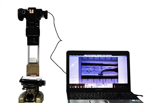 |
Figure 2 Imaging setup of the samples. |
Root Canal Instrumentation
Root canal instrumentation was performed in strict accordance with the manufacturer recommendations for every single system. All files were operated by a 1:16 gear reduction handpiece powered by the X-SmartPlus motor (Dentsply Maillefer, Ballaigues, Switzerland). The ProTaper files were operated at a rotational speed of 250 rpm, the PTN files were manually operated with a persistent alternation at a speed of 300 rpm with light apical compression, and the WO files were used in a reciprocating operational signal produced by the device (WaveOne all mode). Every canal was organized to a working length of 16 mm in a crown-down arrangement, and the last apical preparation was adjusted to a size of 25 in each group. The preparation sequences were as follows:
ProTaperTM System
First, a glide path was created using path file nos. 1, 2, and 3 (Dentsply Maillefer, Ballaigues, Switzerland) at the full working length (WL=16 mm) using Glyde from similar company as a lubricating agent. Then, the coronal and middle thirds of the canal were prepared with ProTaper S1 and S2 files, respectively. Finally, the apical section of the canal was finished with ProTaper F1 and F2 files at full working length.
ProTaper NextTM System
After, a reproducible glide path was made using a small manual (#10) and dedicated mechanical glide path files (sizes P1 and P2) at full working length (WL=16mm) using Glyde as the lubricating agent. Then, preparation of the full length of the canal with PTN files X1 and X2 was conducted.
WaveOneTM System
The glide path was used with the same working length and by using similar lubricating agent. Then, each model was prepared until the final working length with the WO file using a pecking motion, while the file was in a reciprocating working motion. Among each file size, each root canal was irrigated and cleaned with 2 mL of distilled water using a 27-gauge needle (HenrySchein. Inc, Melville, NY, USA). Patency was held utilizing a size#10K file. Every instrument was disposed of after manipulation in five canals.
Photography of Instrumented Post-Operative Resin Blocks
Following the completion of instrumentation, each canal was soaked with red India ink, and post-operative photographs were captured using the same technique for the un-instrumented blocks as shown in Figure 2.
Evaluation of Shaping Ability
Image analysis software OnyxCeph3 DENTAURUM (GmbH & Co. KG Turnstr. 31, 75,228 Ispringen, Germany) was used to analyze the profiles of the prepared canals by superimposing the pre-instrumentation images over the post-instrumentation images, thus creating a composite image. The orientation grooves guaranteed a precise matching of pre- and post-instrumentation images, as shown in Figure 3.
 |
Figure 3 Superimposition of pre-operative and post-operative photos of the simulated root canals. |
Preparation Time
The time for each model preparation, which comprised total active cleaning and shaping, was documented to the closest second. Every instrument was changed within the sequence during the cleaning of the flutes of the instruments and irrigation.
Instrument Failure
The number and size of fractured and permanently deformed instruments in each group during canal preparation were recorded.
Root Canal Aberrations
An assessment was performed on several types of canal aberration19,20 and recorded as follows; Zipping as an over-enlargement of the canal along the outer side of the curve and under-preparation along the inner aspect of the curve at the apical end point, Elbow as a narrow region of the root canal at the point of maximum curvature, Ledge, as an indentation in the outer aspect of the curved portion of the canal, Danger zone, which considered as an area with excessive removal of dentin from the inner aspect of the curve, Perforation as a creation of a false canal that was not confluent with the original canal, and Outer widening which described as an over-preparation and straightening along the outer side of the curve without displacement of the apical foramen.
Width Measurements
The assessment of the amount of resin removed during preparation of the canals was carried out at five points along the canal length by use the method described by Calberson et al 2002.21 The different points were the canal orifice (O), halfway from the beginning of the curve to the orifice (HO), beginning of the curve (BC), and apex of the curve of the original canal (AC). Those were determined by the intersection of two lines, and the inner and outer widths at each of these points were measured perpendicular to the long axis of the original central canal path using the image analysis software on different positions.
Statistical Analysis
The data were gathered, saved, and analyzed using SPSS version 20.0 (SPSS Inc., Chicago, IL, USA) software. Numerical analysis between the various groups was performed using one-way analysis of variance (ANOVA) and Scheffe post-hoc tests. In all analyses, the level of statistical significance was set at p < 0.05.
Results
Preparation Time
The mean time needed for preparation was highest in ProTaper (4.893±0.687 minutes). PTN and WO needed 2.9 minutes each. One-way ANOVA showed a statistically significant difference between time and the system (p=0.000). Post-hoc Scheffe test showed a statistically significant difference between WO and ProTaper (p=0.000) and PTN and ProTaper (p=0.000). The difference between WO and PTN was statistically non-significant (p > 0.05), as shown in Table 1.
 |
Table 1 Mean Canal Preparation Time of the Canals of the Three systemsP |
Canal Aberrations
A total of nine aberrations were noted. Three zips and elbows, one ledge, one outer widening, and one danger zone were noted in ProTaper. In the case of PTN, one outer widening was observed. One ledge and one outer widening were observed in WO. No perforations were found in any of the canals, as shown in Table 2.
 |
Table 2 Incidence of Canal Aberrations and Instrument Failure Among the Three Systems |
Total Width Measurements
The maximum mean total width was obtained by PTN in the orifice of the canal (2.81±.4285 mm), halfway to the orifice (1.12±0.078 mm), and the beginning of the curve (0.84±0.0699). ProTaper (0.66±0.0699 mm) in the apex of the curve and WO (0.48±0.1033) at 0.5 mm from end point were marginally higher than PTN. However, the difference between the groups was statistically non-significant (p > 0.05), as shown in Table 3.
 |
Table 3 Mean Width Measurements Between Groups |
Instrument Failure
Only one primary WO file fractured during preparation, while none of the remaining instruments showed deformation without any significant differences between groups.
Discussions
The ProTaper (Dentsply Maillefer, Ballaigues, Switzerland) rotary system is a thermally treated instrument that has the same features as ProTaper Universal, and it provides a complex heating-cooling proprietary treatment that results in a visible titanium oxide layer on the surface of the instrument, creating a shape memory alloy.21 The PTN (Dentsply Sirona) rotary shaping system delivers the same predictable results as its predecessor PTU (Dentsply Sirona) but reportedly has greater efficiency and fewer files. In 2011, the WO NiTi file system (Dentsply Sirona) was launched as a single-use, single-file system to shape the root canal in a reciprocating motion. The system is constructed of M-Wire and is made up of three files.22 This laboratory study compared the preparation time and canal aberration of the ProTaper, WO, and PTN Ni–Ti systems in simulated curved (L-shaped) root canals. Resin blocks are commonly used to determine the shaping ability of endodontic instruments under the most accurate and precise laboratory rules and conditions. They also allow direct visual to assess the prepared canal spaces.
Overall results show the significance of canal preparation times, which somewhat agrees with previous studies,23–26 and not for total width measurements as mentioned in other studies.24,26 The null hypothesis was partially rejected because a significant difference was documented between the different rotary systems used in relation to preparation time and aberrations, but no significant differences were recorded in the width measurements and fractured files.
The preparation time of the root canal is a major issue in measuring the ability of an endodontic instrument. It depends on a number of reasons, such as experience of the person operating the instrument, the number of the instruments that are in use, the nature of instrument, and the performance being get through.27 In the present study, the preparation time included active instrumentation, time required for exchanges and shifting between different instruments, irrigation, washing, and scrubbing of the tips of the instruments. All these factors were useful in setting the proper preparation time and for accurate comparisons of the documented outcome because the experiments were performed earlier under the same circumstances and conditions.25,26,28
In the present study, the preparation time of PTN was shorter than that of ProTaper and WO. None of the previous studies compared these systems in terms of the preparation time. However, other studies have found that K3 takes less time to prepare canals compared with Profile and ProTaper.15,24,29 A single study reported that WO was significantly faster than Mtwo and ProTaper files and the instrumentation time of ProTaper was shorter than manual instrumentation.28 In the present study, PTN was the fastest, which is in accordance with the findings of a recent study that compared it with other multiple rotary file systems.30
With regard to canal aberrations, this study found that PTN caused less transportation at the apical area and sustained better canal curvature than other file systems did. However, the entire file system straightened the apical curvature, but the PTN system produced more transportation in the straight area compared with ProTaper and WO. This condition can be attributed to the metallurgy of the file systems. Ni–Ti alloy displays strength and hardness in austenite phase and flexibility and ductility in martensite phase. At the microstructure level, ProTaper consists of austenite, while WO and PTN consist of martensite phase. Hence, ProTaper adjusted the canal curve in severely warped canals and did not maintain the original position of the canal. The design of the instrument also plays a vital part in determining the preparation of the canal. The continuing taper of PTN makes it more adaptable than ProTaper and WO at the apical portion. Thus, PTN produced minimum transportation at the apical part in models with severely curved canals.31,32
For Ni–Ti files with the same system, the cross-sectional design is the greatest factor affecting the extrusion of debris, followed by motion mode. In addition, the multifile system could reduce the degree of root canal transportation.33 The management of curved canals can be time consuming, challenging, and frustrating. A sound knowledge of the internal anatomy of the tooth and a thorough assessment of pre-operative radiographs are crucial prerequisites in managing curved canals. Patience is essential in managing challenging mid-root curvatures. The canals should first be negotiated by using hand files, and then rotary files with copious irrigation in between each file should be applied.34 A consistent strategy should be followed for the successful management of curved canals.
Each file organization has its own set of values and limitations. Ni–Ti files are manufactured in cross-section as a rectangle, triangle, square, or slender rectangle. A single study documented that endodontic rotary system files with a square cross-section have the highest screw-in cleaning, shaping, and flexural stiffness, followed by the rectangular, triangular, and the slender rectangular ones.35 ProTaper has a cross-section of rounded or convex triangle.25 WO modified cross sections completed the working length from a modified convex triangle in the tip region to a round triangle comparable to ProTaper adjacent to the shaft.28 PTN has an off-center rectangular cross section, which causes the files to turn in an isolated irregular action like a snake.36 Hence, in PTN, a rectangular geometry together with a diminishing taper at the coronal part had higher screw-in force and flexural rigidity in comparison to ProTaper and WO, thus resulting in more transportation at the straight part in severely curved canals. In this study, few aberrations were observed, and only one WO file was fractured. This condition can be attributed to the reciprocating motion of the WO file, which can cause more fatigue to the instrument when a single file is used to prepare the canal.
Recent works provided necessary principles for the management of curved canals: diagnosis of apical pathosis, pre-operative radiographic interpretation of the pulp chamber and canal anatomy, customized design of the access cavity to fit the underlying anatomy, exploration of the pulp chamber under magnification and coaxial illumination, use of ultrasonics and long-shafted burs to locate and penetrate calcifications, securing and maintaining a reproducible glide path, and safe enlargement of curved canals with highly flexible and fatigue-resistant instruments.37 Also, high-precision computer numerical control milling allows the creation of standardized simulated curved root canals in laboratory dentin. These models may be useful for testing and comparing materials and concepts of chemo-mechanical root canal instrumentation.38
As shown in Table 3 and in relation to the total width measurements of the curved canals after using the three tested rotary systems, no significant differences were recorded at different levels. Similar findings were recorded by Sharma et al,39 while Vakili-Gilani et al40 recorded a significant difference in simulated L-canals, which can be due to the use of recent rotary file systems with advanced mechanical properties. Other study recorded a significant difference, which could be related to the use of cone-beam computed tomography.41
This laboratory study has some drawbacks. First, it compared three old rotary systems. Second, it did not use natural teeth. Third, multiple usage of files can be considered a limitation, along with the software version that was used. Finally, PathFiles did not considered for preparation time analysis. Further studies using a recent rotary system and micro computed-tomography or CBCT with natural teeth are highly recommended.
Conclusion
Within the limitations of this study, the ProTaper next system prepared the root canals faster than the WO and ProTaper systems. ProTaper produced the highest number of canal aberrations, followed by WO, while PTN recorded a single canal aberration. None of the file systems fractured, except one file from the WO system.
Disclosure
The authors report no conflicts of interest in this work.
References
1. Schilder H. Cleaning and shaping the root canal. Dent Clin North Am. 1974;18(2):269–296. doi:10.1016/S0011-8532(22)00677-2
2. Iqbal MK, Maggiore F, Suh B, Edwards KR, Kang J, Kim S. Comparison of apical transportation in four Ni-Ti rotary instrumentation techniques. J Endod. 2003;29(9):587–591. doi:10.1097/00004770-200309000-00011
3. Schäfer E, Erler M, Dammaschke T. Comparative study on the shaping ability and cleaning efficiency of rotary Mtwo instruments. Part 1. Shaping ability in simulated curved canals. Int Endod J. 2006;39(3):196–202. doi:10.1111/j.1365-2591.2006.01074.x
4. Shi L, Yang Y, Wan J, Xie W, Yang R, Yao Y. Shaping ability of rotary and reciprocating single-file systems in combination with and without different glide path techniques in simulated curved canals. J Dent Sci. 2022;17(4):1520–1527. doi:10.1016/j.jds.2022.04.029
5. Weine FS, Kelly RF, Lio PJ. The effect of preparation procedures on original canal shape and on apical foramen shape. J Endod. 1975;1(8):255–262. doi:10.1016/S0099-2399(75)80037-9
6. Al-Omari MA, Dummer PM. Canal blockage and debris extrusion with eight preparation techniques. J Endod. 1995;21(3):154–158. doi:10.1016/s0099-2399(06)80443-7
7. Peters OA, Schönenberger K, Laib A. Effects of four Ni-Ti preparation techniques on root canal geometry assessed by micro computed tomography. Int Endodontic J. 2001;34(3):221–230. doi:10.1046/j.1365-2591.2001.00373.x
8. Tabassum S, Zafar K, Umer F. Nickel-Titanium Rotary File Systems: what’s New? Eur Endod J. 2019;4(3):111–117. doi:10.14744/eej.2019.80664
9. Thompson SA. An overview of nickel-titanium alloys used in dentistry. Int Endod J. 2000;33(4):297–310. doi:10.1046/j.1365-2591.2000.00339.x
10. Parashos P, Messer HH. Rotary NiTi instrument fracture and its consequences. J Endod. 2006;32(11):1031–1043. doi:10.1016/j.joen.2006.06.008
11. Plotino G, Grande NM, Cordaro M, Testarelli L, Gambarini G. A review of cyclic fatigue testing of nickel-titanium rotary instruments. J Endod. 2009;35(11):1469–1476. doi:10.1016/j.joen.2009.06.015
12. Al-Hadlaq SMS, Aljarbou FA, AlThumairy RI. Evaluation of cyclic flexural fatigue of M-wire nickel-titanium rotary instruments. J Endod. 2010;36(2):305–307. doi:10.1016/j.joen.2009.10.032
13. El-Anwar MI, Mandorah AO, Yousief SA, Soliman TA, El-Wahab TMA. A finite element study on the mechanical behavior of reciprocating endodontic files. Br J Oral Sci. 2015;14(1):52–59. doi:10.1590/1677-3225v14n1a11
14. Conceição I, Ferreira I, Braga AC, Pina-Vaz I. Simulated root canals preparation time, comparing ProTaper Next and WaveOne Gold systems, performed by an undergraduate student. J Clin Exp Dent. 2020;12(8):e730–5. doi:10.4317/jced.56981
15. Mohammadzade Akhlaghi N, Delvarani A, Norouzi V, Mohebbi P, Meraji N. Incidence of Dentinal Crack Formation Using ProTaper Universal and WaveOne Systems in Straight and Curved Root Canals. Iran Endod J. 2018;13(4):549–553. doi:10.22037/iej.v13i4.21114
16. Iosif L, Dimitriu B, Ni¸toi DF, Amza O. Endodontic Dentistry: analysis of Dentinal Stress and Strain Development during Shaping of Curved Root Canals. Healthcare. 2023;11:2918. doi:10.3390/healthcare11222918
17. Silva RV, Alcalde MP, Horta MC, et al. Root canal shaping of curved canals by Reciproc Blue system and Pro Taper Gold: a micro-computed tomographic study. J Clin Exp Dent. 2021;13(2):e112–e118. doi:10.4317/jced.57180
18. Zarean P, Özcan M, Zarean P, et al. Micro-Computed Tomographic Assessment of Microcrack Formation before and after Instrumentation of Curved Root Canals with Neoniti Rotary Files. Materials. 2022;15:3002. doi:10.3390/ma15093002
19. Bryant ST, Dummer PM, Pitoni C, Bourba M, Moghal S. Shaping ability of.04 and.06 taper ProFile rotary nickel-titanium instruments in simulated root canals. Int Endod J. 1999;32(3):155–164. doi:10.1046/j.1365-2591.1999.00256.x
20. Jakupovic S, Konjhodzic A, Brankovic LH, et al. Canal Aberration Assessment in Simulated Root Canals: a Comparative Study. Med Arch. 2017;71(3):204–207. doi:10.5455/medarh.2017.71.204-207
21. Calberson FL, Deroose CA, Hommez GM, Raes H, De Moor RJ. Shaping ability of GTTM Rotary Files in simulated resin root canals. Int Endod J. 2002;35(7):607–614. doi:10.1046/j.1365-2591.2002.00540.x
22. van der Vyver PJ, Vorster M, Paleker F, de Wet FA. Root canal preparation: a literature review and clinical case reports of available materials and techniques. SADJ May. 2019;74(4):187–199. doi:10.17159/2519-0105/2019/v74no4a4
23. Schäfer E, Schulz-Bongert U, Tulus G. Comparison of hand stainless steel and nickel titanium rotary instrumentation: a clinical study. J Endod. 2004;30(6):432–435. doi:10.1097/00004770-200406000-00014
24. Mohammadzade Akhlaghi N, Khalilak Z, Baradaran Mohajeri L, Sheikholeslami M, Saedi S. Comparison of Canal Preparation Pattern of K3 and ProTaper Rotary Files in Curved Resin Blocks. Iran Endod J. 2008;3(2):11–16.
25. Schäfer E, Vlassis M. Comparative investigation of two rotary nickel-titanium instruments: proTaper versus RaCe. Part 2. Cleaning effectiveness and shaping ability in severely curved root canals of extracted teeth. Int Endod J. 2004;37(4):239–248. doi:10.1111/j.0143-2885.2004.00783.x
26. Bürklein S, Schäfer E. The influence of various automated devices on the shaping ability of Mtwo rotary nickel-titanium instruments. Int Endod J. 2006;39(12):945–951. doi:10.1111/j.1365-2591.2006.01171.x
27. Hülsmann M, et al. Mechanical preparation of root canals: shaping goals, techniques and means. Endodontic Topics. 2005;10:30–76. doi:10.1111/j.1601-1546.2005.00152.x
28. Bürklein S, Hinschitza K, Dammaschke T, Schäfer E. Shaping ability and cleaning effectiveness of two single-file systems in severely curved root canals of extracted teeth: reciproc and WaveOne versus Mtwo and ProTaper. Int Endod J. 2012;45(5):449–461. doi:10.1111/j.1365-2591.2011.01996.x
29. Al-Omari MA, Aurich T, Wirtti S. Shaping canals with ProFiles and K3 instruments: does operator experience matter? Oral Surg Oral Med Oral Pathol Oral Radiol Endod. 2010;110(3):e50–5. doi:10.1016/j.tripleo.2010.03.003
30. Dhingra A, Ruhal N, Miglani A. Evaluation of Single File Systems Reciproc, Oneshape, and WaveOne using Cone Beam Computed Tomography -An In Vitro Study. J Clin Diagn Res. 2015;9(4):ZC30–4. doi:10.7860/JCDR/2015/12112.5803
31. Shen Y, Zhou HM, Zheng YF, Peng B, Haapasalo M. Current challenges and concepts of the thermomechanical treatment of nickel-titanium instruments. J Endod. 2013;39(2):163–172. doi:10.1016/j.joen.2012.11.005
32. Pereira ES, Gomes RO, Leroy AM, et al. Mechanical behavior of M-Wire and conventional NiTi wire used to manufacture rotary endodontic instruments. Dent Mater. 2013;29(12):e318–24. doi:10.1016/j.dental.2013.10.004
33. Yu D, Guo L, Liu GJ. Evaluation of apical extrusion of debris and centering ability in different nickel-titanium files during curved root canal preparation. BMC Oral Health. 2023;23:395. doi:10.1186/s12903-023-03070-3
34. Mittal R. Endodontic management of curved canals in mandibular molars- A case series. Int J Oral Health Dent. 2023;9(1):48–51.
35. Versluis A, Kim H, Lee W, Kim B, Lee C. Flexural Stiffness and Stresses in Nickel-Titanium Rotary Files for Various Pitch and Cross-sectional Geometries. J Endod. 2012;38:1399–1403.
36. Elnaghy AM, Elsaka SE. Assessment of the Mechanical Properties of ProTaper Next Nickel-Titanium Rotary Files. J Endod. 2014;40:1830–1834.
37. Chaniotis A, Ordinola-Zapata R. Present status and future directions: management of curved and calcified root canals. Int Endod J. 2022;55 Suppl 3:656–684. doi:10.1111/iej.13685
38. Hofpeter K, Zehnder M, Hülsmann M, Al-Jadaa A, Deari S. Precision-milled simulated curved root canals in bovine dentine for the assessment of chemo-mechanical root canal preparation. Int Endod J. 2023. doi:10.1111/iej.13988
39. Sharma Y, Naqvi S, Mishra P, Bishnoi A, Sharma D, Worlikar N. Comparison of three endodontic rotary system shaping in curved canals – an In-vitro study. Eur Chem Bull. 2023;12(issue 8):6361–6367. doi:10.48047/ecb/2023.12.8.513
40. Vakili-Gilani P, Tavanafar S, Saleh ARM, Karimpour H. Shaping ability of three nickel-titanium rotary instruments in simulated L-shaped canals: oneShape, Hero Shaper, and Revo-S. BMC Oral Health. 2021;21:378. doi:10.1186/s12903-021-01734-6
41. Singh T, Kumari M, Kochhar R. Comparative evaluation of canal transportation and centering ability of rotary and reciprocating file systems using cone-beam computed tomography: an in vitro study. J Conserv Dent. 2023;26(3):332–337. doi:10.4103/jcd.jcd_112_23
 © 2024 The Author(s). This work is published and licensed by Dove Medical Press Limited. The full terms of this license are available at https://www.dovepress.com/terms.php and incorporate the Creative Commons Attribution - Non Commercial (unported, v3.0) License.
By accessing the work you hereby accept the Terms. Non-commercial uses of the work are permitted without any further permission from Dove Medical Press Limited, provided the work is properly attributed. For permission for commercial use of this work, please see paragraphs 4.2 and 5 of our Terms.
© 2024 The Author(s). This work is published and licensed by Dove Medical Press Limited. The full terms of this license are available at https://www.dovepress.com/terms.php and incorporate the Creative Commons Attribution - Non Commercial (unported, v3.0) License.
By accessing the work you hereby accept the Terms. Non-commercial uses of the work are permitted without any further permission from Dove Medical Press Limited, provided the work is properly attributed. For permission for commercial use of this work, please see paragraphs 4.2 and 5 of our Terms.