")
Back to Journals » Journal of Experimental Pharmacology » Volume 15
Aloe adigratana Reynolds: Preliminary Phytochemical Screening, Proximate Content, Essential Oil Analysis, and In Vitro Antifungal Activity Studies of Its Leaf Peels and Gel
Authors Gebremariam A, Gebrezgabher BG, Desta KT, Sbhatu DB , Berhe GG, Abdirkadir M , Tsegay E
Received 10 June 2023
Accepted for publication 22 August 2023
Published 28 August 2023 Volume 2023:15 Pages 321—332
DOI https://doi.org/10.2147/JEP.S420990
Checked for plagiarism Yes
Review by Single anonymous peer review
Peer reviewer comments 2
Editor who approved publication: Prof. Dr. Junmin Zhang
Abraha Gebremariam,1 Brhane Gebremedhin Gebrezgabher,2 Kebede Taye Desta,3 Desta Berhe Sbhatu,4 Goitom Gebreyohannes Berhe,4 Mahmud Abdirkadir,4 Ephrem Tsegay4
1Kebri Dehar University, Kebri Dehar, Ethiopia; 2Jigjiga University, Jigjiga, Ethiopia; 3Adama Science and Technology University, Adama, Ethiopia; 4Mekelle University, Mekelle, Ethiopia
Correspondence: Desta Berhe Sbhatu, Mekelle University, PO Box 231/1632, Mekelle, Ethiopia, Tel +251 914735090, Email [email protected]
Background: Aloe species are among the most significant plants with several applications. Many of the species, however, are underexplored, owing to their scarcity and limited geographical distribution. A. adigratana Reynolds, which is common in Ethiopia, is one of the little-studied and endangered Aloe species.
Objective: This preliminary study focuses on the phytochemical screening, proximate analysis, essential oil content, and antifungal activities of A. adigratana leaf peels. Antifungal activities were also tested on the gels of the plant for comparison.
Methods: Standard procedures were used for phytochemical and proximate composition studies. Essential oil analysis was performed using a gas chromatography-mass spectrometry instrument. Using the well-diffusion method, investigations on antifungal activity were performed on three clinically isolated specimens of dandruff-causing fungus; namely, Malassezia furfur, Malassezia restricta, and Malassezia globosa.
Results: The leaf peels of A. adigratana contained alkaloids, flavonoids, tannins, and terpenes. The mean moisture, ash, and crude fat levels were 85.69, 92.20, and 8.00%, respectively, whereas the mean total protein and mean total carbohydrate values were 2.59 and 3.04%. Gas chromatography-mass spectrometry investigation confirmed the presence of fifteen essential oils. The most prevalent essential oil component was discovered to be phytol (33.78%), followed by decane (11.29%). In a dose-dependent way, the leaf latex and gel extracts prevented the growth of three dandruff-causing Malassezia fungal species (M. furfur, M. restricta, and M. globosa). Both the latex and gel demonstrated the maximum activity on M. globosa, the most prevalent fungus in the research area, with minimum inhibitory concentrations of 0.24 and 0.48 mg/mL and minimum fungicidal concentrations of 0.48 and 0.97 mg/mL, respectively.
Conclusion: In general, the proximate and essential oil compositions of A. adigratana leaves were comparable to those of other Aloe species widely used in the food, cosmetic, and pharmaceutical industries, implying that A. adigratana could be a potential future plant for such industries.
Keywords: Aloe adigratana, antifungal activity, dandruff, essential oil, proximate
Introduction
There are an estimated 500–600 species in the genus Aloe, and they are spread over the globe. Fifty of these species were discovered to be endemic to Ethiopia and Eritrea. Aloe adigratana Reynolds is one of these species, which is mostly found in Ethiopia’s Tigrai region, and the species name “adigratana” is derived from the town of Adigrat, where it was first discovered.1–3
A. adigratana is a shrubby perennial plant that grows at elevations ranging from 2000 to 2700 meters. It is distinguished by red flowers that bloom from January to April. It has densely flowered racemes and dull green, crowded leaves. The plant has erect or sprawling stems with long internodes that can reach a height of 2 meters.1,4,5 According to a recent ethnobotanical study conducted by Tefera and Kim,6 the leaves, stems, barks, and roots of A. adigratana are used in traditional medicine to treat gastritis and skin disorders in both humans and livestock. In many areas where it grows, A. adigratana is used to create fences for backyards, farmlands,1,7 churchyards, footpaths, area enclosures, and property demarcations. Unfortunately, the plant is one of Ethiopia’s threatened and endangered species.8
Several Aloe species, such as A. barbadensis (A. vera), A. arborescens, A. sinkatana, A. ferox, and A. pulcherrima, are known to be rich sources of metabolites such as essential oils, fatty acids, alkaloids, and phenolic compounds that have a variety of therapeutic and health benefits for humans.9–14 Furthermore, extracts and isolates of Aloe species were found to have a variety of pharmacological properties, including antioxidant, anticancer, anti-inflammatory, and antimicrobial activities. As a result of these benefits, the economic potential of Aloe species and their applications in the pharmaceutical, cosmetic, and food industries is growing.10,15–17 Despite this, there are still unstudied species in the genus that could play important roles in these industries. One such underexplored species is A. adigratana, and the plant’s ecological and geographical constraints may have contributed to the dearth of scientific research on the plant. So far, only a few studies have attempted to assess the biochemical compositions and pharmacological activities of A. adigratana tissues. Tsegay et al,18 for example, reported the anti-inflammatory activities of aloin (A/B) and microdontin (A/B) from the plant’s leaf latex, and these compounds remained the only known biomolecules reported thus far. Another study on the screening assay of ethanol extracts of its gel reported the presence of flavonoids, phenolics, tannins, and terpenoids with in vitro antioxidant properties.19 Previously, we evaluated the physicochemical properties of lab-based shampoo prepared from A. adigratana gels.20 This study aimed to conduct the first more in-depth preliminary investigation into the phytochemical screening, proximate contents, essential oil compositions, and antifungal activities of A. adigratana leaf peels. Our findings could provide background information and pave the way for future research that improves the plant’s utilization in various industries.
Materials and Methods
Chemicals and Reagents
All the chemicals and reagents used in this study were analytical grade with high purity. They were: methanol and chlorophorm (LOBA CHEME, India); sulfuric acid (H2SO4), dimethylsulfoxide (DMSO), hydrochloric acid (HCl), lead acetate, ammonium solution, ferric chloride (FeCl3), and sodium sulfate (CARLO ERBA, France); Dixon agar, Sabouraud dextrose broth, Sabouraud dextrose agar (Hi Media Laboratories, Mumbai, India); Resazurin dyes (Sisco Research Laboratories, Delhi, India), and Fluconazole, Tween 20, 40, 60 and 80, and olive oil.
Plant Material Collection and Preparation
A. adigratana leaves (Figure 1) were collected in February 2018 from the south side of Mount Mesebo which is located about 8 km north of Mekelle city (lat.: 13.555; long.: 39.518; alt.: 2212 meters) in Tigrai region, Ethiopia. The collected samples were transported using plastic bags to the Organic Chemistry Laboratory at Mekelle University (Ethiopia) for analysis. A curator at the National Herbarium of Ethiopia, Department of Biology, Addis Ababa University, verified the plant specimens. Voucher specimen (Voucher No. 139524/09) was deposited in the Herbarium. The collected leaves were rinsed with tap water to remove dirt and soil. Separation of the outer green skin (i.e., peel) from the gels was achieved by peeling the skin off with a knife. The separated leaf samples were then dried in the shade at room temperature, pulverized in an electric grinder, and stored at –4 °C in sealed plastic bags pending extraction and analysis.
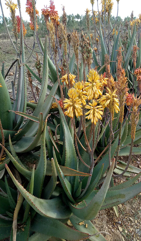 |
Figure 1 A. adigratana (Photo Credit: D.B. Sbhatu at Mesebo, Tigrai, Ethiopia; 20.2.2018). |
Crude Extract Preparation and Phytochemical Screening Tests
For crude extract preparation, 700 g leaf peel was macerated in 1.8 L of methanol with continuous stirring. After 72 hrs, the mixtures were filtered, concentrated and dried using a rotary-evaporator at 40 °C, and the crude methanol extract was used for phytochemicals screening tests and stored in a refrigerator at –4 °C when not used. Phytochemical screening tests for major metabolites such as flavonoids, alkaloids, tannins, anthraquinones, saponins, and terpenoids were performed using previously reported protocols.21–23 The Salkowski reaction was used to determine the presence of terpenoids. In brief, 0.15 g of methanol extract was combined with 2 mL chloroform, followed by the addition of 4 mL concentrated H2SO4. The mixture was allowed to form a layer and a reddish-brown coloration at the interface was inspected for positive results. For the alkaloid test, the Wagner method was used. In the beginning, 10 mg of methanol extract was mixed with 3 mL of 1% HCl in a water bath for 10 min before being filtered. Then 1mL of the filtrate was diluted with 6 drops of Wagner’s reagent and a brownish-red precipitate was inspected for positivity. The lead acetate test was used to screen flavonoids. A 0.2 g methanol extract was dissolved in 2 mL of 10% lead acetate solution and allowed to react at 25 °C. The formation of a yellow precipitate was examined to confirm the presence of flavonoids. A frothing test was used to detect saponins, in which 0.2 g of methanol extract was mixed with 5 mL of distilled water and boiled in a water bath for 10 min. The presence of saponins was determined by inspecting the formation of stable foam. Borntrager’s reaction was used to screen for anthraquinones. Briefly, 0.2 g of methanol extract was mixed with 2 mL of concentrated HCl and allowed to react in a water bath for 10 min. The mixture was then cooled to 25 °C and filtered. Then, 2 mL of chloroform and three drops of NH3 were sequentially added to the filtrate. The final mixture was heated in a water bath for 10 min, and the formation of rose-pink color was examined for anthraquinone presence. The test for tannins was carried out using a ferric chloride test. About 0.2 g of dried extract was mixed with 5 mL water and was allowed to react in a water bath for 10 min. The mixture was cooled, filtered, and 0.5 mg of ferric chloride was added to the filtrate. The appearance of dark-green color was inspected to confirm the presence of tannins.
Proximate Composition
The moisture, ash, crude fat, total protein, and carbohydrate contents of A. adigratana leaf peel were determined using the methods established in the Association of Official Analytical Chemists.24 In each case, measurements were taken in triplicate and the results were reported as mean (±SD) values.
Essential Oils Extraction
The extraction of essential oils from A. adigratana leaf peel followed a previously reported procedure by Sozmen et al25 with some modifications. In brief, 300 g of dried and powdered leaf sample was mixed with 1.5 L water in a Clevenger-type apparatus and distillation was conducted for 5 hrs. The essential oils were separated from the aqueous phase in a 100 mL receiving flask. After drying over anhydrous sodium sulfate, the oil was filtered through Whatman filter paper (No. 40). The retained light yellow liquid extract was transferred to a 5 mL dark brown sample vial and stored at 4 °C until analysis.
Gas Chromatography-Mass Spectrometry (GC-MS) Analysis
The essential oil components were identified and quantified using a 7890B Agilent gas chromatography instrument equipped with a quadrupole mass detector (5977B MSD) as described in our recent study.20 An HP-88 column (30 m × 0.25 mm, 0.25 μm) was used for separation. The oven temperature was set to start at 60 °C and increased to 110 °C at a rate of 3 °C/min followed by a rise to 140 °C at a rate of 10 °C/min for 1 min. It was then increased to 195 °C at a rate of 5 °C/min for 10 min followed by an increase to 225 °C at the same rate for 6 min. For the final 4 min, the temperature was again increased to 250 °C at a rate of 20 °C/min. Throughout the analysis, helium was used as a carrier gas at a flow rate of 0.5 mL/min. Sample (1 µL) was injected in a splitless mode, the inlet temperature was kept at 250 °C and the solvent delay was 5 min. The transfer line and ion source temperatures were set at 250 and 230 °C, respectively. Electron impact (EI) was used as the ionization mode at 70 eV. The acquisition was made through full scan mode between 50–550 amu. The structures of the essential oils were elucidated from the total ion chromatogram (TIC) and the spectra were sketched in terms of counts versus acquisition time and the structures of the essential oils were identified by comparing data from NIST library and literature using MassHunter software. The levels of the identified essential oils were determined as a percentage of the integrated peak areas.
Antifungal Activities of Leaf Latex and Gel Extracts
Sample Collection, Fungal Growth and Identification
This study was started after an approval was secured from the Health Research Ethical Review Committee of the College of Health Sciences, Mekelle University (Ref. No.: ERC 1500/2020). Samples of Malassezia fungi were collected from 217 socio-demographically diverse participants who visited six dermatology clinics in Mekelle, Ethiopia, between February 2019 and June 2020 and formally consented to participate in the study. Participants with dandruff were identified by the clinical appearance of white to silvery powdery scales on their scalps, hairs, and shoulders. Clinical specimens were collected by dividing the hair using a sterile comb and scrapping it with a sterile blunt scalpel. In the process, the participants were handled in accordance with the Case Report guidelines and the Declaration of Helsinki. The materials were put in sterile plastic bags before being delivered to the microbiology lab at the College of Veterinary Medicine, Mekelle University, for further clinical investigation. The collected clinical specimens were cultured on Dixon agar, which was infused with chloramphenicol to inhibit the growth of bacterial contamination. The cultures were incubated at 32 °C for 7 days, and the agar plates were checked every day for fungal development. Before performing the physiological test, pure colonies were generated from plates with positive growth by sub-culturing on Sabouraud dextrose agar (SDA) covered with olive oil. Species were identified using colony morphology, microscopy, and catalase and Tween assimilation assays.26–28
Determination of Antifungal Activities
The antifungal activities were determined using the agar diffusion method of Zoya et al28 with slight modifications. Using sterile cotton swabs, growing test organisms were transferred from modified Dixon’s agar plates into fresh Sabouraud dextrose broth (SDB). The sterile SDA was uniformly swabbed with an inoculum suspension set to McFarland standards of 0.5. In each plate, two millimeter-apart wells (dia. 6 mm) were created using a sterile cork borer. The methanol extracts of A. adigratana leaf latex were prepared by serial dilution in dimethyl sulfoxide (DMSO) at concentrations of 62.5, 125, and 250 mg/mL. Then, 100 µL of each sample was added to each well separately and allowed to diffuse at room temperature for 2 hrs. A commercially available fluconazole standard served as a positive control, and DMSO served as a negative control. The zone of inhibition was measured from triplicate assays after 72 hrs of incubation at 32 °C. A similar approach was performed to record the antifungal activity of A. adigratana gel extracts.
Determining the Minimum Inhibitory and Fungicidal Concentration
The minimum inhibitory concentration (MIC) and minimum fungicidal concentration (MFC) of leaf latex and gel extracts were determined separately in 96-well plates using the Broth micro-dilution method.29 Inbrief, 100 µL SDB with olive oil was placed in each well of a microtiter plate. Then, 100 µL of the target aloe sample was added to the first column, which was serially diluted row by row until the tenth column. The last column’s mixed solution (100 µL) was discarded, and each well was allowed to contain 100 µL of solution in serially descending concentrations. The plates were covered with parafilm aseptically and incubated at 32 °C for 72 hrs. Finally, 30 µL 0.015% w/v resazurin solution was added to each well and incubated again to observe the color change from blue to pink or any other color.30 The concentrations (in the wells) that did not change color due to the leaf latex and gel extract were then recorded as MIC values. MFC was also determined using the same wells that were used for MIC determination. A loop of broth was collected from the treated wells and streaked onto a new sterile SDA overlaid with olive oil plates in triplicate. They were, then, incubated at 32 °C for 72 hrs. The MFC was determined as the lowest concentration with no visible colony formation.31
Statistical Analyses
Required data were collected from all observations, tests, and experiments. All sampling, replicates (measurements), and tests (experiments) were conducted in triplicate. Quantitative data were analyzed using relevant descriptive and inferential statistical methods with SPSS Version 20 software. Inferential (sample) data were analyzed using the analysis of variance (ANOVA), and used for comparisons of means (±SD) at a p-value of ≤ 0.05. Post-hoc comparisons of means (±SD) were carried out using Least Significant Difference (LSD). Qualitative data collected by visual and microscopic observations were used to strengthen the results of the quantitative data analyses.
Results
Phytochemical Screening and Proximate Contents
The phytochemical screening test of leaf peels of A. adigratana revealed the presence of flavonoids, tannins, terpenoids, and alkaloids. Anthraquinones and saponins, on the other hand, were not found (Table 1). The proximate analysis also revealed significant contents in the A. adigratana peels. The moisture level was determined to be considerable (85.69%), whereas the ash content was low (8.00%). Similarly, the crude fat level was 0.68%, the total protein content was 2.59%, and the carbohydrate content was 3.04% (Table 2).
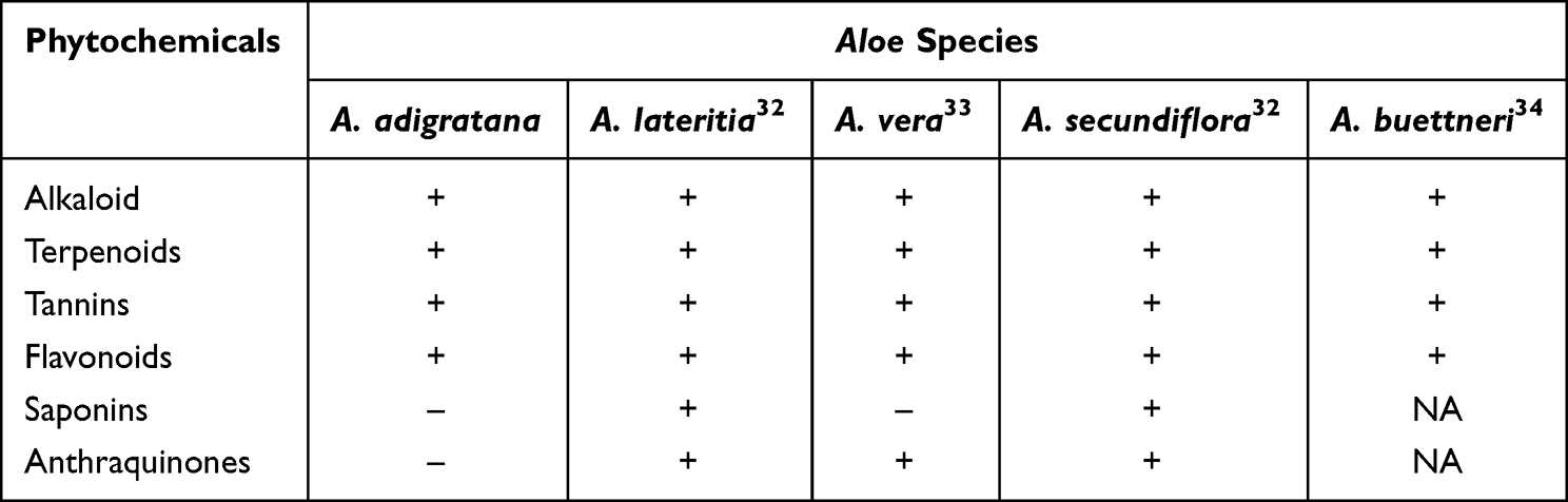 |
Table 1 Phytochemical Screening results from A. adigratana Leaves Compared to Other Aloe Species |
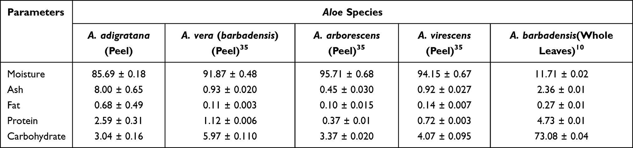 |
Table 2 Proximate Composition of A. adigratana Leaves Compared to Other Aloe Species |
GC-MS Analysis of Essential Oils
The GC-MS analysis revealed fifteen essential oils in A. adigratana leaf peels (Table 3), and their structures were identified using data obtained from the GC-MS, the NIST library, and literature, as previously stated. Among the fifteen essential oils, five (dodecane, nonane, benzene (1,2,4-trimethyl-), decane, and undecane) were hydrocarbons, four (aR-turmerone, tumerone, turlone, 2-Pentadecanone (6,10,14-trimethyl-)) were ketones and other five (methyl tetradecanoate, hexadecanoic acid (methyl ester), 9,12-Octadecadienoic acid (Z,Z)- (methyl ester), 9-Octadecenoic acid (E)- (methyl ester), and methyl stearate) were esters. Phytol was the only Diterpene alcohol detected. Furthermore, except for 2-Pentadecanone (6,10,14-trimethyl-), all of the ketones were sesquiterpenes. In terms of their content, phytol was determined to be the most prevalent component (33.78%), followed by decane (11.29%) and undecane (9.64%). Tumerone, on the other hand, was the least abundant molecule (1.16%), followed by 9,12-Octadecadienoic acid (Z,Z)- (methyl ester) (1.36%), and methyl stearate (1.53%).
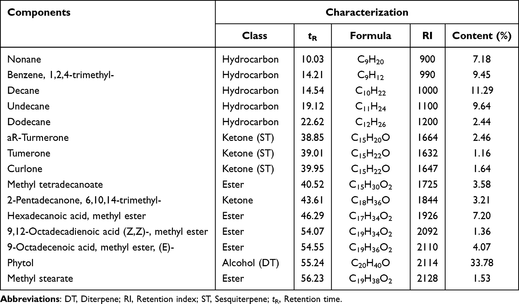 |
Table 3 Essential Oil Components of Leaves of A. adigratana |
Antifungal Activities
As previously stated, the antifungal properties of A. adigratana leaf latex and gel extracts were studied using the agar diffusion method. The study demonstrated that the extracts have good anti-fungal effects against three dandruff-causing Malassezia species common in the study area.
Antifungal Activities of Leaf Latex
Study using the well diffusion method revealed that A. adigratana leaf latex has caused remarkable growth inhibition against all the Malassezia fungi with as low as 62.5 mg/mL concentration (Table 4). Increasing the dose of the leaf latex from 62.5 to 250 mg/mL resulted in statistically significantly higher mean growth inhibitions in all three fungal species (p ≤ 0.05). However, the lowering of the concentrations of the leaf latex by half (ie, from 250 mg/mL to 125 mg/mL and from 125 mg/mL to 62.5 mg/mL) resulted in barely slight reduction in its growth inhibition capacities in all the dandruff-causing fungi. When we look into the effect of the three concentrations on the three fungal species, all the concentrations resulted in statistically significantly higher growth inhibition on M. globosa − the most abundant fungi in the study area. At 250 mg/mL, the leaf latex has a statistically comparable growth inhibition effect on M. globosa as the antifungal drug Fluconazole (at 50 μg/mL). M. restricta looks like the second susceptible species to the leaf latex while M. furfur is relatively less susceptible. In contrast, M. furfur is more resistant to the fluconazole antifungi drug compared to the other two species.
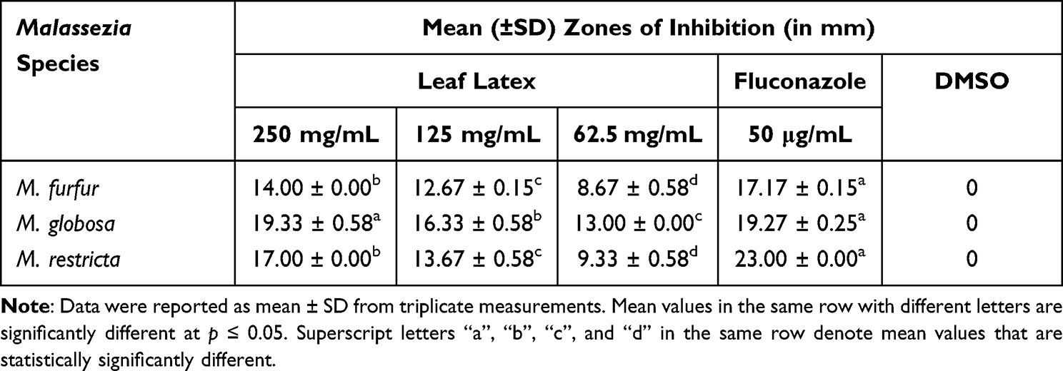 |
Table 4 Zones of Inhibition (in Mm) of A. adigratana Leaf Latex and Fluconazole Against Malassezia Isolates |
Antifungal Activities of Methanol Leaf Gel Extract
The methanol leaf gel extract has a comparable effect on the growth inhibition of the three fungal species with nearly similar patterns (Table 5). Though the differences lacked statistical significance, M. globosa is more susceptible while M. furfur is less susceptible to the leaf gel extract. The leaf gel extract, like the leaf latex, has a comparably more profound effect on the growth inhibition of M. globosa. It seems also relatively more potent against M. furfur compared to the leaf latex.
 |
Table 5 Zones of Inhibition (in Mm) of A. adigratana Gel Extract and Fluconazole Against Malassezia Isolates |
MIC and MFC of Leaf Latex and Gel Extract
Tests on minimum inhibitory concentration (MIC) and minimum fungicidal concentration (MFC) support the findings of the tests on growth inhibition. While M. globosa has the smallest inhibitory concentrations with both extracts, M. furfur required relatively higher concentrations (Table 6). The MIC and MFC of A. adigratana leaf latex and gel extract were similar for M. furfur and M. restricta but leaf latex was twice as effective compared to leaf gel extract for M. globosa. Whereas the MIC values of leaf latex and gel extract against all the species ranged between 0.24 (±0.00) and 7.80 (±0.00) mg/mL, the MFC values of the same ranged between 0.48 (±0.00) and 15.60 (±0.00) mg/mL.
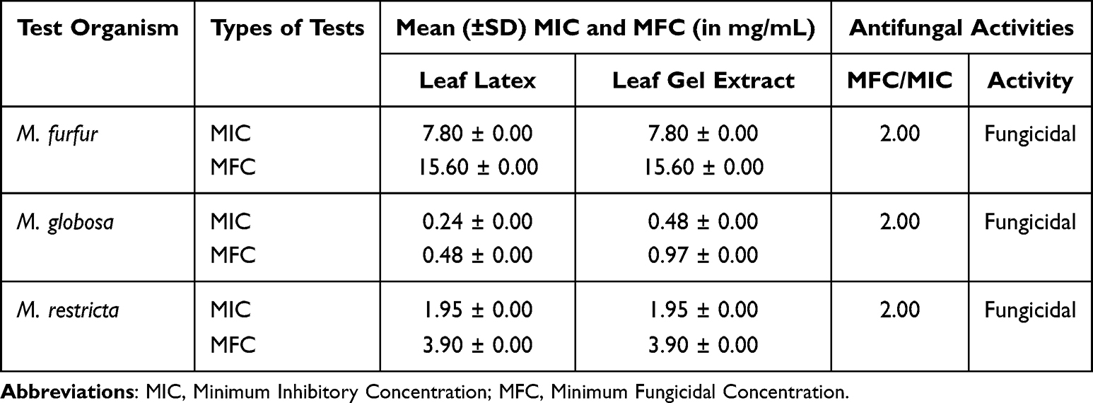 |
Table 6 MIC and MFC of A. adigratana Leaf Latex and Leaf Gel Extract Against Malassezia Isolates |
Discussion
Phytochemical Properties
Because of their biochemical compositions and biological activity, certain Aloe species, particularly A. vera, have substantial use in the pharmaceutical, cosmetic, and food sectors. A. adigratana is one of several additional species that have received little attention.20 Phytochemical screening assays on A. adigratana leaf peel revealed that it contained secondary metabolites such as alkaloids, terpenoids, tannins, and flavonoids. The detected molecules have also been reported from other Aloe species, including A. lateritia, A. vera, A. secundiflora, and A. buettneri, as indicated in Table 1.32–34,36 As a result of such phytochemical screening studies, many medically significant phytochemicals have been identified and extracted from diverse Aloe species.37,38 As a result, A. adigratana leaf peels probably contain beneficial flavonoids, tannins, terpenes, and alkaloids, the identity of which should be examined further in future studies.
Proximate Contents
The proximate study also revealed that A. adigratana could have nutritional properties, as prior research indicated equivalent values from peels of other Aloe species10,35 (Table 2). Adesuyi et al (2012), on the other hand, reported a reduced crude fat content (0.27%) from the entire A. barbadensis leaves.10 The same study discovered that the protein (4.73%) and carbohydrate (73.08%) concentrations were higher than in our study. In addition to the differences in the genetic makeup of the Aloe species, the presence of latex and gels could have resulted in such variances compared to our investigation. Furthermore, carbohydrates and essential amino acids from A. ferox, A. greatheadii, and A. secundiflora have been described.39,40 In summary, the leaf peels of A. adigratana could be good sources of nutritional components used in the development of food products.
Essential Oils
The GC-MS analysis confirmed the existence of numerous essential oil classes, with phytol being the most abundant essential oil component (33.78%), followed by decane (11.29%) (Table 3). Phytol was the second most prevalent component in the gels (26.38%), according to our previous research.20 Nonane, benzene (1,2,4-trimethyl-), decane, undecane, dodecane, and 2-Pentadecanone (6,10,14-Trimethyl-) were detected in the leaves, in contrast to our previous report in the gels of A. adigratana. However, naphthalene (1-Methyl-), decanoic acid (methyl ester), and dodecanoic acid (methyl ester) detected in the gels were not found in the leaf peels, indicating a difference in essential oil distributions among A. adigratana tissues. In general, the detected essential oils were previously reported from other Aloe species such as A. barbadensis and A. ferox. Besides, these essential oils were found to have a wide spectrum of applications in food, pharmaceutical, and cosmetic industries signifying the potential of A. adigratana for similar use.17,36,41,42
Antifungal Activities
Dandruff-causing fungi belong to the genus Malassezia. Malasseziaincludes14 species, of which seven are dandruff-causing, with M. globosa and M. restricta being the most dominant.43 Various studies on the prevalence of these dandruff-causing fungi in different countries reported diverse results. In Bangladesh and Iran, for example, M. furfur has been recognized as the most common cause of dandruff.44,45 Similarly, in a study on seborrhoeic dermatitis patients, M. restricta was shown to be prominent in Korea,46 whilst M. furfur was reported to be the dominant one in Iran.47 M. globosa was the cause of dandruff in 67.15% of the participants in our study, making it the most common dandruff-causing species followed by M. furfur, (20.70%) and M. restricta (12.15%) (Unpublished manuscript).48 Previously, M. globosa was identified as the primary dandruff-causing species in Bosnia and Herzegovina.49
Several Aloe species have been found to be beneficial in the treatment of fungal infections such as dandruff.16,50 The antifungal efficacy of A. adigratana leaf latex and gel extracts against three clinical fungal species, M. globosa, M. furfur, and M. restricta, was examined using well diffusion technique. A. adigratana leaf latex and gel extracts inhibited the development of all Malassezia fungi at concentrations as low as 62.5 mg/mL (Table 4 and 5). In each case, increasing the dose from 62.5 to 250 mg/mL resulted in statistically significantly larger mean growth inhibitions in all three fungal species (p< 0.05). All concentrations of A. adigratana leaf latex and gel extracts showed higher inhibition of M. globosa growth, the most prevalent fungus in the current investigation. At 250 mg/mL, the leaf latex has a statistically comparable growth inhibition effect on M. globosa as the antifungal drug fluconazole (at 50 μg/mL). In contrast, M. furfur was more resistant to the fluconazole antifungi drug compared to the other two species.
The MIC and MFC of both samples were below 0.5 mg/mL against M. globosa signifying a pronounced antifungal activity (Table 6). The findings of the present study are comparable to the empirical observations of Asmerom et al on a yeast fungus called Candida krusei.51 Low MIC and MFC values imply potentially high efficacy of plant extracts as antifungal agents.52 Plant extracts could have two types of activities − fungicidal when the ratio of MFC/MIC is less than 4.0 and fungistatic when the ratio is greater than 4.0.53,54 Fungistatics are constituents that inhibit the growth of fungal species without killing them. This indicates that, according to the findings of the present study, A. adigratana leaf latex and gel extract are sources of phytochemical constituents with fungicidal properties against the three Malassezia fungal species. Such an effect is proposed to be via physiological interference or structural damage.55 As far as we can tell; this is the plant’s first recorded antifungal action against M. globosa, M. furfur, and M. restricta, and the results show A. adigratana’s potential in combating dandruff-causing fungi.
Strengths and Limitations of the Study
This study has both strengths and limitations. First-ever evidence of the antifungal effects of A. adigratana is presented in this paper, suggesting the possibility for this plant to be used as a dandruff remedy. The study also included fungal samples that were directly collected from clinical patients, which has significant implications for the utilization of A. adigratana’s gel and latex for treating fungi. Moreover, the study also paved the path for future research by contrasting the phytochemical composition and essential oil concentration of A. adigratana leaves. This study also has some limitations. Although the essential oil analysis was carried out using GC-FID, future investigations in this sector are advised because no profile of additional metabolites was undertaken. In addition, only the most typical dandruff-causing fungi were considered in this study. Therefore, it is strongly advised to conduct more research on the antibacterial capabilities of A. adigratana.
Conclusion
This study provided the first in-depth analysis of phytochemicals, proximate composition, and antifungal properties of A. adigratana leaf peels and gel. The findings demonstrated that A. adigratana is abundant in a variety of phytochemicals, including alkaloids, flavonoids, tannins, and terpenes; as a result, the plant may supply a broad range of biomolecules if taken into consideration in subsequent studies. Additionally, the extracted proximate contents were equivalent to those of numerous other Aloe species that are widely employed in the food industry. The discovery of several classes of essential oils, such as hydrocarbons, ketones, and esters, suggests that the plant could be a useful source of these metabolites. The identified essential oils are widely used in the beauty, food, and pharmaceutical industries and are known to offer numerous health advantages. More significantly, in vitro antifungal activity revealed that the latex and gel extracts prevented the growth of three common dandruff-causing fungi, M. globosa, M. furfur, and M. restricta. Overall, the findings of our investigation indicated that A. adigratana may be a reliable source of vital nutrients and non-nutritive metabolites as well as anti-dandruff compounds. Thus, this research is significant in two ways. (1) It provides useful insights into the phytochemical and biomedical potentials of the plant. (2) It encourages future large-scale studies toward establishing comprehensive phytochemical and biomedical profile of the plant. These, in turn, encourage future development programs aiming at the conservation and sustainable use of the plant.
Abbreviations
ANOVA, Analysis of variance; DMSO, Dimethyl Sulfoxide; DT, Diterpene; EI, Electron Impact; eV, Electron volt; GC-MS, Gas chromatography-mass spectrometry; MFC, minimum fungicidal concentration; MIC, Minimum inhibitory concentration; NIST, National Institute of Standards and Technology; RI, Retention index; SD, Standard deviation; SDA, Sabouraud dextrose agar; SDB, Sabouraud dextrose broth; SPSS, Statistical package for the social sciences; ST, Sesquiterpene; TIC, Total ion chromatogram; tR, Retention time.
Data Sharing Statement
The datasets used and/or analyzed during the current study are available from the corresponding author on reasonable request.
Approval and Consent to Participate
The institutional review committees of the College of Natural and Computational Sciences and the College of Health Sciences, Mekelle University, approved the studies.
Acknowledgments
The authors are highly indebted to acknowledge Mekelle University for funding this study. They are also thankful to Mr Melaku Wondafrash, a curator at the National Herbarium at the Department of Biology, Addis Ababa University, for verifying A. adigratana specimens. Finally, the authors would like to thank the anonymous reviewers.
Author Contributions
All authors made a significant contribution to the work reported, whether that is in the conception, study design, execution, acquisition of data, analysis and interpretation, or in all these areas; took part in drafting, revising or critically reviewing the article; gave final approval of the version to be published; have agreed on the journal to which the article has been submitted; and agree to be accountable for all aspects of the work.
Funding
This study was supported by Mekelle University, PO Box 231, Mekelle, Ethiopia through the Grant No.: CRPO/MIT/LARGE/001/09.
Disclosure
The authors declare that they do not have any conflicts of interest for this work.
References
1. Demissew S, Nordal I. Lilies and Aloes of Ethiopia and Eritrea.
2. Reynolds T. Aloes: The Genus Aloe. Boca Raton, FL, USA: CRC Press; 2004.
3. Fentaw E, Dagne K, Ronsted N, et al. Karyotypes in Ethiopian Aloe species (Xanthorrhoeaceae: Asphodeloideae). Kew Bull. 2013;68:599–607. doi:10.1007/s12225-013-9475-8
4. Oda BK, Erena BA. Aloes of Ethiopia: a review on uses and importance of aloes in Ethiopia. Int J Plant Biol Res. 2017;5(1):1–6.
5. Kahsay T, Degu HD. Assessment of genetic diversity in two endemic aloe germplasm populations from Ethiopia using morphological markers. Int J Bioresour Stress Manag. 2016;7:080–087.
6. Tefera BN, Kim YD. Ethnobotanical study of medicinal plants in the Hawassa Zuria District, Sidama zone, Southern Ethiopia. J Ethnobiol Ethnomed. 2019;5:1–21.
7. Demissew S, Friis I, Feye TA, et al. Four new species of Aloe (Aloaceae) from Ethiopia, with notes on the ethics of describing new taxa from foreign countries. Kew Bull. 2011;66(1):111–121. doi:10.1007/s12225-011-9263-2
8. Hernick C, Neigh A, Worku A, et al. Tropical Forest and Biodiversity (FAA 118/119) Assessment. Addis Ababa, Ethiopia: USAID/ETHIOPIA; 2016.
9. Periasamy G, Kassa S, Sintayehu B, et al. Cosmetic use of Aloe vera – a review. World J Pharm Pharm Sci. 2014;3(5):342–458.
10. Adesuyi AO, Awosanya OA, Adaramola FB, et al. Nutritional and phytochemical screening of Aloe barbadensis. Curr Res J Biol. 2012;4(1):4–9.
11. Rodríguez ER, Martín JD, Romero CD. Aloe vera as a functional ingredient in foods. Crit Rev Food Sci Nutr. 2010;50(4):305–326. doi:10.1080/10408390802544454
12. Elhassan GOM, Adhikari A, Yousuf S, et al. Phytochemistry and antiglycation activity of Aloe sinkatana Reynolds. Phytochem Lett. 2012;5(1):725–728. doi:10.1016/j.phytol.2012.07.012
13. Chen W, Van Wyk B-E, Vermaak I, et al. Cape aloes - a review of the phytochemistry, pharmacology and commercialisation of Aloe ferox. Phytochem Lett. 2012;5(1):1–12. doi:10.1016/j.phytol.2011.09.001
14. Park MK, Park JH, Kim NY, et al. Analysis of 13 phenolic compounds in Aloe species by high performance liquid chromatography. Phytochem Anal. 1998;9(4):186–191.
15. Sanchez-Machado DI, Lopez-Cervantes J, Sendon R, et al. Aloe vera: ancient knowledge with new frontiers. Trends Food Sci Technol. 2017;61:94–102. doi:10.1016/j.tifs.2016.12.005
16. Maan AA, Nazir A, Kashif M, et al. The therapeutic properties and applications of Aloe vera: a review. J Herb Med. 2018;12:1–10. doi:10.1016/j.hermed.2018.01.002
17. Salehi B, Albayrak S, Antolak H, et al. Aloe genus plants: from farm to food applications and phytopharmacotherapy. Int J Mol Sci. 2018;19(9):1–49. doi:10.3390/ijms19092843
18. Tsegay M, Tewabe Y, Bisrat D, et al. In vivo anti-inflammatory activity of two anthrones from the leaf latexes of Aloe adigratana Reynolds and Aloe elegans Todaro. Ethiop Med J. 2018;34(1):1–8. doi:10.4314/epj.v34i1.1
19. Brhane GH, Gopalakrishnan VK, Hagos Z, et al. Phytochemical screening and in vitro antioxidant activities of ethanolic gel extract of Aloe adigratana Reynolds. J Pharm Technol. 2018;12(1):13–19.
20. Sbhatu DB, Berhe GG, Hndeya AG, et al. Formulation and physicochemical evaluation of lab-based Aloe adigratana Reynolds shampoos. Int J Anal Chem. 2020;2020:1–7. doi:10.1155/2020/6290617
21. Farnsworth NR. Biological and phytochemical screening of plants. J Pharm Sci. 1996;55(3):225–275.
22. Edeoga HO, Okwu DE, Mbaebie BO. Phytochemical constituents of some Nigerian medicinal plants. Afr J Biotechnol. 2005;4(7):685–688. doi:10.5897/AJB2005.000-3127
23. Sofowora A. Medicinal Plants and Traditional Medicine in Africa. Chichester, England: John Wiley and Sons Ltd.; 1982.
24. AOAC (Association of Official Analytical Chemists). Official Methods of Analysis. Arlington, VA: Association of Official Analytical Chemists Inc; 2003.
25. Sozmen F, Uysal B, Oksal BS, Kose EO, Deniz IG. Chemical composition and antibacterial activity of Origanum saccatum P.H. Davis essential oil obtained by solvent-free microwave extraction: comparison with hydrodistillation. J AOAC Int. 2011;94(1):343–350. doi:10.1093/jaoac/94.1.243
26. Ro BI, Dawson TL. The role of sebaceous gland activity and scalp microfloral metabolism in the etiology of seborrheic dermatitis and dandruff. J Invest Dermatol. 2005;10(3):194–197.
27. Guillot J, Guého E, Lesourd M, et al. Identification of Malassezia species. A practical approach. J Mycol Med. 1996;6(3):103–110.
28. Zoya M, Bhikhu M, Shah G. Anti-dandruff activity of synthetic and herbal shampoos on dandruff causing isolate: malassezia. Int J Appl Res. 2016;2(7):80–85.
29. Boudreau MD, Beland FA. An evaluation of the biological and toxicological properties of Aloe barbadensis (Miller), Aloe vera. J Environ Sci Health C. 2006;24(1):103–154. doi:10.1080/10590500600614303
30. Boudreau MD, Mellick PW, Olson GR, et al. Clear evidence of carcinogenic activity by a whole-leaf extract of Aloe barbadensis Miller (Aloe vera) in F344/N rats. Toxicol Sci. 2013;131(1):26–39. doi:10.1093/toxsci/kfs275
31. Anzaku AA, Akyala JI, Juliet A, et al. Antibacterial activity of lauric acid on some selected clinical isolates. Ann Clin Lab Sci. 2017;5(2):1–5. doi:10.21767/2386-5180.1000170
32. Mbithi CM, Matu EN, Maina NW. Phytochemical screening, antioxidant activity and hypoglycemic potential of Kenyan Aloe lateritia and Aloe secundiflora extracts in alloxan-induced diabetic Swiss albino mice. European J Med Plants. 2018;24(1):1–18. doi:10.9734/EJMP/2018/40799
33. Sanmukhiya MR, Soulange JG, Lavergne C, et al. Molecular biology, phytochemistry and bioactivity of three endemic Aloe species from Mauritius and Réunion Islands. Phytochem Anal. 2010;21(6):566–574. doi:10.1002/pca.1234
34. Kombate B, Metowogo K, Kantati YT, et al. Phytochemical screening, antimicrobial and antioxidant activities of Aloe buettneri, Mitracarpus scaber and Hannoa undulata used in Togolese Cosmetopoeia. J Drug Deliv Ther. 2022;12(2–s):19–24. doi:10.22270/jddt.v12i2-S.5409
35. Cha TY, Baek JH, Lee SY. Comparative study on chemical composition of Korean aloes of three species according to different portions. Food Eng Prog. 2007;11(3):175–184.
36. Saljooghianpour M, Javaran TA. Identification of phytochemical components of Aloe plantlets by gas chromatography-mass spectrometry. Afr J Biotechnol. 2013;12(49):6876–6880.
37. Blitzke T, Porzel A, Masaoud M, et al. A chlorinated amide and piperidine alkaloids from Aloe sabaea. Phytochemistry. 2000;55(8):979–982. doi:10.1016/S0031-9422(00)00269-7
38. Dring JV, Nash RJ, Roberts MF, et al. Hemlock alkaloids in aloes: occurrence and distribution of ɤ-coniceine. Planta Med. 1984;50(5):442–443. doi:10.1055/s-2007-969761
39. Human H, Nicolson SW. Nutritional content of fresh, bee-collected and stored pollen of Aloe greatheadii var. davyana (Asphodelaceae). Phytochemistry. 2006;67(14):1486–1492. doi:10.1016/j.phytochem.2006.05.023
40. Mabusela WT, Stephen AM, Botha MC. Carbohydrate polymers from Aloe ferox leaves. Phytochemistry. 1990;29(11):3555–3558. doi:10.1016/0031-9422(90)85275-K
41. Tariq M, Ali S, Ahmad F, et al. Identification, FT-IR, NMR (1H and 13C) and GC/MS studies of fatty acid methyl esters in biodiesel from rocket seed oil. Fuel Process Technol. 2011;92(3):336–341. doi:10.1016/j.fuproc.2010.09.025
42. Ahmad M, Nangyal H, Sherwani S, et al. Effect of heat stress on fatty acids profiles of Aloe vera and Bryophyllum pinnatum leaves. World Appl Sci J. 2103;28(11):1592–1596.
43. Park M, Park S, Jung WH. Skin commensal fungus Malassezia and its lipases. J Microbiol Biotechnol. 2021;31(5):637–644. doi:10.4014/jmb.2012.12048
44. Kindo AJ, Sophia SK, Kalyani JAS, et al. Identification of Malassezia species. Indian J Med Microbiol. 2004;22(3):179–181. doi:10.1016/S0255-0857(21)02832-2
45. Gupta AK, Kohli Y, Faergemann J, et al. Epidemiology of Malassezia yeasts associated with pityriasis versicolor in Ontario, Canada. Med Mycol. 2001;39(2):199–206. doi:10.1080/mmy.39.2.199.206
46. Won-Lee Y, Byun HJ, Kim BJ, et al. Distribution of Malassezia species on the scalp in Korean seborrheic dermatitis patients. Ann Dermatol. 2011;23(2):156–161. doi:10.5021/ad.2011.23.2.156
47. Mahmoudi E, Saeidi M, Marashi MA, et al. In vitro activity of kombucha tea ethyl acetate fraction against Malassezia species isolated from seborrhoeic dermatitis. Curr Med Mycol. 2016;2(4):30–36. doi:10.18869/acadpub.cmm.2.4.30
48. Gebrezgabiher BG, Abdulkadir M, Sbhatu DB, et al. Prevalence, Etiologic Agents, and Associated Risk Factors of Dandruff in Mekelle City, Tigray, Ethiopia [Unpublished Manuscript]; 2023.
49. Orphic A, Simi D, Sadikovic TJ, et al. Distribution of Malassezia species on healthy human skin in Bosnia and Herzegovina: correlation with body part, age and gender. Iran J Microbiol. 2014;6(4):253–262.
50. Singh K, Ajao AA, Sabiu S. Ethnobotanical, phytochemistry, toxicological and pharmacological significance of the underutilized indigenous Aloe species of West Africa. S Afr J Bot. 2015;147:1007–1015. doi:10.1016/j.sajb.2021.12.014
51. Asmerom D, Hailu GS, Yimer EM, et al. Antimicrobial Evaluation of Latex and TLC Fractions from the Leaves of Aloe adigratana Reynolds. Evid Based Complementary Altern Med. 2020;2020:1–10. doi:10.1155/2020/8312471
52. Doughari JH, Ndakidemi PA, Human IS, et al. Antioxidant, antimicrobial and antiverotoxic potentials of extracts of C. dentata. J Ethnopharmacol. 2012;141(3):1041–1050. doi:10.1016/j.jep.2012.03.051
53. Okusa PN, Penge O, Devleeschouwer M, et al. Direct and indirect antimicrobial effects and antioxidant activity of Cordia gilletii De Wild (Boraginaceae). J Ethnopharmacol. 2007;112(3):476–481. doi:10.1016/j.jep.2007.04.003
54. Hazen KC. Fungicidal versus fungistatic activity of terbinafine and itraconazole: an in vitro comparison. J Am Acad Dermatol. 1998;38(5):37–41. doi:10.1016/S0190-9622(98)70482-7
55. Asamenew G, Bisrat D, Mazumder A, et al. In vitro antimicrobial and antioxidant activities of anthrone and chromone from the latex of Aloe harlana Reynolds. Phytother Res. 2011;25(12):1756–1760. doi:10.1002/ptr.3482
 © 2023 The Author(s). This work is published and licensed by Dove Medical Press Limited. The full terms of this license are available at https://www.dovepress.com/terms.php and incorporate the Creative Commons Attribution - Non Commercial (unported, v3.0) License.
By accessing the work you hereby accept the Terms. Non-commercial uses of the work are permitted without any further permission from Dove Medical Press Limited, provided the work is properly attributed. For permission for commercial use of this work, please see paragraphs 4.2 and 5 of our Terms.
© 2023 The Author(s). This work is published and licensed by Dove Medical Press Limited. The full terms of this license are available at https://www.dovepress.com/terms.php and incorporate the Creative Commons Attribution - Non Commercial (unported, v3.0) License.
By accessing the work you hereby accept the Terms. Non-commercial uses of the work are permitted without any further permission from Dove Medical Press Limited, provided the work is properly attributed. For permission for commercial use of this work, please see paragraphs 4.2 and 5 of our Terms.