")
Back to Journals » Research and Reports in Urology » Volume 15
Uncommon Haemorrhagic Cystitis of Infectious Origin: A Narrative Review for Urologists
Authors Ambrosini F, Riccardi N , Occhineri S, Matucci T, Paraboschi I, Calarco A , Berrettini A, Tiseo G, Canetti D , Van Der Merwe A , Terrone C, Mantica G
Received 13 March 2023
Accepted for publication 20 July 2023
Published 4 August 2023 Volume 2023:15 Pages 365—373
DOI https://doi.org/10.2147/RRU.S412278
Checked for plagiarism Yes
Review by Single anonymous peer review
Peer reviewer comments 2
Editor who approved publication: Dr Panagiotis J Vlachostergios
Francesca Ambrosini,1,2 Niccolò Riccardi,3,4 Sara Occhineri,3,4 Tommaso Matucci,3,4 Irene Paraboschi,5 Alessandro Calarco,6 Alfredo Berrettini,5 Giusy Tiseo,3 Diana Canetti,7 André Van Der Merwe,8 Carlo Terrone,1,2 Guglielmo Mantica1,2
1IRCCS Ospedale Policlinico San Martino, Genova, Italy; 2Department of Surgical and Diagnostic Integrated Sciences (DISC), University of Genova, Genova, Italy; 3Infectious Diseases Unit, Department of Clinical and Experimental Medicine, University of Pisa, Pisa, Italy; 4StopTB Italia Onlus, Milan, Italy; 5Department of Pediatric Urology, Fondazione IRCCS Ca’ Granda Ospedale Maggiore Policlinico, Milan, Italy; 6Villa Pia Hospital, Via Folco Portinari 5, Rome, Italy; 7Infectious Diseases Unit, San Raffaele Scientific Institute, Milan, Italy; 8Department of Urology, Faculty of Medicine and Health Sciences, 26697 Stellenbosch University, Cape Town, South Africa
Correspondence: Guglielmo Mantica, Department of Surgical and Diagnostic Intergrated Sciences (DISC), University of Genova, Genova, Italy, Tel +390105555960, Email [email protected]
Purpose: Haemorrhagic cystitis may be due to different etiologies with infectious diseases representing an insidious cause to diagnose. The aim of this narrative review is to provide a comprehensive overview of less common but difficult-to-diagnose causes of infectious haemorrhagic cystitis of bacterial, mycobacterial, and parasitic origin, Moreover, we highlight possible diagnostic tools and currently available treatment options in order to give an updated tool for urologists to use in daily practice.
Patients and Methods: The search engine PubMed was used to select peer-reviewed articles published from 1/Jan/2010 to 31/Aug/2022.
Results: Bacteria, fungal, TB and schistosomiasis are uncommon causes of haemorrhagic cystitis burdened by high morbidity, especially if not promptly diagnosed.
Conclusion: Because haemorrhagic cystitis ranges in severity from mild dysuria associated with pelvic discomfort to severe life-threatening haemorrhage, punctual diagnosis, and immediate treatment are essential to avoid further complications.
Keywords: cystitis, haemorrhagic cystitis, schistosomiasis, tuberculosis, GUTB
Introduction
Cystitis is the inflammation of the urinary bladder; it can be acute or chronic and the degree of severity can range from mild dysuria associated with pelvic discomfort to severe life-threatening haemorrhage.1 Haemorrhagic cystitis, defined as the necrosis of bladder transitional epithelium and associated blood vessels leading to hematuria, may be due different etiologies: it is usually caused by chemical and/or toxin, radiation, as well as mechanical insults; however, infectious diseases (bacterial/mycobacterial, fungal, parasitic or viral) may represent an insidious and difficult-to-diagnose cause of hemorrhagic cystitis.2 In fact, haemorrhagic cystitis can develop in the context of a systemic infectious disease (eg, Cytomegalovirus (CMV), Tuberculosis) or may be isolated to the urinary bladder (eg, uropathogenic Escherichia coli).3,4 While the symptoms are typical (dysuria, frequency and urgency, irritative voiding symptoms, suprapubic pain, and haematuria), they are not specific to a single infectious etiology.5 Indeed, the individual level of immune suppression is related to the infectious agents that can be responsible for hemorrhagic cystitis.6 In this narrative review, we provide a comprehensive overview of less common but difficult-to-diagnose causes of infectious hemorrhagic cystitis of bacterial, mycobacterial, and parasitic origin; moreover, we highlight possible diagnostic tools and currently available treatment options for each pathogen.
Materials and Methods
A narrative review was carried out to retrieve the scientific evidence on bacterial, Candida spp, Tuberculosis (TB), and Schistosoma spp. causes of haemorrhagic cystitis. The search engine PubMed was used to select peer-reviewed articles published from 1/Jan/2010 to 31/Aug/2022. The references of the selected manuscripts were carefully assessed to collect articles not included in the primary search by two different authors (NR and GM). The following keywords were used to broaden the spectrum of the article: (haemorrhagic cystitis) AND (infection); (haemorrhagic cystitis) AND (infection). A total of 584 records were found. Based on titles, abstracts, and the content of the full texts, a total of 29 studies were deemed suitable (Figure 1).
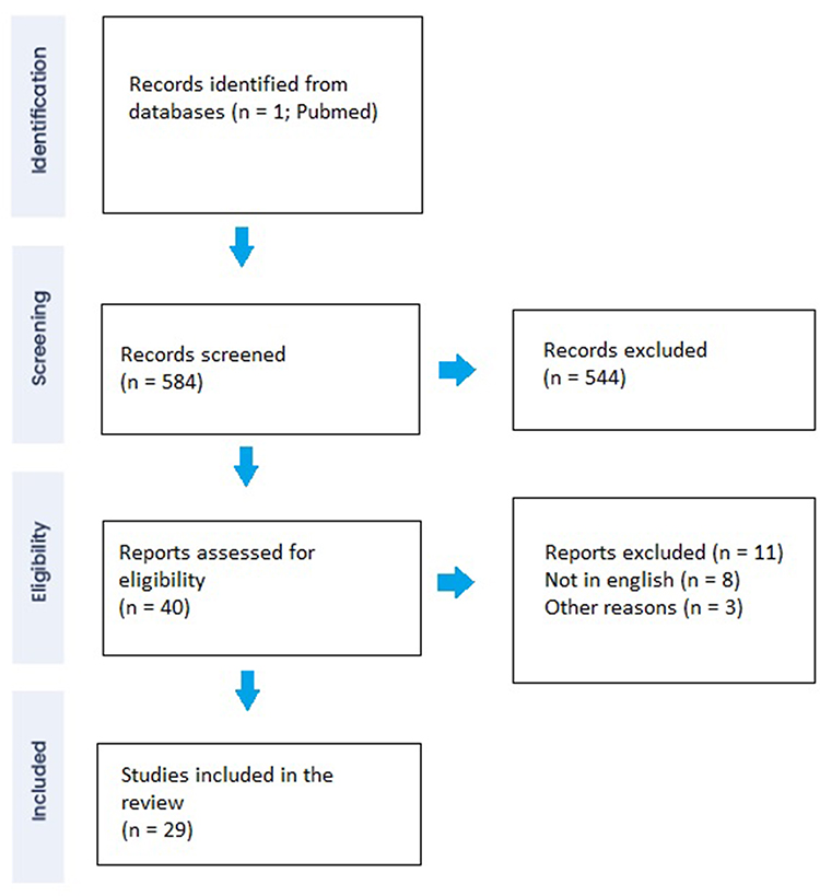 |
Figure 1 Flowchart. Adapted from Page MJ, McKenzie JE, Bossuyt PM, Boutron I, Hoffmann TC, Mulrow CD, et al. The PRISMA 2020 statement: an updated guideline for reporting systematic reviews. BMJ. 2021;372:n71. Creative Commons. |
Results
Fungal Infection
Fungal infections of the urinary tract are a relatively rare condition with an incidence of 1–2.2%.7 Candida spp. is the most common organism that can cause urinary tract infections (UTIs) among fungi.8,9 Other fungi include Cryptococcus neoformans, Aspergillus spp., and Mucorales. Blastomyces dermatitidis can cause acute prostatitis, and both B. dermatitidis and Histoplasma capsulatum can be responsible for epididymal-orchitis, but they are not usually involved in hemorrhagic cystitis.9 Fungal infections associated with hemorrhagic cystitis include Candida spp., Aspergillus spp., and Mucorales.
Candida Species
Epidemiology
Candida spp. is a fungal pathogen that can be part of the healthy human microbiome but can also cause mucosal infections or invasive candidiasis.10 Candida urinary tract infections (UTIs) are less common in the community, whereas they are more frequently reported in hospitalized and immunocompromised patients.8 Literature lacks standardized and generalizable criteria for defining infection and colonization.8 The prevalence of candiduria (≥104 cfu/mL in women and ≥5 × 10³ cfu/mL in men) in urine samples from both outpatients and inpatients is high, accounting for 1% of urine cultures positive for any pathogen in hospital laboratories (0.2% of all cultures evaluated).11 The incidence of candiduria can increase up to 22% in intensive care units,12 17% in catheterized patients with spinal cord injury,13 3.4–11% in renal transplant recipients,14,15 and 10% in patients with type 2 diabetes patients.16 However, the clinical significance of candiduria is controversial. In a prospective multicenter observational study, only 2–4% of patients with candiduria had lower urinary tract symptoms (LUTS) or hematuria suggestive of UTIs.17 Some risk factors for Candida urinary tract infections have been identified, including diabetes mellitus, indwelling catheters, renal transplantation, and immunosuppression.8,12,18
Clinical Presentation
Most patients diagnosed with candiduria are asymptomatic and do not need any medical medicalt. If symptoms are present, clinical features are usually indistinguishable from bacterial UTIs.8 Hemorrhagic cystitis due to Candida species is uncommon and usually belongs to high-risk patients such as HIV-infected patients with persistent low CD4+ cell count, hematological, and/or oncological patients with persistent neutropenia.19 Greene et al reported a case of hemorrhagic emphysematous cystitis (EC) secondary to Candida albicans in a patient with agnogenic myeloid metaplasia and treated with prolonged intermittent corticosteroids.20
Tran et al reported a unique case of haemorrhagic Candida glabrata cystitis in an immunocompetent patient after three months of therapy with empagliflozin.21 The patient had a history of type 2 diabetes and neurogenic bladder after transurethral resection of the prostate. The Authors did not provide information on diabetes control. They hypothesized that empagliflozin-induced glycosuria may have played a role in the development of the infection. It is well established that empagliflozin is associated with a higher rate of genital mycotic infections22 and in some rare cases has been associated with disruptive clinical conditions, such as Fournier’s gangrene.23
Nonetheless, a pooled analysis of four Phase 3 trials did not demonstrate an increased risk of UTIs for empagliflozin compared with placebo. Therefore, the evidence for an etiologic link between empagliflozin and hemorrhagic cystitis remains controversial.24
Anecdotal cases of coinfections with multiple fungal organisms have been reported in the literature. A rare double fungal infection of the bladder due to Candida albicans and Cladosporium presented with lower urinary tract symptoms (LUTS) and hematuria was reported by Kandaswami et al.20
Diagnosis
Although detection of Candida in urine culture is essential for diagnosis, colony counts do not help to distinguish contamination from colonization or infection.18 Cystoscopic examination may reveal pseudo membranes or pale plaques overlying the urothelium. Ultrasonography or computed tomography may be used in selected cases to detect complications (fungus ball, abscesses, pyelonephritis) or abnormalities of the urinary tract.8 Imaging studies may be recommended to find any risk factor (for example, urinary retention) in case of failure of antifungal therapy.25
Treatment
Fluconazole is usually the drug of choice, pending antifungal susceptibility test (400 mg loading dose, 200 mg (3 mg/kg) daily for 14 days) due to its good penetration in the urine and in the bladder. In patients with Candida spp resistant to fluconazole, liposomal amphotericin B, 3–5 mg/kg daily for 1–7 days, or oral flucytosine, 25 mg/kg 4 times daily for 7–10 days may be considered.8,26 In selected patients (intolerance to systemic therapy, adverse reactions, or azole-resistant organisms) amphotericin B deoxycholate bladder irrigation (50 mg/L sterile water daily for 5 days) may be considered.27
Other Fungal Infections
The literature on other fungal organisms as etiologic factors of hemorrhagic cystitis is scarce and only a few case reports are available. Mucorales, Cryptococcus neoformans, Aspergillus fumigatus, and Torulopsis glabrata may be associated with haemorrhagic cystitis.28,29 Muthe et al reported the case of a 60-year-old diabetic woman diagnosed with a mucormycosis infection of the urinary tract presenting with LUTS and recurrent hematuria during SARS-CoV-2 infection.20 Rhizopus species was detected in a fungus-ball-specimen retrieved during cystoscopy in a patient with haemorrhagic cystitis. The patient received intravenous liposomal amphotericin B for 6 weeks followed by posaconazole 300 mg daily for 4 weeks. Complete recovery of symptoms was reported after 10 days. According to the guidelines of the Third European Confederation of Medical Mycology, and the Mycoses Study Group Education and Research Consortium, the first-line therapy for mucormycosis is liposomal amphotericin B (L-AMB) and surgical resection.30 However, despite effective antifungal treatment combined with surgery, mucormycosis maintains a poor prognosis with a mortality rate of 60–90%, particularly in immunocompromised patients.31,32
Bacterial Infection
Epidemiology
Uropathogenic Escherichia coli (UPEC) is by far the most common cause of UTI, accounting for 80% of outpatient infections and 25% of nosocomial infections.33 Among the most common bacterial causes of hemorrhagic cystitis Escherichia coli, Staphylococcus saprophyticus, Proteus spp, and Klebsiella spp can be found.29,34 Non-typhoidal Salmonella (NST)35,36 and Chryseobacterium indologenes are anecdotally reported as bacterial causes of haemorrhagic cystitis.37
Clinical Presentation
Bacterial hemorrhagic cystitis presents with LUTS and hematuria and is indistinguishable from other infectious causes. Chronic and recurrent bacterial cystitis can rarely lead to life-threatening complications, such as spontaneous bladder rupture. Olanipekun et al reported a case of a 57-year-old woman with diabetes mellitus and end-stage renal disease on hemodialysis who presented with bladder necrosis and perforation due to E. coli.38
A rare form of complicated bacterial UTIs is emphysematous cystitis (EC) which is characterized by the presence of gas within the bladder wall and the lumen.39 EC is more frequently reported in elderly women with uncontrolled diabetes mellitus. Other risk factors for EC include recurrent UTIs, neurogenic bladder, and urinary stasis due to bladder outlet obstructions.39 The most common gas-producing bacteria are E. coli (60%) and Klebsiella pneumonia (10–20%).40,41
Clinical symptoms of EC range from asymptomatic conditions (up to 7%) to severe sepsis.39 Gross haematuria occurs in 60% of cases.39,42 The most common manifestation is abdominal pain (80%), whereas pneumaturia is usually less frequent.39
A few anecdotal cases of hemorrhagic cystitis due to less common bacteria have been reported in the literature. Teh et al described the case of an immunocompetent 27-year-old male who presented with gross hematuria and LUTS after a five-month trip through South America. He also complained of sweating, fever, nausea, vomiting, abdominal cramps, and diarrhea. A urine culture at presentation revealed Salmonella Oranienburg. The patient reported complete symptomatic recovery after the administration of intravenous ampicillin and gentamicin and subsequent oral switch to amoxicillin–clavulanate.35 Another case of hemorrhagic cystitis due to NST that caused massive bleeding and shock was described by Na et al.36 He presented with disorientation, high temperature (38°C), pyuria, hematuria, and watery diarrhea. He suffered from not well-controlled diabetes.
Diagnosis
The most important investigation in the evaluation of a patient with bacterial haemorrhagic cystitis is midstream urine culture with antimicrobial susceptibility testing (AST) in order to guide appropriate treatment.43 Ultrasound (US) should be performed to rule out upper urinary tract obstruction or renal stone disease in patients with a history of urolithiasis, renal dysfunction, or high urine pH.44 In patients with persistent hematuria or pathogens other than E. coli, urethrocystoscopy and further imaging, such as a contrast-enhanced computed tomography (CT) scan, or excretory urography, may be recommended to detect underlying conditions and to estimate the severity of infection.1 In case of symptomatic EC, abdominal CT could be useful to assess the presence of ascending emphysematous infections, intraabdominal abscesses, or adjacent neoplastic disease.39
Treatment
Antibiotics, bladder drainage, and treatment of predisposing conditions are the most important aspect of the treatment of hemorrhagic cystitis. Antimicrobial treatment is always guided by AST, while empiric antibiotic treatment should cover Gram negative pathogens and should be guided by local epidemiological resistance assessment.45
Tuberculosis
Epidemiology
According to 2022 WHO Global Tuberculosis (TB) Report, TB cases are increasing across the world, and 10.6 million people fell ill with TB in 2021.46 Pulmonary dis-ease is the most common presentation of TB,47 however between 5% and 45% of disease localization have features of extrapulmonary TB (EPTB), and genito-urinary TB (GUTB) is among the most frequent localization.48 Moreover, between 2% and 20% of cases of pulmonary TB show concurrent genitourinary involvement through haematogenous dissemination of Mycobacterium tuberculosis from the lungs to kidneys, ureters, bladder, prostate, epididymis, urethra, ejaculatory ducts, and genital organs, often well before a diagnosis of pulmonary disease has been established.49,50 Due to its frequently asymptomatic presentation, the prevalence of GUTB is hard to estimate. However, 15–20% of the 10 million new TB diagnoses presented with extrapulmonary TB annually.50 GUTB is most frequently described in middle-aged (40 to 50 years old) and males (2-to-1 ratio).50 Nation-wide TB prevalence, TB-HIV co-infection rates, or individual-specific characteristics, such as immunosuppression, malnutrition, renal transplantation, chronic renal disease and dialysis, liver disease, alcohol or substance abuse, and homelessness may further increase the risk of GUTB.50
Clinical Presentation
Symptoms of GUTB, such as pollakiuria and incontinence, appear when the chronic inflammatory insult to the bladder wall epithelium causes fibrosis of the tissue and ureterovesical junction dysfunction, resulting in reduced bladder capacity (usually less than 100mL).49 Other described symptoms of GUTB are low-back pain (34.4%) and hematuria (35.6% overall, 24.5% in developed countries, 44.3% in developing countries).49 Furthermore, GUTB may cause symptoms only in advanced stages of the disease48 when vesicoureteral reflux has already determined chronic renal failure.51
Diagnosis
The gold standard for the diagnosis of GUTB is the identification of the mycobacterium on culture of urine samples; however, this method is often untimely (with from 4to8 weeks required), requires multiple samples (3–6 morning urine samples), with a reported sensitivity varying from 10.7 to 90%.52 Direct microscopy of Ziehl-Neelsen staining of urine samples for the detection of mycobacteria is a rapid test with a specificity of 96.7% and sensitivity of 42.1–52.1%, but it cannot discriminate between Mycobacterium tuberculosis and other mycobacterial species.53 The sensitivity and specificity of off-label use of molecular testing (GenXpert) on urine samples ranged from 89% and 95% in culture-confirmed cases, while to 55% and 99% in clinically diagnosed GUTB cases with a composite reference standard;54–56 although not validated for extrapulmonary samples, this test combines the possibility of an early diagnosis with the detection of genotypic resistance to rifampicin; conversely, a negative test may not exclude GUTB. In the subgroup of people living with HIV (PLWH), lateral flow lipoarabinomannan (LF-LAM) assay on urinary specimens may be considered to consolidate the diagnosis of TB in combination with other methods.57 Radiological testing such as intravenous urography and abdominal computerized tomography scan (CT-scan) may support initial clinical suspect, demonstrating the presence of calyceal irregularities, infundibular stenosis, pseudotumor or renal scarring, renal cavitation, urinary tract calcification, collecting system thickening, stenosis, or dilatation, and contracted bladder:58 these findings have shown sensitivity up to 91.4% for GUTB.52 Invasive procedures (cystoscopy, transurethral biopsy) may reveal the presence of histological alteration compatible with mycobacterial infection and support clinical suspicion when other methods are inconclusive.59
Treatment
The treatment regimen for GUTB is unaltered from the regimen used to treat pulmonary TB and is guided by drug-susceptibility-testing (DST); for drug-susceptible TB (DS-TB) 2 months of rifampicin (R), isoniazid (H), pyrazinamide (Z) and ethambutol (E), followed by 4 months of HR alone are indicated. Resistance patterns may vary and may comprehend first-line drugs as H and R (multi-drug-resistant TB or MDR-TB), or first-line drugs and any second-line drug as fluoroquinolones (FLQ) (pre-extensively DR-TB or pre-XDR-TB) and at least one of bedaquiline (BDQ) and linezolid (LZD) (ex-tensively-drug-resistant TB or XDR-TB); expert specialists in the management of drug-resistant TB (DR-TB) should select the adequate regimen in these cases.60 Surgical interventions to restore adequate bladder capacities, such as augmentation or orthotopic neobladder via devulgarized bowel segments, have been performed in case of severe fibrosis to avoid vesicoureteral reflux and subsequent kidney injury. Late diagnosis of GUTB may result in progression of disease to the point that ablative surgery represents the only available approach.61,62
Schistosomiasis
Epidemiology
Schistosoma haematobium, a trematode with a widespread presence in developing countries’ fresh waters,63 responsible for infecting 250 million people across the world and for 280.000 deaths each year, is a common cause of genito-urinary bleeding.64 However, schistosomiasis may be underdiagnosed in non-endemic countries.65,66
Clinical Presentation
Although mostly asymptomatic, haematuria and dysuria may be present in the early and post-acute phases of schistosomiasis alongside fever, and nonspecific systemic symptoms.67 Hydronephrosis secondary to urinary tract obstruction with or without kidney involvement, bladder calcification, urothelial damage, bladder squamous cell carcinoma, as well as fertility decline may follow the progression of the disease.67
Diagnosis
Although lacking a gold standard definition, the detection of viable eggs through urine filtration microscopy is the mainstay for the diagnosis of schistosomiasis,68 while serological assays may represent a valuable option for screening of imported cases in low-burden countries, with a reported sensitivity ranging from 21.4 to 71.4% and specificity ranging from 48.8% to 94.3%.69,70 Further diagnostic methods, including anti-genic and molecular tests, are under study.71
Treatment
Praziquantel (PZQ) is considered the best available drug for schistosomiasis although its mechanism of action is not fully understood.71,72 In the absence of standardized regimen, a single-day, oral dose of 40 mg/kg of PZQ is indicated by most authors as safe and effective for all Schistosoma spp, either in one or divided in two administrations,71 with a reported cure rate of 77.1% for S. haematobium.73 No correlation between cure rates and dosage of PZQ has been described for S. hematobium,73 how-ever, especially in low-burden settings, higher dosages (up to 80 mg/kg), or a second PZQ dose after 15 days from the first, have been safely and effectively used.71 Reports of decreased PZQ sensitivity in some Schistosoma spp. strains are concerning;72 however, new therapeutic strategies are in development.72,74,75
Conclusion
Bacteria, fungal, TB, and schistosomiasis are uncommon causes of haemorrhagic cystitis burdened by high morbidity, especially if not promptly diagnosed. Due to the paucity of literature and the difficult-to-diagnose presentation, especially in Western countries, urologists are not well aware of which infectious disease agent may be the cause of haemorrhagic cystitis.
Because haemorrhagic cystitis ranges in severity from mild dysuria associated with pelvic discomfort to severe life-threatening haemorrhage, punctual diagnosis, and immediate treatment are essential to avoid further complications.
Disclosure
The authors report no conflicts of interest in this work.
References
1. Laguna MP, Albers P, Algaba F. EAU guidelines. Edn. Presented at the EAU Annual Congress Amsterdam 2022. ISBN 978-94-92671-16-5; 2022.
2. Haldar S, Dru C, Bhowmick NA. Mechanisms of hemorrhagic cystitis. Am J Clin Exp Urol. 2014;2(3):199–208.
3. Mantica G, Ambrosini F, Riccardi N, et al. Genitourinary tuberculosis: a comprehensive review of a neglected manifestation in low-endemic countries. Antibiot Basel Switz. 2021;10(11):1399.
4. Furukawa R, Homma H, Inoue T, Horiuchi H, Usui K. Cytomegalovirus hemorrhagic cystitis in a malignant glioma patient treated with temozolomide. Intern Med Tokyo Jpn. 2018;57(20):3047–3050. doi:10.2169/internalmedicine.1005-18
5. Visintini E, Visintini C, Venturini M, Palese A. Patients’ experience of haemorrhagic cystitis after Haematopoietic Stem Cell Transplantation: findings from a phenomenological study. Eur J Oncol Nurs. 2021;51:101926. doi:10.1016/j.ejon.2021.101926
6. Mori Y, Miyamoto T, Kato K, et al. Different risk factors related to adenovirus- or BK virus-associated hemorrhagic cystitis following allogeneic stem cell transplantation. Biol Blood Marrow Transplant. 2012;18(3):458–465. doi:10.1016/j.bbmt.2011.07.025
7. Ismail M, Hashim H. Complications of fungal cystitis. Curr Bladder Dysfunct Rep. 2013;8(3):212–216. doi:10.1007/s11884-013-0191-x
8. Odabasi Z, Mert A. Candida urinary tract infections in adults. World J Urol. 2020;38(11):2699–2707. doi:10.1007/s00345-019-02991-5
9. Kauffman CA. Diagnosis and management of fungal urinary tract infection. Infect Dis Clin North Am. 2014;28(1):61–74. doi:10.1016/j.idc.2013.09.004
10. World Health Organization (WHO). Fungal Priority Pathogens List to Guide Research, Development and Public Health Action. Geneva: World Health Organization; 2022. Licence: CC BY-NC-SA 3.0 IGO.
11. Colodner R, Nuri Y, Chazan B, Raz R. Community-acquired and hospital-acquired candiduria: comparison of prevalence and clinical characteristics. Eur J Clin Microbiol Infect Dis. 2008;27(4):301–305. doi:10.1007/s10096-007-0438-6
12. Álvarez-Lerma F, Nolla-Salas J, León C, et al. Candiduria in critically ill patients admitted to intensive care medical units. Intensive Care Med. 2003;29(7):1069–1076. doi:10.1007/s00134-003-1807-y
13. Goetz LL, Howard M, Cipher D, Revankar SG. Occurrence of candiduria in a population of chronically catheterized patients with spinal cord injury. Spinal Cord. 2010;48(1):51–54. doi:10.1038/sc.2009.81
14. Delgado J, Calvo N, Gomis A, et al. Candiduria in renal transplant recipients: incidence, clinical repercussion, and treatment indication. Transplant Proc. 2010;42(8):2944–2946. doi:10.1016/j.transproceed.2010.08.019
15. Safdar N, Slattery WR, Knasinski V, et al. Predictors and outcomes of candiduria in renal transplant recipients. Clin Infect Dis. 2005;40(10):1413–1421. doi:10.1086/429620
16. Esmailzadeh A, Zarrinfar H, Fata A, Sen T. High prevalence of candiduria due to non- albicans Candida species among diabetic patients: a matter of concern? J Clin Lab Anal. 2018;32(4):e22343. doi:10.1002/jcla.22343
17. Kauffman CA, Vazquez JA, Sobel JD, et al. Prospective multicenter surveillance study of funguria in hospitalized patients. Clin Infect Dis. 2000;30(1):14–18. doi:10.1086/313583
18. Padawer D, Pastukh N, Nitzan O, et al. Catheter-associated candiduria: risk factors, medical interventions, and antifungal susceptibility. Am J Infect Control. 2015;43(7):e19–e22. doi:10.1016/j.ajic.2015.03.013
19. Duong DT, Goodman HS. Eosinophilic cystitis caused by Candida glabrata: a case report. Urol Case Rep. 2019;26:100970. doi:10.1016/j.eucr.2019.100970
20. Greene MH. Emphysematous cystitis due toClostridium perfringens and Candida albicans in Two patients with hematologic malignant conditions. Cancer. 1992;70(11):2658–2663. doi:10.1002/1097-0142(19921201)70:11<2658::AID-CNCR2820701115>3.0.CO;2-B
21. Tran L, Thomas M, Harvey J, Sampath R, Rose R. 1700. A rare case of candida glabrata hemorrhagic cystitis with empagliflozin use. Open Forum Infect Dis. 2019;6(Supplement_2):S622–S623. doi:10.1093/ofid/ofz360.1564
22. Levine MJ. Empagliflozin for type 2 diabetes mellitus: an overview of Phase 3 clinical trials. Curr Diabetes Rev. 2017;13(4). doi: 10.2174/1573399812666160613113556
23. Khokhar F, Hernandez C, Mahapatra R. Fournier’s gangrene in an HIV-positive patient on empagliflozin for the treatment of diabetes mellitus. Cureus. 2022. doi:10.7759/cureus.26083
24. Kim G, Gerich JE, Salsali A, et al. Empagliflozin (EMPA) increases genital infections but not urinary tract infections (UTIs) in pooled data from four pivotal Phase III trials. Diabetes. 2013;62(Suppl 1):LB21 (Abstract 74–LB).
25. Thomas L, Tracy CR. Treatment of fungal urinary tract infection. Urol Clin North Am. 2015;42(4):473–483. doi:10.1016/j.ucl.2015.05.010
26. D’Amico MJ, Foss H, Uhr A, Rudnick B, Kloniecke E, Gomella LG. Hemorrhagic cystitis: a review of the literature and treatment options. Can J Urol. 2022;29(5):11276–11283.
27. Pappas PG, Kauffman CA, Andes DR, et al. Clinical practice guideline for the management of candidiasis: 2016 update by the Infectious Diseases Society of America. Clin Infect Dis. 2016;62(4):e1–e50.
28. Muthe MM, Shirsath SD, Shirsath RD, Firke VP. A rare case of isolated vesical mucormycosis in a patient with COVID-19 pneumonitis. Indian J Radiol Imaging. 2022;32(03):408–410. doi:10.1055/s-0042-1744137
29. Manikandan R, Kumar S, Dorairajan LN. Hemorrhagic cystitis: a challenge to the urologist. Indian J Urol. 2010;26(2):159–166. doi:10.4103/0970-1591.65380
30. Cornely OA, Alastruey-Izquierdo A, Arenz D, et al. Global guideline for the diagnosis and management of mucormycosis: an initiative of the European Confederation of Medical Mycology in cooperation with the Mycoses Study Group Education and Research Consortium. Lancet Infect Dis. 2019;19(12):e405–e421. doi:10.1016/S1473-3099(19)30312-3
31. Lanternier F, Sun HY, Ribaud P, Singh N, Kontoyiannis DP, Lortholary O. Mucormycosis in organ and stem cell transplant recipients. Clin Infect Dis. 2012;54(11):1–8. doi:10.1093/cid/cis195
32. Bader MS, Hawboldt J, Brooks A. Management of complicated urinary tract infections in the era of antimicrobial resistance. Postgrad Med. 2010;122(6):7–15. doi:10.3810/pgm.2010.11.2217
33. Ronald A. The etiology of urinary tract infection: traditional and emerging pathogens. Am J Med. 2002;113(1):14–19. doi:10.1016/S0002-9343(02)01055-0
34. Sabih A, Leslie SW. Complicated urinary tract infections. In: StatPearls. Treasure Island (FL): StatPearls Publishing; 2022. Available from: http://www.ncbi.nlm.nih.gov/books/NBK436013/.
35. Teh J, Quinlan M, Bolton D. Salmonella Oranienburg haemorrhagic cystitis in an immunocompetent young male. JMM Case Rep. 2017;4(8):e005105. doi:10.1099/jmmcr.0.005105
36. Na SK, Jung HK, Kim YS, et al. Hemorrhagic cystitis with massive bleeding from nontyphoidal Salmonella infection: a case report. Kidney Res Clin Pract. 2013;32(2):84–86. doi:10.1016/j.krcp.2013.04.005
37. Mehta R, Pathak A. Emerging chryseobacterium indologenes infection in Indian neonatal intensive care units: a case report. Antibiotics. 2018;7(4):109. doi:10.3390/antibiotics7040109
38. Olanipekun T, Effoe V, Turner J, Flood M. Bladder necrosis and perforation in end-stage renal disease and recurrent urinary tract infection: a rare medical emergency. Int J Crit Illn Inj Sci. 2019;9(2):101–104. doi:10.4103/IJCIIS.IJCIIS_72_18
39. Amano M, Shimizu T. Emphysematous cystitis: a review of the literature. Intern Med. 2014;53(2):79–82. doi:10.2169/internalmedicine.53.1121
40. Mokabberi R, Ravakhah K. Emphysematous urinary tract infections: diagnosis, treatment and survival (Case Review Series). Am J Med Sci. 2007;333(2):111–116. doi:10.1097/00000441-200702000-00009
41. Bjurlin MA, Hurley SD, Kim DY, et al. Clinical outcomes of nonoperative management in emphysematous urinary tract infections. Urology. 2012;79(6):1281–1285. doi:10.1016/j.urology.2012.02.023
42. Yoshida K, Murao K, Fukuda N, Tamura Y, Ishida T. Emphysematous cystitis with diabetic neurogenic bladder. Intern Med. 2010;49(17):1879–1883. doi:10.2169/internalmedicine.49.3247
43. Hooton TM, Bradley SF, Cardenas DD, et al. Diagnosis, prevention, and treatment of catheter-associated urinary tract infection in adults: 2009 international clinical practice guidelines from the Infectious Diseases Society of America. Clin Infect Dis. 2010;50(5):625–663.
44. van Nieuwkoop C, Hoppe BPC, Bonten TN, et al. Predicting the need for radiologic imaging in adults with febrile urinary tract infection. Clin Infect Dis. 2010;51(11):1266–1272. doi:10.1086/657071
45. Wagenlehner FME, Hoyme U, Kaase M, Fünfstück R, Naber KG, Schmiemann G. Uncomplicated Urinary Tract Infections. Dtsch Ärztebl Int. 2011. doi:10.3238/arztebl.2011.0415
46. World Health organization (WHO). Global Tuberculosis Report 2022. Geneva: World Health Organization; 2022. Licence: cc bY-Nc-sa 3.0 iGo.
47. Gow J. Genitourinary tuberculosis. In: Walsh PC, Retik AB, Vaughan ED, Wein AJ, editors. Campbell’s Urology.
48. Kulchavenya E. Extrapulmonary tuberculosis: are statistical reports accurate? Ther Adv Infect Dis. 2014;2(2):61–70. doi:10.1177/2049936114528173
49. Figueiredo AA, Lucon AM, Srougi M, Schlossberg D. Urogenital tuberculosis. Microbiol Spectr. 2017;5(1). doi:10.1128/microbiolspec.TNMI7-0015-2016
50. Muneer A, Macrae B, Krishnamoorthy S, Zumla A. Urogenital tuberculosis - epidemiology, pathogenesis and clinical features. Nat Rev Urol. 2019;16(10):573–598. doi:10.1038/s41585-019-0228-9
51. Figueiredo AA, Lucon AM, Gomes CM, Srougi M. Urogenital tuberculosis: patient classification in seven different groups according to clinical and radiological presentation. Int Braz J Urol. 2008;34(4):422–32; discussion 432. doi:10.1590/S1677-55382008000400004
52. Hemal AK, Gupta NP, Rajeev TP, Kumar R, Dar L, Seth P. Polymerase chain reaction in clinically suspected genitourinary tuberculosis: comparison with intravenous urography, bladder biopsy, and urine acid fast bacilli culture. Urology. 2000;56(4):570–574. doi:10.1016/S0090-4295(00)00668-3
53. Moussa OM, Eraky I, El-Far MA, Osman HG, Ghoneim MA. Rapid diagnosis of genitourinary tuberculosis by polymerase chain reaction and non-radioactive DNA hybridization. J Urol. 2000;164(2):584–588. doi:10.1016/S0022-5347(05)67427-7
54. World Health Organization (WHO). The use of next-generation sequencing technologies for the detection of mutations associated with drug; 2018. Available from: https://apps.who.int/iris/bitstream/handle/10665/274443/WHO-CDS-TB-2018.19-eng.pdf.
55. World Health Organization (WHO). Automated Real-Time Nucleic Acid Amplification Technology for Rapid and Simultaneous Detec-Tion of Tuberculosis and Rifampicin Resistance: Xpert MTB/RIF Assay for the Diagnosis of Pul-Monary and Extrapulmonary TB in Adults and Children: Policy Update. Geneva, Switzerland: World Health Organization; 2013.
56. Chen K, Malik AA, Nantasenamat C, et al. Clinical validation of urine-based Xpert® MTB/RIF assay for the diagnosis of urogenital tuberculosis: a systematic review and meta-analysis. Int J Infect Dis. 2020;95:15–21.
57. World Health Organization (WHO). The Use of Lateral Flow Urine Lipoarabinomannan Assay (LF-LAM) for the Diagnosis and Screening of Active Tuberculosis in People Living with HIV: Policy Guidance. World Health Organization; 2015. Available from: https://apps.who.int/iris/handle/10665/193633.
58. Figueiredo AA, Lucon AM, Arvellos AN, et al. A better understanding of urogenital tuberculosis pathophysiology based on radiological findings. Eur J Radiol. 2010;76(2):246–257. doi:10.1016/j.ejrad.2009.05.049
59. Shapiro AL, Viter VI. Цистоскопия и эндовезикальная биопсия при туберкулезе почек [Cystoscopy and endovesical biopsy in renal tuberculosis]. Urol Nefrol. 1989;1:12–15. Russian.
60. World Health Organization (WHO). Consolidated Guidelines on Tuberculosis. Module 4: Treatment - Drug-Susceptible Tuberculosis Treatment. Geneva: World Health Organization; 2022. Licence: CC BY-NC-SA 3.0 IGO.
61. Figueiredo AA, Lucon AM, Junior RF, Srougi M. Epidemiology of urogenital tuberculosis worldwide: urogenital tuberculosis epidemiology. Int J Urol. 2008;15(9):827–832. doi:10.1111/j.1442-2042.2008.02099.x
62. de Figueiredo AA, Lucon AM, Srougi M. Bladder augmentation for the treatment of chronic tuberculous cystitis. Clinical and urodynamic evaluation of 25 patients after long term follow-up. Neurourol Urodyn. 2006;25(5):433–440. doi:10.1002/nau.20264
63. Colley DG, Bustinduy AL, Secor WE, King CH. Human schistosomiasis. Lancet Lond Engl. 2014;383(9936):2253–2264. doi:10.1016/S0140-6736(13)61949-2
64. LoVerde PT. Schistosomiasis. Adv Exp Med Biol. 2019;1154:45–70.
65. Riccardi N, Nosenzo F, Peraldo F, et al. Increasing prevalence of genitourinary schistosomiasis in Europe in the Migrant Era: neglected no more? PLoS Negl Trop Dis. 2017;11(3):e0005237. doi:10.1371/journal.pntd.0005237
66. Mantica G, Van der Merwe A, Terrone C, et al. Awareness of European practitioners toward uncommon tropical diseases: are we prepared to deal with mass migration? Results of an international survey. World J Urol. 2020;38(7):1773–1786. doi:10.1007/s00345-019-02957-7
67. Chiamah OC, Ubachukwu PO, Anorue CO, Ebi S. Urinary schistosomiasis in Ebonyi State, Nigeria from 2006 to 2017. J Vector Borne Dis. 2019;56(2):87–91. doi:10.4103/0972-9062.263721
68. World Health Organization (WHO). Guideline on Control and Elimination of Human Schistosomiasis. Geneva: World Health Organization; 2022. Licence: CC BY-NC-SA 3.0 IGO.
69. Comelli A, Riccardi N, Canetti D, et al. Delay in schistosomiasis diagnosis and treatment: a multicenter cohort study in Italy. J Travel Med. 2020;27(1):taz075. doi:10.1093/jtm/taz075
70. Kinkel HF, Dittrich S, Bäumer B, Weitzel T. Evaluation of eight serological tests for diagnosis of imported schistosomiasis. Clin Vaccine Immunol. 2012;19(6):948–953. doi:10.1128/CVI.05680-11
71. Cucchetto G, Buonfrate D, Marchese V, et al. High-dose or multi-day praziquantel for imported schistosomiasis? A systematic review. J Travel Med. 2019;26(7):taz050. doi:10.1093/jtm/taz050
72. Ndamse CC, Masamba P, Kappo AP. Bioorganometallic compounds as novel drug targets against schistosomiasis in Sub-Saharan Africa: an alternative to praziquantel? Adv Pharm Bull. 2022;12(2):283–297. doi:10.34172/apb.2022.029
73. Zwang J, Olliaro PL, Jones MK. Clinical efficacy and tolerability of praziquantel for intestinal and urinary schistosomiasis-a meta-analysis of comparative and non-comparative clinical trials. PLoS Negl Trop Dis. 2014;8(11):e3286. doi:10.1371/journal.pntd.0003286
74. Molehin AJ, McManus DP, You H. Vaccines for human schistosomiasis: recent progress, new developments and future prospects. Int J Mol Sci. 2022;23(4):2255. doi:10.3390/ijms23042255
75. Rennar GA, Gallinger TL, Mäder P, et al. Disulfiram and dithiocarbamate analogues demonstrate promising antischistosomal effects. Eur J Med Chem. 2022;242:114641. doi:10.1016/j.ejmech.2022.114641
 © 2023 The Author(s). This work is published and licensed by Dove Medical Press Limited. The full terms of this license are available at https://www.dovepress.com/terms.php and incorporate the Creative Commons Attribution - Non Commercial (unported, v3.0) License.
By accessing the work you hereby accept the Terms. Non-commercial uses of the work are permitted without any further permission from Dove Medical Press Limited, provided the work is properly attributed. For permission for commercial use of this work, please see paragraphs 4.2 and 5 of our Terms.
© 2023 The Author(s). This work is published and licensed by Dove Medical Press Limited. The full terms of this license are available at https://www.dovepress.com/terms.php and incorporate the Creative Commons Attribution - Non Commercial (unported, v3.0) License.
By accessing the work you hereby accept the Terms. Non-commercial uses of the work are permitted without any further permission from Dove Medical Press Limited, provided the work is properly attributed. For permission for commercial use of this work, please see paragraphs 4.2 and 5 of our Terms.