")
Back to Journals » Research and Reports in Urology » Volume 15
The Utility of Renal Mass Biopsy in Large Renal Masses
Authors Chau M , Thia I , Saluja M
Received 26 January 2023
Accepted for publication 15 March 2023
Published 29 August 2023 Volume 2023:15 Pages 403—408
DOI https://doi.org/10.2147/RRU.S404998
Checked for plagiarism Yes
Review by Single anonymous peer review
Peer reviewer comments 2
Editor who approved publication: Dr Panagiotis J Vlachostergios
Matthew Chau,* Ivan Thia,* Manmeet Saluja
Royal Perth Hospital, Perth, Western Australia, Australia
*These authors contributed equally to this work
Correspondence: Matthew Chau, Department of Urology, Level 11. Royal Perth Hospital, Victoria Square, Perth, Western Australia, 6000, Australia, Tel +61 8 9224 2244, Email [email protected]
Objectives: The role of needle core renal biopsy in large renal masses, defined as lesions larger than 4 cm, is debatable, as larger renal masses are associated with malignant histology. We aim to review the safety and impact of renal biopsy on the management of large renal masses.
Methods: A retrospective, single-center review of all renal biopsies performed between January 2011 and December 2020 at Royal Perth Hospital was conducted. Indications for biopsy, complications and final management plans were correlated to assess the value of biopsies in large renal masses.
Results: In total, 126 biopsies were performed. Indeterminate imaging findings and comorbidities were the main indications for biopsies. We identified 116 (92.1%) diagnostic biopsies and 10 (8.0%) non-diagnostic biopsies due to insufficient samples or inflammatory tissue. Of the diagnostic biopsies, 99 (78.6%) were malignant and 17 (13.5%) were benign. Unnecessary extirpative surgery was avoided in 17 patients. Histology included renal cell carcinoma (96%) and other malignancies such as urothelial carcinoma (3%) and non-Hodgkin’s lymphoma (1%). Benign biopsies identified histology including angiomyolipoma (35.3%) and oncocytoma (52.5%). The median follow-up time was 68 months (range 19– 132 months).
Conclusion: Renal biopsies in large renal masses may aid in preventing unnecessary surgery, especially in situations where imaging findings are equivocal or in patients with many comorbidities.
Keywords: biopsy, kidney neoplasms, nephrectomy, renal mass
Introduction
Percutaneous biopsies are increasingly being utilized in patients with renal masses. Renal mass biopsy aids in reducing unnecessary interventions and has been proven to be a safe diagnostic tool.1–4 Previous observations from large centers, such as the Memorial Sloan–Kettering Cancer Center, have shown a strong correlation between the risk of malignancy and high-grade tumors, increasing with tumor size.1 Large renal masses are defined as any lesions larger than 4 cm. Given the correlation between the risk of malignancy and lesion size, surgical extirpation by means of a radical nephrectomy or partial nephrectomy remains the gold-standard therapeutic strategy.2,3 Advances in biopsy techniques and technology have resulted in the increased safety and efficacy of renal mass biopsies. However, there are several concerns over the use of renal mass biopsies. For example, the rate of non-diagnostic yield is up to 22.6%, and the risk of tumor seeding along the tract is rare, but of significant concern.4–8 The impact of increasing numbers of such biopsies being performed, especially for large renal masses, has not been well studied.
Published research has established that renal mass biopsy has a 96.2% specificity, 97.5% sensitivity and 99.8% positive predictive value.4 Renal mass biopsies are associated with significantly reduced rates of overtreatment of benign masses, and offer more information to patients making choices between management options. Concordance rates in the literature have recorded histological type concordance of 90.3% but only 62.5% concordance with tumor grade.9 Complications such as hematuria, pseudoaneurysms, sepsis and tumor seeding all need to be considered.9
Literature regarding the use of biopsy in large renal masses (>4 cm) is scarce, probably because of the strong correlation between tumor size and malignant potential. However, as in small renal masses, biopsy in large renal masses can assist in distinguishing between histological subtypes and guide management. The histological subtyping can not only guide decision making for extirpative surgery but also assist with systemic therapy and trials. Asghar et al published the first series looking at biopsy in large renal masses for pT1b–2 renal masses.10 They reported that benign or low-risk disease was identified in 12.8% of their patients. When multivariate analysis of clinicopathological variables, mass anatomic complexity and patient comorbidities was performed, only younger age was associated with indolent tumors (p=0.04, OR 0.97). They recommended the greater utilization of renal mass biopsies for patients in whom the case for extirpative surgery was unclear.
As such, we aimed to determine the value of biopsies in large renal masses (>4 cm) and determine whether they should be performed routinely prior to proceeding with surgical or other therapies.
Materials and Methods
After institutional permission was obtained from a single tertiary hospital, Royal Perth Hospital, a retrospective review was performed of all patients who had undergone a computed tomography (CT)-guided percutaneous core renal biopsy of a large solid renal mass between 2011 and 2020. Information on patients who underwent radical or partial nephrectomy without prior biopsies of large renal masses during the same period were also collected. Data were collated using medical records, imaging (AFGA Impax) and theater management software. Patients who had undergone biopsies for known or suspected medical renal or non-malignant pathologies were excluded. Further exclusion criteria included age younger than 18 years, patients lost to follow-up and patients who had had multiple biopsies.
All available clinicopathological data were assessed for patient demographics, including age, sex and radiological size of the renal lesion. Renal mass biopsies were performed by trained interventional radiologists and discussed at a multidisciplinary team meeting, with consensus being reached prior to the biopsy being performed.
The histopathology of each biopsy was evaluated and correlated with the final histopathology in patients where extirpative surgery was performed. Histological assessment was performed by accredited anatomical pathology laboratory technicians and urological anatomical pathologists. Biopsy samples were fixed in a solution and embedded in paraffin wax, and thin slices were cut and mounted on glass slides. To facilitate visualization of cellular and extracellular components, the slides were stained using a variety of dyes, including hematoxylin and eosin. In addition, immunohistochemical staining can be used to identify specific antigens and assist with differential diagnosis. The stained slides were examined by a board-certified pathologist with specialized training in urological pathology. For patients who had undergone extirpative surgery, concordance with grading and renal cell carcinoma (RCC) subtype were evaluated. Non-diagnostic biopsies were determined as those with inadequate tissue sample, non-diagnostic material or no renal parenchyma in the sample. Descriptive analysis was applied to our dataset on the use of renal biopsies in large renal masses to gain insights into the distribution of patient demographics, tumor size and biopsy success rates.
Biopsy Technique
This technique can be performed using local anesthesia alone, or under general anesthesia or sedation. Prior to performing the biopsy, CT imaging is reviewed to determine the location of the lesion. When renal biopsy is considered necessary and the patient has consented to CT-directed biopsy, the standard CT-guided biopsy protocol at our institution is applied. This includes an assessment for coagulopathy and bleeding risk.
The patient is placed on the CT table and a non-contrast CT scan is performed for planning. A radio-opaque marker is placed on the skin to assist with the puncture. The skin including the biopsy site is prepared with iodine solution and a drape is applied using sterile techniques. The cannula of an 18-gauge coaxial core biopsy needle is advanced to the biopsy position. Repeat CT scanning and needle manipulation is performed until the desired position is reached. Then, 18-gauge core biopsies are performed. Patients are observed for 1–4 hours following the biopsy to assess them for early post-procedure complications.
Results
Between 2011 and 2020, 335 patients underwent renal mass biopsies in interventional radiology. Of these, 209 patients were excluded as the renal masses were smaller than 4 cm. A total of 126 patients had large renal masses biopsied. Patient characteristics are summarized in Table 1. The mean age of the biopsied patients was 64.5 years (range 28–90 years), with 84 (66.7%) males and 42 (33.3%) females. The size of the renal lesion ranged between 40 and 110 mm (mean 52.4 mm). The indication for biopsy was documented for all patients, with indeterminate imaging findings being the most prevalent, in 80 patients (63.5%). The presence of multiple comorbidities that made surgical intervention unsuitable was the second most common indication for biopsy, in 20 patients (15.9%). Other indications included bilateral renal masses (n=14, 11.1%), metastatic disease (n=7, 5.6%) and other suspected malignancy, not RCC (n=5, 4.0%). Of the 126 biopsies performed, only 10 (7.9%) were non-diagnostic. Moreover, 99 (78.6%) of the diagnostic biopsies were malignant, 95 (96.0%) of these being RCC (with clear cell, papillary and chromophobe variants). Seventeen patients were identified as having benign pathologies, including angiomyolipoma (AML), inflammatory tissue and oncocytoma (Table 2). Of the patients with non-diagnostic biopsies, three were placed on surveillance, three underwent repeat biopsy (one oncocytoma and two clear cell RCC, but second biopsy excluded from study), two proceeded to radiofrequency ablation, one proceeded to extirpative surgery and one patient died from an unrelated condition (sepsis due to aspiration pneumonia). Only two complications (1.6%) were encountered or recorded during this period; both patients experienced visible hematuria that required readmission for overnight observation, but no intervention.
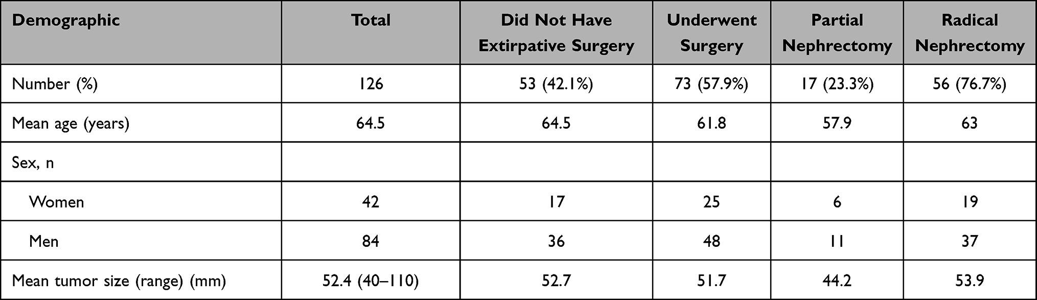 |
Table 1 Patient Demographics and Tumor Size |
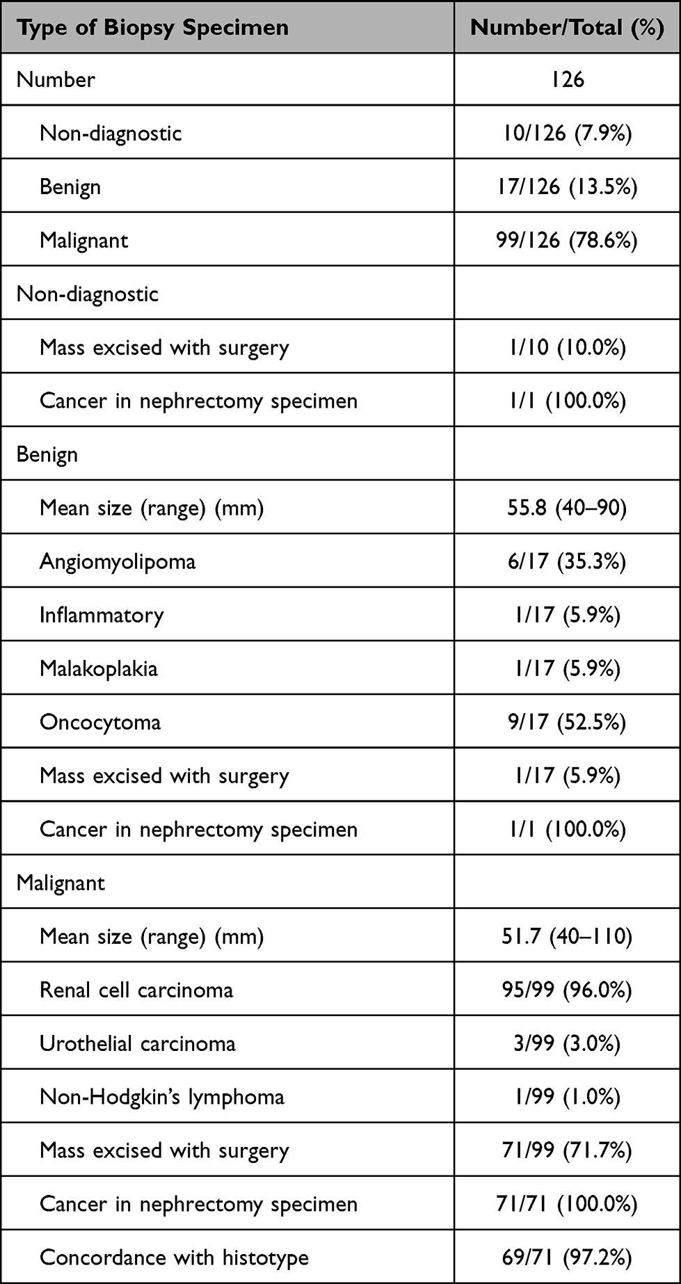 |
Table 2 Biopsy Histopathology, Management Following Non-Diagnostic Biopsy, and Concordance of Biopsy With Final Histology in Patients Who Underwent Extirpative Surgery |
During this period, 289 radical and partial nephrectomies were performed for large renal masses, without prior biopsies. Eighteen (6.2%) of these patients had benign pathologies, including AML, oncocytoma and renal cysts. Of the 126 patients who had biopsies, 56 underwent radical nephrectomy, 17 had a partial nephrectomy and 53 did not undergo extirpative surgery. Histotype concordance was present in 69 (94.5%) of the 73 patients who underwent extirpative surgery (Figure 1). Two of the 73 patients had benign and non-diagnostic biopsies initially, but the final histology identified cancer. In those with concordant histotyping, the histological grading was upgraded in 29 patients (42.0%) and downgraded in two (2.9%).
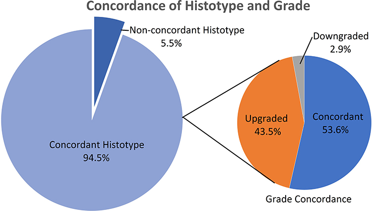 |
Figure 1 Descriptions of positive biopsy and extirpative specimens grade concordance. |
Discussion
This paper is one of the first of its kind looking at large renal mass biopsies and their impact on the management of large renal masses. Apart from a study conducted by Asghar et al, published in 2021, there has been little research in this area, with pathological diagnoses being targeted predominantly at small renal masses. This is because the risk of malignancy increases with size, and therefore a renal biopsy is a potentially redundant procedure.
Power analysis shows that the numbers recruited in the current study are sufficient to detect statistically significant endpoints, as demonstrated, reinforcing the internal validity of our findings. As the procedure was also performed by a small number of highly experienced interventional radiologists, using a rigorous and reproducible biopsy technique, the results of this study can be extrapolated to other tertiary centers, improving the external validity.
Our series supports the recommendation for greater utilization of renal mass biopsy in patients who may have unclear benefits from extirpative surgery. Our study identified 17 patients (13.5%) who had large renal masses with benign conditions, of whom 16 (12.7%) avoided extirpative surgery. A histotype concordance rate of 97.2% supports the established literature on the accuracy of renal mass biopsies.11–13 Renal mass biopsies have been extensively reviewed in the literature regarding their safety and the minimal risks involved.6,7,12,13 In our series, only two patients experienced minor complications of hematuria. Patient selection for biopsy will be crucial to avoid overtreatment with biopsy. Patients such as those with significant comorbidities, or who are not safe surgical candidates, or have bilateral renal masses or indeterminate imaging findings, would be deemed appropriate for consideration of a renal mass biopsy. For patients in whom surgical intervention may pose a significant morbidity and mortality risk, the use of a biopsy may assist them in making an informed decision. Biopsy results may also open up opportunities for clinical trials in advanced disease, such as the CheckMate-214, CheckMate-9ER and KEYNOTE-426 trials.14 Multidisciplinary team meetings to discuss imaging consensus, patient factors and management options remain necessary for patient selection.
While the role of renal biopsy in large renal masses (>4 cm) remains controversial, our study shows that it may also be a useful tool in the management of these tumors, particularly in certain patient populations. The use of renal biopsy as a diagnostic tool for renal masses has become standard practice, particularly in the management of small renal masses (<4 cm).2–5 The high levels of accuracy and safety of renal biopsy in small renal masses, combined with the potential for avoiding unnecessary surgical intervention, make it an essential component of the diagnostic algorithm for these tumors. This may be due to the lower malignant potential of small renal masses.1 Overall, renal biopsy plays a critical role in the management of small renal masses and should be considered on a case-by-case basis in the context of larger renal masses.
There are several avenues of research into renal mass biopsies that are yet to be explored. Apart from qualitative data analyses, as described above, a cost/benefit analysis could be conducted to determine whether renal mass biopsies as a standard investigative tool could help to reduce the overall cost to each individual patient, the hospital, and the healthcare system as a whole, with a reduction in the number of procedures and admissions. Improvements in imaging modalities as well as prediction models and tumor markers in diagnosing and prognosticating renal malignancies should also be explored as further adjuncts to avoid exposing patients to unnecessary risks. For example, increased experience in reading and interpreting sestamibi scans, PSMA scans, DMSA scans, and agents such as zirconium-89-girentuximab, zirconium-89-bevacizumab and zirconium-89-atezolizumab in RCC expressing PD-L1 may open up other avenues of research.15,16 Collaboration with interventional radiologists to offer same-day consultations and CT-guided renal biopsies could also make this a truly one-stop renal cancer clinic, which seems to work well for many other cancers, such as breast cancer and prostate cancer.17,18
The current study is limited by its retrospective nature. A larger sample size with prospective database collection would improve on this issue and aid in the risk stratification of large renal masses to help clinicians in determining which lesions should be biopsied.
Conclusion
Renal mass biopsy is a low-morbidity procedure that provides the treating clinician with a means of obtaining a diagnosis that may obviate the need for a major operation. To date, no other investigation has been shown to be as conclusive as a formal tissue diagnosis. Given that a significant proportion of patients have had changes to their management following this invasive investigation, we conclude that a renal mass biopsy should always be considered, even for large renal masses, and should be incorporated into multidisciplinary team discussions when possible.
Ethics and Consent Statements
Institutional approval was obtained from the Royal Perth Bentley Group ethics committee (GEKO ID: 47539). Informed consent was waived by the ethics committee owing to the retrospective nature of the study, which had no impact on patient confidentiality or management. The guidelines outlined in the Declaration of Helsinki were followed.
Disclosure
The authors report no conflicts of interest in this work.
References
1. Kurta JM, Thompson RH, Kaag M, et al. Tumor size is associated with malignant potential in renal cell carcinoma. J Urol. 2009;181(4S):215. doi:10.1016/S0022-5347(09)60616-9
2. Silverman SG, Israel GM, Herts BR, Richie JP. Management of the incidental renal mass. Radiology. 2008;249(1):16. doi:10.1148/radiol.2491070783
3. Chen DY, Uzzo RG. Evaluation and management of the renal mass. Med Clin. 2011;95(1):179–189. doi:10.1016/j.mcna.2010.08.021
4. Maturen KE, Nghiem HV, Caoili EM, Higgins EG, Wolf JS Jr, Wood DP Jr. Renal mass core biopsy: accuracy and impact on clinical management. Am J Roentgenol. 2007;188(2):563–570. doi:10.2214/AJR.06.0220
5. Ball MW, Bezerra SM, Gorin MA, et al. Grade heterogeneity in small renal masses: potential implications for renal mass biopsy. J Urol. 2015;193(1):36–40. doi:10.1016/j.juro.2014.06.067
6. Kutikov A, Smaldone MC, Uzzo RG, Haifler M, Bratslavsky G, Leibovich BC. Renal mass biopsy: always, sometimes, or never? Eur Urol. 2016;70(3):403–406. doi:10.1016/j.eururo.2016.04.001
7. Lane BR, Samplaski MK, Herts BR, Zhou M, Novick AC, Campbell SC. Renal mass biopsy—a renaissance? J Urol. 2008;179(1):20–27. doi:10.1016/j.juro.2007.08.124
8. Mullins JK, Rodriguez R. Renal cell carcinoma seeding of a percutaneous biopsy tract. Can Urol Assoc J. 2013;7(3–4):E176. doi:10.5489/cuaj.499
9. Patel HD, Johnson MH, Pierorazio PM, et al. Diagnostic accuracy and risks of biopsy in the diagnosis of a renal mass suspicious for localized renal cell carcinoma: systematic review of the literature. J Urol. 2016;195(5):1340–1347. doi:10.1016/j.juro.2015.11.029
10. Asghar AM, McIntosh AG, Smith ZL, et al. Pathological characteristics of the large renal mass: potential implication for clinical role of renal biopsy. Can J Urol. 2021;28(2):10620–10624.
11. Richter F, Kasabian N, Irwin R, Watson R, Lang E. Accuracy of diagnosis by guided biopsy of renal mass lesions classified indeterminate by imaging studies. Urology. 2000;55(3):348–352. doi:10.1016/S0090-4295(99)00468-9
12. Herrera-Caceres JO, Finelli A, Jewett MA. Renal tumor biopsy: indicators, technique, safety, accuracy results, and impact on treatment decision management. World J Urol. 2019;37(3):437–443. doi:10.1007/s00345-018-2373-9
13. Volpe A, Kachura JR, Geddie WR, et al. Techniques, safety and accuracy of sampling of renal tumors by fine needle aspiration and core biopsy. J Urol. 2007;178(2):379–386. doi:10.1016/j.juro.2007.03.131
14. George DJ, Lee C-H, Heng D. New approaches to first-line treatment of advanced renal cell carcinoma. Ther Adv Med Oncol. 2021;13:17588359211034708. doi:10.1177/17588359211034708
15. Hekman M, Rijpkema M, Oosterwijk E, Boerman O, Oyen W, Mulders P. Zirconium-89-girentuximab PET/CT imaging in renal cell carcinoma: first in man results. Eur Urol Suppl. 2016;12(15):e1501. doi:10.1016/S1569-9056(16)30351-7
16. Gorin MA, Rowe SP, Baras AS, et al. Prospective evaluation of 99mTc-sestamibi SPECT/CT for the diagnosis of renal oncocytomas and hybrid oncocytic/chromophobe tumors. Eur Urol. 2016;69(3):413–416. doi:10.1016/j.eururo.2015.08.056
17. Hawks C, Moe A, McCombie S, Hamid A, Brown M, Hayne D. ‘One Stop Prostate Clinic’: prospective analysis of 1000 men attending a public same‐day prostate cancer assessment and/or diagnostic clinic. Anz J Surg. 2021;91(4):558–564. doi:10.1111/ans.16329
18. Dey P, Dixon JM, Bundred N, et al. Costs and benefits of a one stop clinic compared with a dedicated breast clinic: randomised controlled trialCommentary: one stop clinics should not be abandoned. BMJ. 2002;324(7336):507–510. doi:10.1136/bmj.324.7336.507
 © 2023 The Author(s). This work is published and licensed by Dove Medical Press Limited. The full terms of this license are available at https://www.dovepress.com/terms.php and incorporate the Creative Commons Attribution - Non Commercial (unported, v3.0) License.
By accessing the work you hereby accept the Terms. Non-commercial uses of the work are permitted without any further permission from Dove Medical Press Limited, provided the work is properly attributed. For permission for commercial use of this work, please see paragraphs 4.2 and 5 of our Terms.
© 2023 The Author(s). This work is published and licensed by Dove Medical Press Limited. The full terms of this license are available at https://www.dovepress.com/terms.php and incorporate the Creative Commons Attribution - Non Commercial (unported, v3.0) License.
By accessing the work you hereby accept the Terms. Non-commercial uses of the work are permitted without any further permission from Dove Medical Press Limited, provided the work is properly attributed. For permission for commercial use of this work, please see paragraphs 4.2 and 5 of our Terms.