")
Back to Journals » International Medical Case Reports Journal » Volume 17
Second Trimester Spontaneous Fundal Rupture of Unscarred Bicornuate Uterus in Primipara: A Case Report and Literature Review; Jigjiga University Sheik Hassen Yabare Comprehensive Specialized Hospital, Jigjiga, Ethiopia
Authors Areys HM , Omer NH, Osman OA
Received 1 November 2023
Accepted for publication 15 March 2024
Published 19 March 2024 Volume 2024:17 Pages 181—185
DOI https://doi.org/10.2147/IMCRJ.S446718
Checked for plagiarism Yes
Review by Single anonymous peer review
Peer reviewer comments 2
Editor who approved publication: Dr Xudong Zhu
Hassen Mohammed Areys,1 Nour Hies Omer,1 Osman Ali Osman2
1Department of Gynecology and Obstetrics, College of Medicine and Health Science, Jigjiga University, Jigjiga, Ethiopia; 2College of Medicine and Health Science, Jigjiga University, Jigjiga, Ethiopia
Correspondence: Hassen Mohammed Areys, Department of Gynecology and Obstetrics, College of Medicine and Health Science, Jigjiga University, P.O. Box 1020, Jigjiga, Ethiopia, Tel +251-915218641, Email [email protected] Osman Ali Osman, College of Medicine and Health Science, Jigjiga University, P.O. Box 1020, Jigjiga, Ethiopia, Tel +251-915003112, Email [email protected]; [email protected]
Background: Primary rupture of an unscarred uterus is rare. Spontaneous rupture of an unscarred bicornuate uterus is a life-threatening obstetric emergency with high morbidity and mortality in the mother and fetus; however, it most commonly occurs in the first trimester of pregnancy.
Case: A 20-year-old primigravid woman at 22 weeks of gestation, with no prior surgery, presented with severe abdominal pain, anemia, and hemodynamic instability. With a preoperative diagnosis of uterine rupture, she was transfused with three units of cross-matched whole blood and underwent emergency laparotomy. Intraoperative findings showed a ruptured bicornuate uterus and a dead fetus in the abdomen with huge hemoperitoneum. Postoperative recovery was smooth, and the patient was discharged after being counselled on family planning and subsequent pregnancy.
Conclusion: A bicornuate uterus may be an independent risk factor for uterine rupture, which can occur in primigravid women at any stage of pregnancy. Each obstetrician should have a high index of suspicion for a rare condition like ruptured bicornuate uterus, especially for a pregnant woman presenting with acute abdominal pain and hemodynamic instability. Early ultrasonography plays a key role in the evaluation, follow-up, and management of these patients.
Keywords: uterine rupture, bicornuate uterus, unscarred
Introduction
A bicornuate uterus is a rare uterine anomaly resulting from incomplete fusion of the two Müllerian ducts in the sixth to seventh week of embryogenesis. The two Müllerian ducts form the uterus, cervix, and upper third of the vagina.1,2 Uterine rupture in pregnant women is a life-threatening complication that is associated with obstetric care. It is more common in women who have undergone cesarean section or are multiparous and usually occurs during labor.3 Unscarred uterine rupture is relatively rare in the first and second trimesters of pregnancy and is associated with high mortality and morbidity both in the fetus and mother.3,4 The risk factors for unscarred uterine rupture include uterine anomalies, high parity, and placental anomalies.5 This case report describes spontaneous unscarred uterine rupture in a woman with a bicornuate uterus in the second trimester of pregnancy.
Case Presentation
A 20-year-old primigravida woman who did not remember her last menstrual period but stated that she had been amenorrheic for the past 6 months was referred to our hospital with a diagnosis of second trimester pregnancy plus disseminated intravascular coagulation (DIC) plus missed abortion and complained of lower abdominal pain for 1-week duration. Associated with this, she experienced easy fatigability, dizziness, blurred vision, and vertigo of the same duration.
Physical examination revealed blood pressure of 85/50 mmHg, with a pulse rate of 126 beats per minute (bpm). The patient had pale conjunctiva and tender abdomen. Obstetric ultrasound examination revealed a 22-week consistent pregnancy in the peritoneum, negative fetal heartbeat, empty uterus, and free fluid in Douglas and Morrison’s pouch. Laboratory results showed a hemoglobin concentration of 4.3 g/dL.
Owing to unstable vital signs, the patient was transfused with three units of screened, cross-matched whole blood. After informed consent was obtained, the patient was rushed to the operating theatre with a preoperative diagnosis of uterine rupture.
Intraoperative findings revealed hemoperitoneum of 2L, 500gm freshly dead male fetus and placenta, respectively (Figure 1). Further inspection revealed a bicornuate uterus and fundal uterine rupture in the left uterine segment (Figure 2). The hemoperitoneum was suctioned, the placenta and fetus were removed, and the uterus was repaired using a size 1 Vicryl suture (Figure 3).
 |
Figure 1 Shows freshly dead fetus with attached placenta and hemoperitoneum being suctioned. |
 |
Figure 2 Shows left coronal fundal rupture. |
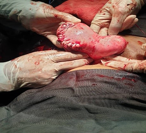 |
Figure 3 Shows repaired left coronal uterine fundus with intact right horn. |
The patient’s postoperative recovery was uneventful, and she was discharged on her fifth postoperative day. The patient was counselled on family planning for at least one year, and any future pregnancy would require elective cesarean delivery.
Discussion
A bicornuate uterus is a rare form of uterine anomaly with an incidence rate of 0.4%.6 Occasionally, the presence of a bicornuate uterus can be detected during pregnancy or childbirth using ultrasound.7,8 Pregnancy in a bicornuate uterus is associated with a poor prognosis both for the mother and fetus.1 In our case, the mother presented with a near-miss condition and a dead fetus.
Uterine rupture is often associated with previous cesarean section and high parity. Other risk factors include uterine anomalies, obstetric maneuvers, abnormal placentation, malpresentation, curettage, injudicious use of oxytocin, and chronic corticosteroid use.9–12 In our case, a bicornuate uterus was identified as a risk factor for uterine rupture.
The classic clinical symptoms of uterine rupture include abdominal pain, vaginal bleeding, and vomiting. These clinical signs in early pregnancy are nonspecific and must be differentiated from acute abdominal conditions, particularly ectopic pregnancy.3,11 Other differential diagnoses include luteal bleeding and molar pregnancy with secondary invasion.12 Early diagnosis and appropriate treatment are essential to reduce the maternal and fetal morbidity and mortality associated with uterine rupture. In the present case, the patient presented with abdominal pain, signs and symptoms of anemia, and unstable vital signs.
Unscarred uterine rupture usually occurs in the lower part of the uterus, but in bicornuate uterus the thickness of anomalous uterine wall is inconsistent and tends to become abnormally thin as pregnancy advance. Fundal rupture, as in this case, occurs in one of the fundal rudimentary horns because of the inability of the malformed uterus to expand as a normal uterus; this led to accumulation of hemorrhage in the peritoneal cavity.13–15 This often leads to delays in diagnosis and treatment.
Immediate surgical intervention is mandatory for the successful treatment of uterine rupture. The type of procedure depends on the extent of the lesion, parity, clinical condition of the patient, and the experience of the obstetrician. Recent studies have shown that the uterus can be repaired, especially in women who want to preserve fertility.5,16 The patient should be counselled about family planning and informed that she must undergo an elective cesarean section in future pregnancies. In the present case, uterine repair was performed because the patient was primiparous.
Conclusion
A bicornuate uterus may be an independent risk factor for uterine rupture, which can occur in primigravid women at any stage of pregnancy. Each obstetrician should have a high index of suspicion for a rare condition like ruptured bicornuate uterus, especially for a pregnant woman presenting with acute abdominal pain and hemodynamic instability. Early ultrasonography plays a key role in the evaluation, follow-up, and management of these patients.
Consent and Ethics
Written informed consent was provided by the patient for publication of case details and accompanying images. This case report was approved by the Institutional Review Board (Jigjiga).
Acknowledgment
We thank the family for their interest and cooperation with the publication of this report.
Funding
The authors did not receive any funding for this work.
Disclosure
The authors declare no conflicts of interest regarding the publication of this study.
References
1. Chan Y, Jayaprakasan K, Tan A, Thornton J, Coomarasamy A, Raine‐Fenning N. Reproductive outcomes in women with congenital uterine anomalies: a systematic review. Ultrasound Obstet Gynecol. 2011;38(4):371–382. doi:10.1002/uog.10056
2. Reichman DE, Laufer MR. Congenital uterine anomalies affecting reproduction. Best Pract Res Clin Obstet Gynaecol. 2010;24(2):193–208. doi:10.1016/j.bpobgyn.2009.09.006
3. Singh N, Singh U, Verma ML. Ruptured bicornuate uterus mimicking ectopic pregnancy: a case report. J Obstetrics Gynaecol Res. 2013;39(1):364–366. doi:10.1111/j.1447-0756.2012.01914.x
4. Hingora MA, Pandya M. Rare case of rupture bicornuate uterus. Indian J Obstetr Gynecol Res. 2017;4(4):460–462.
5. Tola EN. First trimester spontaneous uterine rupture in a young woman with uterine anomaly. Case Rep Obstetr Gynecol. 2014;2014:1–3. doi:10.1155/2014/967386
6. Chan Y, Jayaprakasan K, Zamora J, Thornton J, Raine-Fenning N, Coomarasamy A. The prevalence of congenital uterine anomalies in unselected and high-risk populations: a systematic review. Human Reproduction Update. 2011;17(6):761–771. doi:10.1093/humupd/dmr028
7. Jayaprakash S, Muralidhar L, Sampathkumar G, Sexsena R. Rupture of bicornuate uterus. Case Rep. 2011;2011:bcr0820114633.
8. Nicolini U, Bellotti M, Bonazzi B, Zamberletti D, Candiani GB. Can ultrasound be used to screen uterine malformations? Fertil Sterility. 1987;47(1):89–93. doi:10.1016/S0015-0282(16)49941-3
9. Walsh CA, Baxi LV. Rupture of the primigravid uterus: a review of the literature. Obstetrical Gynecol Surv. 2007;62(5):327–334. doi:10.1097/01.ogx.0000261643.11301.56
10. Nitzsche B, Dwiggins M, Catt S. Uterine rupture in a primigravid patient with an unscarred bicornuate uterus at term. Case Rep Women’s Health. 2017;15:1–2. doi:10.1016/j.crwh.2017.03.004
11. Singh A, Jain S. Spontaneous rupture of unscarred uterus in early pregnancy:-A rare entity. Acta obstetricia et gynecologica Scandinavica. 2000;79(5):431–432.
12. Schrinsky DC, Benson RC. Rupture of the pregnant uterus: a review. Obstetrical Gynecol Surv. 1978;33(4):217–232. doi:10.1097/00006254-197804000-00001
13. Misra M, Roychowdhury R, Sarkar NC, Koley MM. The spontaneous prelabour rupture of an unscarred uterus at 34 weeks of pregnancy. J Clin Diagn Res. 2013;7(3):548. doi:10.7860/JCDR/2013/4496.2820
14. Gordon CA. Ruptured pregnancy in the closed rudimentary horn of a bicornate uterus: external Migration of the spermatazoon; urinary suppression following transfusion. Am J Clin Exp Obstet Gynecol. 1935;29(2):279–282. doi:10.1016/S0002-9378(15)30666-9
15. Daniel ST. Pregnancy in the rudimentary horn of the uterus: review of literature and report of one case. Chinese Med J. 1949;67(09):485–488.
16. Ahmadi S, Nouira M, Bibi M, et al. Uterine rupture of the unscarred uterus. About 28 cases. Gynecol Obstet Fertil. 2003;31(9):713–717. doi:10.1016/S1297-9589(03)00212-1
 © 2024 The Author(s). This work is published and licensed by Dove Medical Press Limited. The full terms of this license are available at https://www.dovepress.com/terms.php and incorporate the Creative Commons Attribution - Non Commercial (unported, v3.0) License.
By accessing the work you hereby accept the Terms. Non-commercial uses of the work are permitted without any further permission from Dove Medical Press Limited, provided the work is properly attributed. For permission for commercial use of this work, please see paragraphs 4.2 and 5 of our Terms.
© 2024 The Author(s). This work is published and licensed by Dove Medical Press Limited. The full terms of this license are available at https://www.dovepress.com/terms.php and incorporate the Creative Commons Attribution - Non Commercial (unported, v3.0) License.
By accessing the work you hereby accept the Terms. Non-commercial uses of the work are permitted without any further permission from Dove Medical Press Limited, provided the work is properly attributed. For permission for commercial use of this work, please see paragraphs 4.2 and 5 of our Terms.