")
Back to Journals » Therapeutics and Clinical Risk Management » Volume 18
Quantitative Evaluation of Biceps Brachii Muscle by Shear Wave Elastography in Stroke Patients
Authors Wei HQ, Gan M, Li GY, Ma SH, Liu JH
Received 9 February 2022
Accepted for publication 11 July 2022
Published 3 October 2022 Volume 2022:18 Pages 879—887
DOI https://doi.org/10.2147/TCRM.S361664
Checked for plagiarism Yes
Review by Single anonymous peer review
Peer reviewer comments 2
Editor who approved publication: Dr Deyun Wang
Hong-Qin Wei,1 Man Gan,1 Guo-Yan Li,2 Sui-Hong Ma,1 Jian-Hua Liu1
1Department of Ultrasound, Guangzhou First People’s Hospital, Guangzhou, Guandong, People’s Republic of China; 2Department of Rehabilitation Medicine, Guangzhou First People’s Hospital, Guangzhou, Guandong, People’s Republic of China
Correspondence: Sui-Hong Ma; Jian-Hua Liu, Tel +86 13824420620 ; +86 13622888381, Fax +86 020 81332620, Email [email protected]; [email protected]
Purpose: The present study aimed to investigate the differences in muscle size and shear wave speed (SWS) values of biceps brachii muscle (BBM) between stroke survivors and healthy controls.
Methods: This study comprised 61 stroke survivors and 24 healthy subjects, examined at Guangzhou First People’s Hospital within one year. Each participant underwent ultrasonic examinations for recording some specific measurement indicators, including muscle thickness, cross-sectional area (CSA), and shear wave speed (SWS) of BBM. The muscular tension of the paretic arm was scored using the modified Ashworth scale (MAS). These above-mentioned indexes were compared between stroke survivors and healthy controls. Also, the correlations among SWS and MAS scores were assessed.
Results: When the lifting arm angle was set for 45°, the CSA and muscle thickness of BBM were obviously decreased in the paretic arms of stroke subjects compared to the non-paretic arms as well as the arms of healthy controls. Moreover, the paretic arms had obviously higher SWS than the non-paretic arms and the healthy arms at 45° or 90°. When the angles of paretic arms were lifted at 90° and 45°, respectively, a positive correlation was established between MAS and SWS.
Conclusion: Ultrasonic examination assessing muscle thickness, CSA, and SWS of the BBM could be used as a means of assessment of the paretic arms of stroke survivors.
Keywords: stroke, muscle, shear wave elastography, ultrasound, thickness, cross-sectional area
Introduction
Stroke is one of the common cerebrovascular diseases, which is a major cause of death and a leading cause of adult disability.1 As the population moves toward an aging society, stroke rates are expected to increase. After the stroke, the central nervous system and the motor conduction pathway are damaged, triggering the disorder correlated with the damage of the muscle structure and properties, such as muscle atrophy.2,3 Limb spasm is a common complication of stroke, which is clinically characterized by increased muscle hardness, declined motor function, and abnormal posture and motion mode, resulting in a heavy burden on the patient’s quality of life.4,5 Moreover, disuse muscular atrophy is another common sequela, and stroke survivors exhibit changes in muscle quantity, such as loss in muscle mass, reduction in cross-sectional area (CSA), and increased intramuscular fat deposition, which leads to joint contracture and loss of muscle strength and limb dysfunction, affecting the patient’s abilities and daily living.6,7 Some studies confirmed that the evaluation of muscle size and muscle stiffness, which was quantified by measuring shear wave elastography (SWE), in paralysis patients after stroke was beneficial for improving motor function and adjustment of rehabilitation treatment plans.8,9
In clinical practice, the severity of spasticity after stroke was assessed by the modified Ashworth scale (MAS)10 or the modified Tardieu scale (MTS).11 However, these methods are affected by subjective factors of the evaluator and do not accurately measure the relevant data of muscle mechanical properties, such as stiffness and elasticity, after stroke, and they lack objective quantitative indicators. Presently, the imaging methods for evaluating muscle volume were magnetic resonance imaging (MRI), computed tomography (CT), and ultrasound (US).12 However, apart from its high cost, CT causes radiation exposure to people, and neither CT nor MRI could observe muscle dynamics.
In recent years, US has gained increasing attention in the assessment of skeletal muscle quality as it is easy to operate in real-time, convenient, and nonradiative.13 It is able to distinguish muscle tissue from subcutaneous fat and measure the thickness and CSA of muscle, and is widely used to diagnose and follow-up the muscle condition of stroke patients.14 Monjo et al15 showed that the thickness of the quadriceps femoris on the paretic side was thinner than that on the non-paretic side in the stroke survivors. Shear wave elastography (SWE) is a quantitative imaging technology that allows the measurement of the hardness of human tissues, and it has already been applied clinically to evaluate muscle stiffness.16,17 Several studies have confirmed the application of SWE for determining muscular states, including muscle injury, stroke spasms, and Parkinson’s disease.18 Liu et al19 showed a significant difference in shear wave propagation speed and Young’s modulus (YM) between the normal and spastic biceps brachii. Wu et al20 showed that the SWV was significantly higher on the paretic side than the non-paretic side in stroke survivors.
During the process of stroke rehabilitation, the assessment of muscle size and stiffness is crucial for the selection and adjustment of rehabilitation treatment plans and monitoring the therapeutic effects and disease prognosis in stroke patients. Although CT and MRI were traditional methods for evaluating muscle CSA in stroke survivors, US had the advantage of real-time measuring the thickness and CSA of the muscle, as well as the muscle stiffness which was evaluated by quantifying SWS as reported previously.1 Therefore, this study aimed to determine the potential clinical value of US examination for quantitative assessing of the muscle function in stroke survivors.
Materials and Methods
Patients
This prospective study was approved by the Institutional Review Committee of Guangzhou First People’s Hospital (K-2021-033-01), Guangzhou, Guangdong. Written informed consent was obtained from all participants before collecting data. This study was conducted from September 2020 to May 2021. The cohort comprised 61 stroke survivors with one-sided limb spasms and 24 healthy volunteers as controls. The height and weight were measured, and the body mass index (BMI) was calculated accordingly.
The inclusion criteria for the stroke group were as follows: (1) patients with unilateral hemiplegia due to stroke for <6 months; (2) cerebral hemorrhage or cerebral infarction confirmed by CT or MRI; (3) able to provide written informed consent and follow basic verbal commands; (4) patients with elevated upper extremity muscle tone and the ability to maintain a stretch position.
The exclusion criteria were as follows: (1) abnormality of muscle tone unrelated to stroke; (2) patients with stiff muscles unable to stretch and flex; (3) patients who cannot cooperate with examination; (4) patients with MAS score of 4. Healthy controls were recruited through community advertising, and those age-matched with stroke patients and normal upper limb movement were selected. The exclusion criteria for healthy volunteers were orthopedic and neurological condition or chronic pain status and history of surgery.
Clinical Evaluation
Before US testing, MAS was evaluated by a licensed, experienced, physical therapist with respect to the upper limb function and spasticity for all patients. MAS scores were as follows: level 0 (no increase in muscle tone): 0 points; level 1 (slight increase in muscle tone, minimal resistance, or sudden release at the end of the joint range of motion): 1 point; level 1+ (slight increase in muscle tone, sudden stuck within 50% of joint motion and then minimal resistance at 50% of joint motion): 2 points; level 2 (a marked increase in muscle tone through most of the region of movement, but easily moved): 3 points; level 3 (considerable increase in muscle tone, passive movement difficult): 4 points; and level 4 (affected part rigid in flexion or extension): 5 points.21
US Examination
All patients underwent conventional US and SWE by an ultrasound scanner (Mindray Bio-Medical Electronics Company, Shenzhen, China) with a multifrequency linear transducer (L11-3U; frequency range = 3–11 MHz). We selected the examination conditions for muscle, the mechanical index (MI) was 1.2, the thermal index (TIS) was 0.2, and the depth was 3 cm. The gain could be adjusted according to the subject’s BBM image to make the image display in the best state. All examinations were performed by one US physician with >5 years of experience in order to decrease the measuring error. All participants lay supine on a flatbed, the upper limbs were positioned at 45° relative to the body with the elbow at 0° flexion. The US transducer was placed mid-substance of the long head of biceps brachii muscle (BBM), and the cross-sectional area and thickness were measured three times to obtain an average value for statistical analysis (Figure 1). After conventional US images were obtained, the transducer was held manually, and oriented perpendicular to the skin and parallel to the BBM fiber bundle, and the US system was switched to the SWE penetration mode. Copious amounts of gel were placed between US transducer and the skin to avoid the pressure of the transducer on the skin to obtain SWE images of adequate quality. The probe was maintained for a few seconds to allow stabilization of the elastography image, while the color was constant in the color elastic map for 3–5 s (Figure 1). Then, the position was adjusted such that the spasticity side of the upper limb was 90° relative to the body with the elbow at 0° flexion, the biceps were extended, and the image was captured. Subsequently, the US images were assessed on both the paretic and non-paretic sides in the stroke group and only on the dominant side in the healthy controls.
 |
Figure 1 Shear wave quality is estimated using a shear wave quality map. Homogeneous green of the left image in the region of interest indicates good quality of shear wave speed estimation; the credibility index is 100%. |
Measurement of the Shear Modulus and SWS
High-quality elastic images were selected, and a 5-mm circular region of interest was marked in the elastography box, avoiding the tendons, aponeuroses, and fascial tissue.22 When the sample box was determined, the system automatically displayed the SWS of the muscle, including the maximum, average, and minimum. The SWS was measured three times for each subject, and the average values were calculated for statistical analysis. For each subject, three images of muscle in the same arm were used to calculate the average values of the related indicators, including the cross-sectional area, thickness, and SWS. Moreover, the reliabilities of the related measurement indicators were assessed by calculating the intraclass correlation coefficient (ICC) using the above-mentioned three muscle images, according to the method described previously.14
Statistical Analysis
Statistical analyses were performed using SPSS 23.0 (SPSS, Inc., Chicago, USA). The muscle size and muscle SWS were presented as mean±standard deviation (SD). A chi-square test was used to compare the proportion of genders between the stroke group and healthy controls. Independent-samples t-test was used to compare age, height, weight, and BMI between the stroke group and healthy controls. One-way analysis of variance (ANOVA) was used to compare the cross-sectional area, thickness, and SWS values between the stroke group (paretic or non-paretic side) and healthy controls with Bonferroni post-hoc tests for multiple comparisons. Spearman’s test was used for the correlation analysis of SWS with MAS. P<0.05 indicated statistically significant difference.
Results
The current research is part of our on-going study to search for muscle function in stroke survivors by conventional ultrasound and SWE. In the present study, we measured the CSA, the thickness and the SWS of biceps brachii muscle and analyzed their correlation with clinical indexes.
Clinical Evaluation
The study enrolled 38 males and 23 females in the stroke group, and the mean age was 63.5±12.7 years. Among these, 45 cases were cerebral infarction and 16 were brain bleeding. The age-matched healthy controls consisted of 13 males and 11 females, aged 62.1±10.3 years. A total of 61 patients had different degrees of MAS: 10 patients with MAS level 0, 17 patients with level 1, 9 patients with level 1+, 21 patients with level 2, and 4 patients with level 3. The clinical information of the patients with stroke and of healthy controls is listed in Table 1.
 |
Table 1 Clinical Characteristics of Stroke Patients and Healthy Controls |
Measurement of the CSA and Thickness of BBM in Stroke Patients and Healthy Controls
As shown in Figure 2, the CSA and the thickness of BBM were obviously smaller on the paretic side, when compared with non-paretic side or healthy control. The paretic side exhibited significantly smaller CSA and thickness than the non-paretic side (both P<0.05). Moreover, the CSA and thickness were found to be smaller in the paretic side versus healthy control (both P<0.05). No significant differences regarding the CSA and thickness were indicated between the non-paretic side and healthy control (both P>0.05) (Table 2).
 |
Table 2 Cross-Sectional Area (CSA) and Thickness Values of BBM Between Stroke Patients (Paretic or Non-Paretic Side) and Healthy Controls |
 |
Figure 2 Ultrasound images showed CSA (cross-sectional area) and thickness on the paretic side and non-paretic side of a stroke survivor and a healthy person. |
Measurement of the SWS of BBM in Stroke Patients and Healthy Controls
The SWS of BBM was measured when the lifting arm angle was set for 90° or 45°. At these two positions, the SWS was shown to be significantly higher on the paretic side, when compared with the non-paretic side or healthy control (Figure 3). The quantification of the SWS is shown in Table 3. As demonstrated in Table 3, when the angle was set as 45°, the paretic side showed obviously higher SWS than the non-paretic side (both P<0.05) and healthy control (both P<0.05). No significant differences regarding these two indexes were found between the non-paretic side and healthy control at the position of 45° (both P>0.05). At the angle of 90°, these two indexes were also increased on the paretic side, when compared with the non-paretic side (both P<0.05) or healthy control (both P<0.05). In addition, SWS was increased on the non-paretic side versus healthy control (both P<0.05).
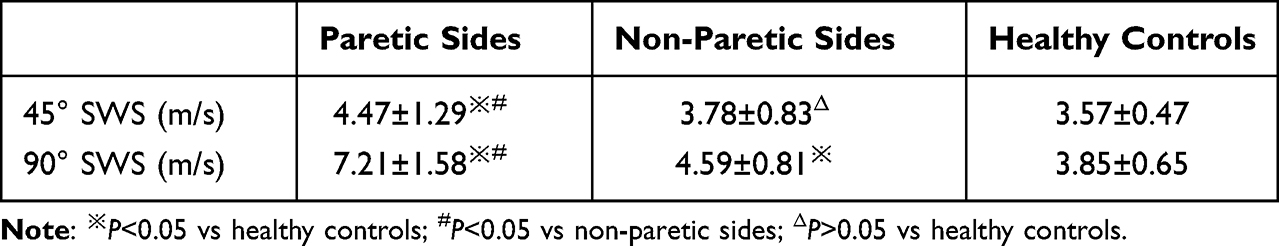 |
Table 3 Shear Wave Velocity (SWS) Values of BBM Between Stroke Patients (Paretic or Non-Paretic Side) and Healthy Controls |
 |
Figure 3 Shear wave elastography of the bilateral biceps brachii muscles (BBM) of stroke patients and healthy individuals, when the upper limbs were positioned at 90° (A) or 45° (B). |
Correlation Analysis Between SWS and MAS
Correlation analysis between SWS and clinical indexes (MAS) in paretic sides was also performed. As shown in Table 4, SWS correlated positively with MAS at 90° and 45°, and the correlation coefficients were 0.399 and 0.382, respectively.
 |
Table 4 SWS Correlation Analysis Using MAS on the Spastic Side |
Discussion
The present study evaluated the BBM size change by measuring muscle CSA and thickness, and also evaluated the stiffness by measuring SWS, respectively, in order to evaluate the modifications in muscle quantity and quality of stroke survivors. It is demonstrated in this current study that the CSA and thickness of BBM were obviously decreased in the paretic sides of stroke patients. Moreover, SWS was increased in the paretic sides. To the best of our knowledge, this is the first study using CSA, muscle thickness, and SWS to reflect the muscle changes in stroke survivors. In addition, the measurement of size and SWS of muscle could be used to quantify the biomechanical properties of skeletal muscle in stroke survivors.
Some studies reported that the muscle mass was decreased in stroke survivors, which led to the decline of muscle and motor function.6,23 Therefore, it is important to quantify muscle SWS in the daily therapeutic management of stroke survivors. US is capable of measuring the thickness and cross-sectional area of muscle, therefore expanding its application in the evaluation of skeletal muscle mass. The commonly used indicators for assessing muscle mass include muscle thickness, CSA, and muscle echo. Li et al24 demonstrated that the CSA of biceps brachii measured by US is a critical indicator associated with sarcopenia, and was significantly higher in the non-sarcopenia group than sarcopenia group. Takai et al25 used ultrasound and dual-energy X-ray absorptiometry-measured muscle thickness and fat-free mass at nine sites of the body in 77 healthy elderly to indicate that ultrasound muscle thickness measurement is useful to predict fat-free mass in the elderly. Wang26 showed that the thickness of the gastrocnemius muscle effectively predicted the decrease in muscle mass. The studies above indicated that ultrasound muscle thickness measurement could predict the decrease in muscle mass, which was consistent with our research. In addition, some studies showed that muscle loss of non-paretic limbs was likely to occur in stroke patients due to a sedentary lifestyle.27 Since this study chose the patients within 6 months of having the first stroke, the results did not find any significant differences in CSA and thickness between the healthy controls and the non-paretic sides. This phenomenon could be attributed to the duration of the disease. For additional evidence, we should examine the muscle mass in the different stages of stroke.
In recent years, the SWE of skeletal muscle uses an acoustic radiation force impulse, which is less dependent on the operator and is a repeatable tool for quantifying muscle stiffness.28,29 Since the muscle is an anisotropic, non-linearly viscoelastic, compressive, deformable, and active tissue, the most appropriate stiffness unit for muscle should be SWS.30 In addition, the shear modulus (kPa) can also be used to represent the elastic characteristics of the tissue, and the correlation between the shear modulus (SM) and the SWS (C) is measured by using the computational formula of SM=ρC2.31 The Mindray machine can display the SM and SWS directly. In this study, we chose SWS as the indicator of muscle hardness. SWS was increased in the paretic side of stroke survivors. In line with the previous studies, our results confirmed the capability of SWE in quantitative assessing of muscle stiffness by measuring SWS in stroke patients with hemiplegia.19,32 Moreover, no significant differences were detected in SWS between the non-paretic side in stroke survivors and healthy people at 45° position, but both values were higher in the non-paretic side in stroke survivors than in healthy people at 90° Position. Some studies showed that adaptive structural changes in the muscle tissue start as early as 4 h after cerebral infarction, and muscle weakness also develops in the unaffected contralateral limb within 1 week after paretic stroke.33 At 3 weeks to 6 months after stroke, muscle mass loss occurred in both paralyzed and non-paralyzed limbs.34 Berenpas et al found that changes in muscle architecture were not solely restricted to the muscles on the paretic side; both paretic and non-paretic side muscles showed deviations from the reference values in healthy individuals.23 The current findings demonstrated that the difference regarding the muscle SWS became significant between the non-paretic side and healthy individuals, when the muscle reached a certain degree of stretching.
Muscle stiffness altered along with the state of the muscle, including muscle contraction, stretching, and resting conditions.35,36 Several studies measured the passive stiffness of various healthy skeletal muscles using US elastography and reported that skeletal muscle became stiffer when it was stretched.22 Liu et al22 collected the shear moduli of gastrocnemius medialis during passive stretching induced by ankle rotation from plantarflexion to dorsiflexion and found that the passive muscle stiffness increased with the augmentation of ankle dorsiflexion. Some studies also reported passive stiffness of muscles in stroke survivors. Eby et al22,37 described the increased SWE of biceps brachii during the process of passive elbow extension in stroke patients, which demonstrated that the passive stiffness increased with elbow extension. We measured two different arm lifting positions (45° and 90°) in this study and observed that the BBM were in different states of stretch and the SWS was increased at 90° versus 45°. These results reflected the effect of stretching on passive stiffness of BBM.
Some recent studies showed controversial ideas concerning the correlation between MAS and elasticity changes of skeletal muscle. Liu et al19 suggested the stiffness of BBM of 60 stroke patients with hemiplegia and one-sided upper limb spasms, with the upper limbs of the spastic side positioned at 90° relative to the body to ensure that biceps brachii were in a stretched position. The results from the above literature concluded that YM and SWS of the biceps brachii were correlated with MAS, and the correlation coefficient was 0.563 and 0.605 for SWS and YM, respectively. Wu et al20 measured the SWS of BBM at 90° and 0° elbow flexion angle in 31 stroke patients and concluded that the paretic-side SWS was positively correlated with MAS at both 90° and 0°; however, the correlation coefficients were higher at 90° than 0°. Lee et al measured the SWS of biceps brachii at the elbow position of 90° in 16 stroke patients, but did not find any correlations between SWS and MAS.38 Gao et al measured the SWS of biceps brachii at 90° elbow flexion and found a correlation between mean SWS and MAS parameters.39 In the current study, we found that SWS was positively correlated with MAS at both 90° and 45°. Harris et al33 proposed that MAS scores were not associated with any relevant results of muscle property changes, including electromyogram, torque, or stiffness, indicating that the current standard clinical evaluation tools need to be improved or supplemented.
The present study has some limitations. First, only one skeletal muscle unit was examined in this study, therefore, the examinations of other muscles need to be carried out. Second, this study recruited stroke patients with a disease course within 6 months, thus different stages of stroke patients also need to be recruited. Lastly it should be mentioned that only a single observer performed all examinations. The difference between observers might affect the elastic measurement result, and we will improve and perfect the research in the future.
Conclusion
The conventional US can only measure the muscle size, while SWE has the capability of assessing the BBM stiffness by measuring SWS. The current study quantified the changes in muscle quantity and quality in stroke survivors of BBM. Therefore, conventional US combined with SWE may be a new quantitative imaging technique to assess the paretic arms of stroke survivors.
Abbreviations
BBM, biceps brachii muscle; CSA, cross-sectional area; SWS, shear wave speed; MAS, modified Ashworth scale; ICC, intraclass correlation coefficient; MTS, modified Tardieu scale; MRI, magnetic resonance imaging; CT, computed tomography; US, ultrasound; SWE, shear wave elastography; YM, Young’s modulus; BMI, body mass index.
Data Sharing Statement
No additional data are available.
Ethical Approval
The study was approved by the Institutional Review Board (IRB) of Guangzhou First People’s Hospital with an approval number of K-2021-033-01. The data confidentiality and compliance with the Declaration of Helsinki were maintained.
Author Contributions
All authors made a significant contribution to the work reported, whether that is in the conception, study design, execution, acquisition of date, analysis and interpretation, took part in drafting, revising and critically reviewing the article; gave final approval of the version to be published; have agreed on the journal to which the article has been submitted; and agree to be accountable for all aspects of the work.
Funding
This work was supported by Science and technology project of Guangzhou Health Commission (20222A010007).
Disclosure
The authors report no conflicts of interest in this work.
References
1. Lehoux MC, Sobczak S, Cloutier F, Charest S, Bertrand-Grenier A. Shear wave elastography potential to characterize spastic muscles in stroke survivors: literature review. Clin Biomech. 2020;72:84–93. doi:10.1016/j.clinbiomech.2019.11.025
2. Sommerfeld DK, Gripenstedt U, Welmer AK. Spasticity after stroke: an overview of prevalence, test instruments, and treatments. Am J Phys Med Rehabil. 2012;91(9):814–820. doi:10.1097/PHM.0b013e31825f13a3
3. Doan QV, Brashear A, Gillard PJ, et al. Relationship between disability and health-related quality of life and caregiver burden in patients with upper limb poststroke spasticity. PM R. 2012;4(1):4–10. doi:10.1016/j.pmrj.2011.10.001
4. Jin Y, Zhao Y. Post-stroke upper limb spasticity incidence for different cerebral infarction site. Open Med. 2018;13:227–231. doi:10.1515/med-2018-0035
5. Bethoux F. Spasticity management after stroke. Phys Med Rehabil Clin N Am. 2015;26(4):625–639. doi:10.1016/j.pmr.2015.07.003
6. Scherbakov N, von Haehling S, Anker SD, Dirnagl U, Doehner W. Stroke induced Sarcopenia: muscle wasting and disability after stroke. Int J Cardiol. 2013;170(2):89–94. doi:10.1016/j.ijcard.2013.10.031
7. English C, McLennan H, Thoirs K, Coates A, Bernhardt J. Loss of skeletal muscle mass after stroke: a systematic review. Int J Stroke. 2010;5(5):395–402. doi:10.1111/j.1747-4949.2010.00467.x
8. Ochi A, Fukumoto M, Takami R, Ohko H, Hayashi T, Yamada K. Effect of ankle stretching combined with arm cycling on the improvement of calf muscle stiffness in patients with stroke: a pilot study. J Phys Ther Sci. 2018;30(10):1305–1309. doi:10.1589/jpts.30.1305
9. Thielman G, Yourey L. Ultrasound imaging of upper extremity spastic muscle post-stroke and the correlation with function: a pilot study. Neuro Rehabilitation. 2019;45(2):213–220. doi:10.3233/nre-192742
10. Bohannon RW, Smith MB. Interrater reliability of a modified Ashworth scale of muscle spasticity. Phys Ther. 1987;67(2):206–207. doi:10.1093/ptj/67.2.206
11. Ansari NN, Naghdi S, Hasson S, Azarsa MH, Azarnia S. The Modified Tardieu Scale for the measurement of elbow flexor spasticity in adult patients with hemiplegia. Brain Inj. 2008;22(13–14):1007–1012. doi:10.1080/02699050802530557
12. Koo TK, Guo JY, Cohen JH, Parker KJ. Relationship between shear elastic modulus and passive muscle force: an ex-vivo study. J Biomech. 2013;46(12):2053–2059. doi:10.1016/j.jbiomech.2013.05.016
13. Brandenburg JE, Eby SF, Song P, et al. Ultrasound elastography: the new frontier in direct measurement of muscle stiffness. Arch Phys Med Rehabil. 2014;95(11):2207–2219. doi:10.1016/j.apmr.2014.07.007
14. Morse CI, Smith J, Denny A, Tweedale J, Searle ND. Gastrocnemius medialis muscle architecture and physiological cross sectional area in adult males with Duchenne muscular dystrophy. J Musculoskelet Neuronal Interact. 2015;15(2):154–160.
15. Monjo H, Fukumoto Y, Asai T, et al. Differences in muscle thickness and echo intensity between stroke survivors and age- and sex-matched healthy older adults. Phys Ther Res. 2020;23(2):188–194. doi:10.1298/ptr.E10018
16. Hobson-Webb LD. Emerging technologies in neuromuscular ultrasound. Muscle Nerve. 2020;61(6):719–725. doi:10.1002/mus.26819
17. Liu J, Qian Z, Wang K, et al. Non-invasive quantitative assessment of muscle force based on ultrasonic shear wave elastography. Ultrasound Med Biol. 2019;45(2):440–451. doi:10.1016/j.ultrasmedbio.2018.07.009
18. Yanagisawa O, Niitsu M, Kurihara T, Fukubayashi T. Evaluation of human muscle hardness after dynamic exercise with ultrasound real-time tissue elastography: a feasibility study. Clin Radiol. 2011;66(9):815–819. doi:10.1016/j.crad.2011.03.012
19. Liu J, Pan H, Bao Y, Zhao Y, Huang L, Zhan W. The value of real-time shear wave elastography before and after rehabilitation of upper limb spasm in stroke patients. Biomed Res Int. 2020;2020:6472456. doi:10.1155/2020/6472456
20. Wu CH, Ho YC, Hsiao MY, Chen WS, Wang TG. Evaluation of post-stroke spastic muscle stiffness using shear wave ultrasound elastography. Ultrasound Med Biol. 2017;43(6):1105–1111. doi:10.1016/j.ultrasmedbio.2016.12.008
21. Şendur HN, Cindil E, Cerit MN, Kılıç P, Gültekin I, Oktar S. Evaluation of effects of aging on skeletal muscle elasticity using shear wave elastography. Eur J Radiol. 2020;128:109038. doi:10.1016/j.ejrad.2020.109038
22. Liu X, Yu HK, Sheng SY, et al. Quantitative evaluation of passive muscle stiffness by shear wave elastography in healthy individuals of different ages. Eur Radiol. 2021;31(5):3187–3194. doi:10.1007/s00330-020-07367-7
23. Berenpas F, Martens AM, Weerdesteyn V, Geurts AC, van Alfen N. Bilateral changes in muscle architecture of physically active people with chronic stroke: a quantitative muscle ultrasound study. Clin Neurophysiol. 2017;128(1):115–122. doi:10.1016/j.clinph.2016.10.096
24. Li S, Li H, Hu Y, et al. Ultrasound for measuring the cross-sectional area of biceps brachii muscle in sarcopenia. Int J Med Sci. 2020;17(18):2947–2953. doi:10.7150/ijms.49637
25. Takai Y, Ohta M, Akagi R, et al. Applicability of ultrasound muscle thickness measurements for predicting fat-free mass in elderly population. J Nutr Health Aging. 2014;18(6):579–585. doi:10.1007/s12603-013-0419-7
26. Wang J, Hu Y, Tian G. Ultrasound measurements of gastrocnemius muscle thickness in older people with sarcopenia. Clin Interv Aging. 2018;13:2193–2199. doi:10.2147/cia.S179445
27. Michael K, Macko RF. Ambulatory activity intensity profiles, fitness, and fatigue in chronic stroke. Top Stroke Rehabil. 2007;14(2):5–12. doi:10.1310/tsr1402-5
28. Alfuraih AM, O’Connor P, Hensor E, Tan AL, Emery P, Wakefield RJ. The effect of unit, depth, and probe load on the reliability of muscle shear wave elastography: variables affecting reliability of SWE. J Clin Ultrasound. 2018;46(2):108–115. doi:10.1002/jcu.22534
29. Tang X, Wang L, Guo R, Huang S, Tang Y, Qiu L. Application of ultrasound elastography in the evaluation of muscle strength in a healthy population. Quant Imaging Med Surg. 2020;10(10):1961–1972. doi:10.21037/qims-20-439
30. Creze M, Nordez A, Soubeyrand M, Rocher L, Maître X, Bellin MF. Shear wave sonoelastography of skeletal muscle: basic principles, biomechanical concepts, clinical applications, and future perspectives. Skeletal Radiol. 2018;47(4):457–471. doi:10.1007/s00256-017-2843-y
31. Couade M, Pernot M, Messas E, et al. In vivo quantitative mapping of myocardial stiffening and transmural anisotropy during the cardiac cycle. IEEE Trans Med Imaging. 2011;30(2):295–305. doi:10.1109/tmi.2010.2076829
32. Lee SSM, Jakubowski KL, Spear SC, Rymer WZ. Muscle material properties in passive and active stroke-impaired muscle. J Biomech. 2019;83:197–204. doi:10.1016/j.jbiomech.2018.11.043
33. Harris ML, Polkey MI, Bath PM, Moxham J. Quadriceps muscle weakness following acute hemiplegic stroke. Clin rehabil. 2001;15(3):274–281. doi:10.1191/026921501669958740
34. Carin-Levy G, Greig C, Young A, Lewis S, Hannan J, Mead G. Longitudinal changes in muscle strength and mass after acute stroke. Cerebrovasc Dis. 2006;21(3):201–207. doi:10.1159/000090792
35. Hug F, Lacourpaille L, Maïsetti O, Nordez A. Slack length of gastrocnemius medialis and Achilles tendon occurs at different ankle angles. J Biomech. 2013;46(14):2534–2538. doi:10.1016/j.jbiomech.2013.07.015
36. Koo TK, Hug F. Factors that influence muscle shear modulus during passive stretch. J Biomech. 2015;48(12):3539–3542. doi:10.1016/j.jbiomech.2015.05.038
37. Eby S, Zhao H, Song P, et al. Quantitative evaluation of passive muscle stiffness in chronic stroke. Am J Phys Med Rehabil. 2016;95(12):899–910. doi:10.1097/phm.0000000000000516
38. Lee SS, Spear S, Rymer WZ. Quantifying changes in material properties of stroke-impaired muscle. Clin Biomech. 2015;30(3):269–275. doi:10.1016/j.clinbiomech.2015.01.004
39. Gao J, He W, Du LJ, et al. Quantitative ultrasound imaging to assess the biceps brachii muscle in chronic post-stroke spasticity: preliminary observation. Ultrasound Med Biol. 2018;44(9):1931–1940. doi:10.1016/j.ultrasmedbio.2017.12.012
 © 2022 The Author(s). This work is published and licensed by Dove Medical Press Limited. The full terms of this license are available at https://www.dovepress.com/terms.php and incorporate the Creative Commons Attribution - Non Commercial (unported, v3.0) License.
By accessing the work you hereby accept the Terms. Non-commercial uses of the work are permitted without any further permission from Dove Medical Press Limited, provided the work is properly attributed. For permission for commercial use of this work, please see paragraphs 4.2 and 5 of our Terms.
© 2022 The Author(s). This work is published and licensed by Dove Medical Press Limited. The full terms of this license are available at https://www.dovepress.com/terms.php and incorporate the Creative Commons Attribution - Non Commercial (unported, v3.0) License.
By accessing the work you hereby accept the Terms. Non-commercial uses of the work are permitted without any further permission from Dove Medical Press Limited, provided the work is properly attributed. For permission for commercial use of this work, please see paragraphs 4.2 and 5 of our Terms.