")
Back to Journals » Drug Design, Development and Therapy » Volume 18
Molecular Mechanism of Salvia miltiorrhiza in the Treatment of Colorectal Cancer Based on Network Pharmacology and Molecular Docking Technology
Received 28 October 2023
Accepted for publication 1 February 2024
Published 12 February 2024 Volume 2024:18 Pages 425—441
DOI https://doi.org/10.2147/DDDT.S443102
Checked for plagiarism Yes
Review by Single anonymous peer review
Peer reviewer comments 2
Editor who approved publication: Dr Tuo Deng
Yi-Ling Jiang, Yi Xun
Department of Oncology, The First Affiliated Hospital, Hengyang Medical School, University of South China, Hengyang, Hunan, People’s Republic of China
Correspondence: Yi Xun, Department of Oncology, The First Affiliated Hospital, Hengyang Medical School, University of South China, Hengyang, Hunan, People’s Republic of China, Tel +8618773486975, Email [email protected]
Purpose: This study aimed to investigate the effect of Salvia miltiorrhiza on colorectal cancer, as well as the mechanisms involved.
Methods: The active compounds of Salvia miltiorrhiza and the associated genes in colorectal cancer were sourced from publicly available databases. Targets associated with colorectal cancer were identified by searching the GeneCards and OMIM databases. Subsequently, the Cytoscape 3.6.0 software was employed to create a regulatory network that illustrates the relationships among active ingredients, colorectal cancer, and their corresponding targets. The String database was utilized to generate a PPI network. Molecular docking studies, conducted with AutoDock Vina, verified the binding capabilities of these active components to core targets. The findings from network pharmacology analysis were corroborated through in vitro experiments.
Results: In this study, we identified 39 active components derived from Salvia miltiorrhiza that are predicted to target 544 genes associated with colorectal cancer through network pharmacology. Through a combined analysis of network pharmacology, we isolated three key targets: SRC, IL6, and INS. Molecular docking results convincingly demonstrated Salvia miltiorrhiza’s strong binding affinity to these targets. Additionally, in vitro experiments confirmed that Salvia miltiorrhiza effectively inhibited the progression of colorectal cancer via regulating the INS/SRC/IL6 pathway.
Conclusion: Salvia miltiorrhiza emerges as a compelling herbal intervention for colorectal cancer. This study lays the foundation for potential future clinical trials assessing the efficacy of Salvia miltiorrhiza in the management of colorectal cancer.
Keywords: Salvia miltiorrhiza, network pharmacology, traditional Chinese medicine, docking technology, colorectal cancer
Introduction
Colorectal cancer is one of the leading causes of death related to cancer worldwide, ranking third and fourth in cancer-related morbidity and mortality, respectively.1,2 The global burden of colorectal cancer is predicted to increase by 60% by 2030, with 2.2 million new cases and 1.1 million cancer deaths.3,4 Around 40–50% of colorectal cancer patients will eventually progress to metastatic disease, with approximately 25% of newly diagnosed patients having distant metastasis at the time of diagnosis.5,6 Moreover, the majority of colorectal cancer patients presented with an advanced stage diagnosis, and it has been reported that the five-year survival rate for individuals diagnosed with colorectal cancer is 14%.7–9 With a history spanning over 2500 years in China’s healthcare system, traditional Chinese medicine boasts a rich heritage. The extensive array of bioactive compounds present in Chinese herbs serves as compelling evidence for their efficacy in managing diverse health conditions. Notably, traditional Chinese medicine has emerged as a promising modality for the treatment of colorectal cancer.10,11
Salvia miltiorrhiza ranks among the most renowned species within the Salvia genus, which belongs to the Lamiaceae family.12,13 Salvia miltiorrhiza, first documented in the “Shen-Nong Herbal Classic”, is characterized by its bitter taste and slightly cold nature. This herb exhibits properties that promote blood circulation, alleviate blood stasis, provide relief from menstrual pain, cool the blood, and aid in abscess elimination. Clinical and pharmacological studies have shown that Salvia miltiorrhiza has anti-inflammatory and anti-tumor effects.14–16 However, the precise role of Salvia miltiorrhiza in colorectal cancer and its underlying mechanisms remain elusive, necessitating further investigation. Network pharmacology, as an emerging technology, systematically investigates the potential effects of drugs on diseases by constructing drug-disease-target-pathway interaction networks, thereby providing novel insights into the material basis and mechanism underlying traditional Chinese medicine treatment of diseases.17,18 The molecular docking method employs computer technology to study the geometric structure and spatial interactions between drug molecules and targets, facilitating the identification of more targeted drugs.19,20 This study aimed to comprehensively explore the impact of Salvia miltiorrhiza on colorectal cancer using network pharmacology and molecular docking techniques. In addition, in vitro experiments were conducted to further validate the efficacy of Salvia miltiorrhiza in treating colorectal cancer, elucidating its potential mechanisms of action and establishing a theoretical foundation for subsequent experimental research and clinical applications.
Materials and Methods
Screening of Active Ingredients of Salvia miltiorrhiza and Prediction of Related Targets
The TCM Systems Pharmacology Database and Analysis Platform (TCMSP) (https://llold.tcmsp-e.com/tcmsp.php) and Bioinformatics Analysis Tool for Molecular Mechanism of Traditional Chinese Medicine (BATMAN-TCM) (http://bionet.ncpsb.org.cn/batman-tcm/index.php) databases were employed to analyze the effective components and genetic targets of drug candidates. The search was performed using “Salvia miltiorrhiza” as the keyword, with criteria set for oral bioavailability (OB) ≥ 30%, drug-likeness (DL) ≥ 0.18, Score > 20, and p-value < 0.05 to identify active ingredients of Salvia miltiorrhiza, followed by collection of gene targets associated with these active ingredients. The UniProt database (https://www.uniprot.org) was utilized to query gene names and abbreviations for all corresponding targets, specifically selecting Homo sapiens as the biological species.
Screening of Targets for Colorectal Cancer
To identify target genes for colorectal cancer, a comprehensive search was conducted using the keyword “colorectal cancer” through GeneCards (https://www.genecards.org/) and Online Mendelian Inheritance in Man (OMIM) (https://omim.org/) databases. Duplicate values were removed to ensure accuracy. Subsequently, UniProt database (https://www.uniprot.org) was utilized to retrieve gene abbreviations specifically for Homo sapiens.
Construction of Drug-Ingredient-Target Network
The Venn program package in R4.0.3 software was utilized to identify the overlapping genes between active ingredients of Salvia miltiorrhiza and colorectal cancer, enabling the construction of a drug-disease relationship map and identification of effective targets for Salvia miltiorrhiza. Additionally, Cytoscape 3.7.2 software was employed to construct a gene network diagram illustrating the interactions among active ingredients of Salvia miltiorrhiza, core targets, and colorectal cancer, facilitating an analysis of the potential mechanisms underlying the therapeutic effects of Salvia miltiorrhiza on colorectal cancer.
Construction of PPI Network and Screening of Core Genes
The common target of Salvia miltiorrhiza- colorectal cancer was queried in the STRING database (https://cn.stringdb.org/) with species restricted to “Homo sapiens”. Subsequently, the obtained PPI information TSV file was imported into Cytoscape 3.8.2 software for network visualization. The Analyze Network plugin was employed to assess network topology parameters, and key targets were identified based on criteria where their degree centrality, betweenness centrality, and closeness centrality exceeded the median.
GO and KEGG Enrichment Analysis
The identified target genes underwent online analysis utilizing the DAVID database (https://david.ncifcrf.gov/), followed by importation into Omicsbean software. Gene Symbol was selected as the type, while “Homo sapiens” served as the specified species. Consequently, GO enrichment and KEGG pathway analyses were performed on these target proteins. The resultant output file was subsequently imported into an Excel worksheet for additional filtering of significant functions and pathways (P < 0.05), which were ultimately presented in a bar chart.
Molecular Docking
Salvia miltiorrhiza’s structural formula was obtained from the PubChem database, and the corresponding 3D structure was created using Chem3D software. The protein domain was downloaded in pdb format from the PDB database. To search for active pockets, the AutoDockTools 1.5.6 software was used to convert pdb format gene files of active ingredients and core proteins into pdb format. Finally, the Vina script was run to calculate molecular binding energy and display the results of molecular docking. The Discovery Studio 2019 software was used to find docking sites and calculate the flexible binding LibDockScore, and the output molecular docking results were imported into PyMOL software to display molecular docking conformation.
Preparation of Salvia miltiorrhiza Extract
Salvia miltiorrhiza was provided by the School of Pharmacy, Guangxi University of Traditional Chinese Medicine for further in vitro experiments. The samples were authenticated by Prof. Weihua Tan (Hengyang Medical School, University of South China), and voucher specimens (No. Ps20230821) were stored at Hengyang Medical School, University of South China. Salvia miltiorrhiza was dissolved in 100% dimethyl sulfoxide (DMSO) to prepare a 20 mg/mL storage solution and was stored at −20°C, diluted with medium before each experiment. All of the control groups in our investigation contained 0.1% DMSO as the final DMSO concentration, which was kept below 0.1% throughout the study. 5-fluorouracil (5-Fu) was used as a positive drug, supplied by SigmaAldrich (USA). According to agency/local regulations, no additional approvals are required to conduct research with plant material.
Cell Culture
The HCT116 and DLD-1 human colorectal cancer cell lines and NCM-460 human normal colonic mucosal epithelial cells were purchased from ATCC. Cells were grown in RPMI-1640 medium supplemented with 10% fetal bovine serum, 1% penicillin, and 1% streptomycin at 37°C in a humidified atmosphere of 5% CO2. The cells were treated with Salvia miltiorrhiza at different concentrations, 5-Fu at 20 μM, or 0.1% DMSO as the vehicle control for 24 h.
CCK-8 Assay
The HCT116, DLD-1, and NCM-460 cells at logarithmic growth stages were selected, and the cell concentration was adjusted to 4×10⁵/mL and then inoculated into 96-well plates. After cell adhesion, the CCK-8 reagent was added for 24 h, 48 h and 72 h. Finally, the OD value at 450 nm wavelength was determined using an enzyme label, according to the CCK-8 kit’s instructions.
Cell Invasion and Migration Experiment
The Transwell assay was used to test cells’ ability to migrate and invade. Logarithmic growth cells were taken for migration experiments, and the cell density of digestive cells was adjusted to 2×10⁶/mL in serum-free medium. The upper chamber received 200uL of cell suspension, while the lower chamber received 700 uL of medium containing 20% fetal bovine serum (FBS). The cells in the upper compartment were wiped with cotton swabs, cleaned with phosphate-buffered saline (PBS), and fixed after 48 h in the incubator. For observation and statistics, the cells were stained with 0.1% crystal violet. Matrigel glue was mixed with serum-free medium and spread in a cold chamber for invasion experiments. In a constant temperature incubator, the chamber was placed in an orifice plate and solidified into a gel overnight. The following experimental procedure is consistent with the transfer experiment.
Scratch Test
Cells at the logarithmic growth stage were inoculated in 6-well plates until they reached 80–90% growth. With a 200 uL gun head, a horizontal line was drawn in the middle of plates, and the drug was rinsed with PBS. They were observed and photographed again 24 h later.
TUNEL Assay
Cell apoptosis was assessed using the In Situ Cell Death Detection Kit (11684817910, Roche, Switzerland) following the manufacturer’s instructions. In summary, the process included paraffin sectioning, dewaxing, hydration, cell permeabilization, addition of TUNEL reaction solution, introduction of converter-POD, reaction with 3,3′-diaminobenzidine (DAB) substrate, cell counting, and image capture (FV1000, Olympus, Japan). Subsequently, Image J software was employed to analyze the images, determine the count of positive cells and total cells in each image, calculate the apoptosis percentage, and perform statistical analysis.
Plate Clone Formation Assay
The HCT116 and DLD-1 cells were inoculated into 96-well plates at logarithmic growth stages after the cell concentration was adjusted to 2×10³/mL. After cell adhesion, various concentrations of breviscapine were added and the cells were incubated for 7 days. After being fixed with 4% paraformaldehyde, the cells were stained with 0.1% crystal violet. To detect clonal formation, the number of stained colonies was counted.
qRT-PCR
The total RNA extraction reagent, Trizol, was purchased from Invitrogen, and the qRT-PCR kit and related gene primers were purchased from Shanghai Bioengineering Co., LTD. in China. Total RNA was extracted from cells using Trizol and its purity was determined. The extracted RNA was reverse-transcribed into cDNA, and the reaction system included the extracted RNA, 5RNA cache solution, M-MLV reverse transcriptase, RNase inhibitor, deoxyribonucleotide (dNTP), and double distilled water (ddH2O) of RNase removal. The reverse transcription was performed in a PCR apparatus set to 37°C for 5 minutes, 85°C for 5 seconds, and 4°C to stop the reaction. Then, the reverse transcription reaction products cDNA, Taq DNA polymerase, dNTP, ddH2O, MgCl2, and buffer were all amplified on PCR plates. The initial denaturation was set at 94 °C for 30 seconds, followed by annealing at 58 °C for 15 seconds and extension at 72 °C for 30 seconds. Ct values were recorded for statistical analysis after 40 cycles were completed. The sequences of qRT-PCR primers are as follows: Bcl-2 forward: 5′-CAGCTGCACCTGACGCCCTT-3′, reverse: 5′-GCCTCCGTTATCCTGGATCC-3′; Bax forward: 5′-CCAGCTCTGAGCAGATCATG-3′, reverse: 5′-TGCTGGCAAAGTAGAAAAGG-3′; caspase-3 forward: 5’-GACCATACATGGGAGCAAGT-3′, reverse: 5’-ATCCGTACCAGAGCGAGA-3′; caspase-9 forward: 5’-ATGGTCACGCTTTGATGGA-3’, reverse: 5’-TTCTCAATGGACACGGAGCA-3’; SRC forward: 5′-AGAGTGCCCTATCCTGGGAT-3′, reverse: 5′-AAAGTAGTCTTCCAGGAAGGCC-3′; IL6 forward: 5′-CCTGAACCTTCCAAAGATGG-3′, reverse: 5′-GGTCAGGGGTGGTTATTGC-3′; INS forward: 5’-GATATAGTAGGGGTTTTTTT-3’, reverse: 5’-AACCTACTTACTAACAACCT-3’; β-actin forward: 5′-TGAGGCCCAGAGCAAGAGA-3′, reverse: 5′-TCGTCCCAGTTGGTGACGAT-3′.
Small Interfering RNA Transfection
Specific small interfering RNAs (siRNAs) against INS (forward, 5′-GGGACCACAAAGATGCTGTT-3′; reverse, 5′-TGGGCTCCTCTCTTACATGG-3′), IL-6 (forward, 5′-AAGGAGGACCTTGTGGCATCT-3′; reverse, 5′- AACAGTGACCTCTG TTGGGCA-3′), and SRC (sense, 5′-GAGACUGACCUGUCCUUCATT-3′ and antisense, 5′-UGAAGGACAGGUCAGUCUCTT-3′) were synthesized by the GenePharma Company (Shanghai, China). The HCT116 cells (60–80% confluent monolayer) were seeded in 12-well plates with 1 mL of standard medium. The cells were transfected with siRNA duplexes (final concentration of 20 nM) the following day, using Lipofectamine™ RNAiMAX reagent (Invitrogen), in accordance with the manufacturer’s instructions. After 72 hours, qRT-PCR analysis was conducted to determine transfection efficiency.
Data Analysis
Data from three separate experiments are presented as the mean and standard deviation (SD). The Student’s t-test was used to compare the means of two groups, and the differences between several groups were evaluated using a one-way ANOVA. All statistical analyses were carried out with the help of GraphPad software (version 8.0.1) and SPSS 24.0. A P value < 0.05 was considered statistically significant.
Results
Acquisition of Active Ingredients of Salvia miltiorrhiza and Corresponding Targets
The TCMSP and BATMAN-TCM databases yielded a list of 39 potential active ingredients from Salvia miltiorrhiza (Table 1). The target genes corresponding to the active ingredients of Salvia miltiorrhiza were retrieved from the TCMSP, SwissTargetPrediction, and PharmMapper databases. After removing duplicate values, 770 target genes and their corresponding GeneSymbols were identified by the UniPort database.
 |
Table 1 Active Ingredients of Salvia miltiorrhiza |
Screening results for Targets Associated with Colorectal Cancer
13675 colorectal cancer-related genes were obtained from OMIM and GeneCards databases. The intersection between genes associated with colorectal cancer and targets linked to active ingredients was identified by the Venn.R software package, followed by the creation of a Venn diagram. As shown in Figure 1, there are a total of 544 intersections between active ingredients and diseases.
 |
Figure 1 Overlap between the target genes of Salvia miltiorrhiza’s active components and those associated with colorectal cancer. |
Construction of Active Ingredients-Target-Disease Network
A network was constructed to illustrate the relationship between potential therapeutic targets for colorectal cancer and the active ingredients found in Salvia miltiorrhiza. As shown in Figure 2, the active components and drug targets were imported into Cytoscape software to visualize the network. Additionally, we expanded upon the primary compounds of Salvia miltiorrhiza by referencing the existing literature. We employed the NetworkAnalyzer tool embedded within Cytoscape 3.7.1 software to establish the active ingredients-target-disease network and assess the node degree values. Within Salvia miltiorrhiza, the active components with a notable number of interconnected targets were Dehydromiltirone (degree = 199), Neocryptotanshinone Ii (degree = 195), and Miltionone I (degree = 195) (Figure 2).
 |
Figure 2 Network construction for ingredient-target interactions. The light pink circular nodes represent the targets, while the light purple prism nodes represent the components of Salvia miltiorrhiza. |
KEGG Pathway and GO Enrichment Analyses
The active ingredient-disease gene targets were inputted into R software and subjected to KEGG enrichment analysis using the Cluster Profiler package. After screening with a confidence interval of P < 0.01, 198 relevant KEGG entries were obtained. Unrelated diseases were eliminated, leaving only the signal transduction pathways of related diseases for comparison. Based on the number of gene enrichments, signaling pathways with smaller P-values and more enriched genes in the top 10 were selected as main influencing pathways, including but not limited to pathways in cancer, MAPK signaling pathway, cAMP signaling pathway, Calcium signaling pathway, and Rap1 signaling pathway (Figure 3). The active ingredients-disease gene targets were input into the R software for GO enrichment analysis using the Cluster Profiler package. A significance level of P < 0.001 was applied to filter the confidence interval, resulting in a total of 120 relevant GO entries. These entries were rearranged based on the number of gene enrichments, and the top 10 entries with smaller P-values and more enriched genes were selected. The identified biological and molecular functions mainly include inflammatory response, cytokine activity, enzyme binding, and RNA polymerase 1 specific DNA-binding transcription factor binding (Figure 4). Taken together, these findings suggest that active ingredients of Salvia miltiorrhiza may inhibit colorectal cancer by the regulation of oncogenes and cytokines.
 |
Figure 3 KEGG enrichment analysis. |
 |
Figure 4 GO enrichment analysis. |
Construction of PPI Network
The intersection genes were inputted into the String database to construct the PPI network. Unlinked nodes were hidden, and both protein interaction diagram and tab-separated values (TSV) files were generated. The resulting file was visualized using Cytoscape 3.7 software. Topological analysis of protein interactions was conducted using R software. As shown in Figure 5A and B, PPI network were established. The higher the target score, the more likely the active ingredient is to play an important role in the treatment of colorectal cancer through that target. The top 10 genes represent key targets of Salvia miltiorrhiza in the treatment of colorectal cancer. Among them, SRC proto-oncogene (SRC),21 interleukin-6 (IL-6),22 and insulin gene (INS)23 are closely related to the occurrence and development of colorectal cancer and are potential therapeutic targets (Figure 5C and Table 2).
 |
Table 2 Screening of Key Targets of PPI Network |
 |
Figure 5 Protein-protein interaction network. (A) PPI network generated through STRING. (B) PPI network generated through Cytoscape. (C) Key target was further screened by betweenness centrality (BC), closeness centrality (CC), and degree centrality (DC). |
Molecular Docking Results
To investigate the interaction between active ingredients of Salvia miltiorrhiza and their target genes, we employed Autodock and Pymol software for molecular docking analysis. Specifically, docking simulations were conducted involving three crucial targets and three active components from Salvia miltiorrhiza, with corresponding binding energies recorded (Figure 6). The results of molecular docking are illustrated in Figures 7–9. These findings provide further validation that the identified three targets through network pharmacology play a significant role in the treatment of colorectal cancer.
 |
Figure 6 Heat map illustrating the binding affinities from molecular docking. |
 |
Figure 7 Molecular docking results of SRC-active components. (A)SRC-Neocryptotanshinone Ii. (B) SRC-Dehydromiltirone. (C) SRC-Miltionone I. |
 |
Figure 8 Molecular docking results of INS-active components. (A) INS-Neocryptotanshinone Ii. (B) INS-Dehydromiltirone. (C) INS-Miltionone I. |
 |
Figure 9 Molecular docking results of IL-6-active components. (A) IL-6-Neocryptotanshinone Ii. (B) IL-6-Dehydromiltirone. (C) IL-6-Miltionone I. |
Salvia miltiorrhiza Suppresses Malignant Phenotype of Human Colorectal Cancer Cells
To investigate the effects of Salvia miltiorrhiza on colorectal cancer, we exposed HCT116, DLD-1, and NCM-460 cells to varying concentrations of Salvia miltiorrhiza. The CCK-8 assay showed that the Salvia miltiorrhiza significantly suppressed the proliferative activity of HCT116 and DLD-1 cells. The IC50 values at 24 h, 48 h, and 72 h for the HCT116 cell line were measured at 16.89 μg/mL, 12.35 μg/mL, and 9.68 μg/mL, respectively (Figure 10A). Similarly, the IC50 values for DLD-1 cells were found to be 15.54 μg/mL (24 h), 11.80 μg/mL (48 h), and 9.17 μg/mL (72 h) as shown in Supplementary figure 1A. Subsequently, the Salvia miltiorrhiza freeze-dried powder solution was categorized into low concentration (5 μg/mL), medium concentration (10 μg/mL), and high concentration (20 μg/mL) groups. Then, cell scratch and transwell experiments corroborated the inhibitory effects of Salvia miltiorrhiza on the proliferation, invasion, and migration of HTC116 and DLD-1 cells (Figure 10B–D and Supplementary figure 1B–D). Furthermore, cloning experiments revealed that Salvia miltiorrhiza significantly impeded cell colony formation (Figure 10E and Supplementary figure 1E). Additionally, the cytotoxicity of Salvia miltiorrhiza to NCM-460 human normal colonic epithelial cells were also measured (Supplementary figure 2), which showed that treatment with Salvia miltiorrhiza at 5 μg/mL, 10 μg/mL and 20 μg/mL for 24 h, 48 h and 72 h did not significantly dampen the viability of NCM-460 cells.
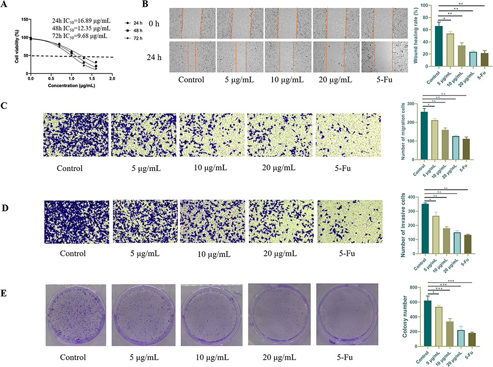 |
Figure 10 Effects of Salvia miltiorrhiza on the proliferation, invasion, and migration of human HCT116 colorectal cancer cells. HCT116 colorectal cancer cells underwent treatment with Salvia miltiorrhiza at different concentrations or 5-Fu at 20 μM. Subsequently, we conducted various assessments, including cell proliferation, viability, migration, invasion, and colony formation, following the methods outlined in the materials and methods section. (A) Cell viability was measured by the CCK-8 assay. (B) Cell proliferation was detected by the scratch experiment. Original magnification: 10×. (C) Cell migration was determined by the transwell assay. Original magnification: 20×. (D) Cell invasion was detected by the transwell experiment. Original magnification: 20×. (E) Cell colony formation. The data presented here are the combined results of three separate experiments. Scale bar: 50 μm. *p < 0.05, **p < 0.01, ***p < 0.001 vs Control. |
Salvia miltiorrhiza Promotes the Apoptosis of Human Colorectal Cancer Cells
Next, we investigated the effect of Salvia miltiorrhiza on apoptosis in human colorectal cancer cells. As illustrated in Figures 11A and B and Supplementary figure 3A and B, the Salvia miltiorrhiza treatment group exhibited a higher number of TUNEL-positive colorectal cancer cells compared to the control model group. Notably, higher doses of Salvia miltiorrhiza led to a more pronounced promotion of cell apoptosis in vitro. Our analysis confirmed a concentration-dependent increase in apoptotic rates induced by Salvia miltiorrhiza. The apoptotic characteristics observed in cancer cells are often a reflection of changes in apoptosis-related genes. The qRT-PCR results provided further evidence that the anti-apoptotic Bcl-2 mRNA levels were reduced, while the proapoptotic Bax mRNA expression was heightened in Salvia miltiorrhiza-treated HCT116 cells when compared to the control (Figure 11C and D). Additionally, caspase-9 and caspase-3 are pivotal components of the apoptotic process. Our findings indicated that Salvia miltiorrhiza augmented the activities of caspase-9 and caspase-3 in HCT116 cells, thus consolidating the proapoptotic effect of Salvia miltiorrhiza in colorectal cancer cells (Figure 11E and F).
 |
Figure 11 Effects of Salvia miltiorrhiza on the apoptosis of human HCT116 colorectal cancer cells and core targets. HCT116 colorectal cancer cells were treated with Salvia miltiorrhiza at 5μg/mL, 10 μg/mL and 20 μg/mL, or 5-Fu at 20 μM, followed by the analyses of TUNEL and qRT-PCR. (A and B) TUNEL analysis of the apoptosis of HCT116 cells treated with Salvia miltiorrhiza or 5-Fu. Original magnification: 10×. (C and D) The real-time qRT-PCR analysis of the mRNA levels of Bax and Bcl-2 in HCT116 cells treated with Salvia miltiorrhiza or 5-Fu. (E and F). The real-time qRT-PCR analysis of the mRNA levels of caspase-9 and caspase-3 in HCT116 cells treated with Salvia miltiorrhiza or 5-Fu. (G–I) The real-time qRT-PCR analysis of the mRNA levels of SRC, IL-6, and INS in HCT116 cells treated with Salvia miltiorrhiza or 5-Fu. Scale bar: 50 μm. *p < 0.05, **p < 0.01, ***p < 0.001, ****p < 0.0001 vs Control. Abbreviation: ns, not significant. |
Salvia miltiorrhiza Inhibited the INS/SRC/IL-6 Pathway in Human Colorectal Cancer Cells
Network pharmacology and molecular docking experiments showed the strong affinity between the active components of Salvia miltiorrhiza and key target genes (SRC, IL-6, and INS). Of note, many studies have demonstrated the important role of SRC, IL-6, and INS in the proliferation and migration of colorectal cancer cells.24–26 Thus, we conducted qRT-PCR assays to validate the effect of Salvia miltiorrhiza on the expression of these core targets. The results demonstrated that Salvia miltiorrhiza markedly suppressed SRC, IL-6, and INS expression (Figure 11G–I). Additionally, our results also showed that Salvia miltiorrhiza decreased SRC and IL-6 levels by inhibiting INS expression (Supplementary figure 3C–E). In summary, these findings collectively suggest that Salvia miltiorrhiza hinders the proliferation, migration, and invasion of human colorectal cancer cells by inhibiting the INS/SRC/IL-6 pathway.
Discussion
The composition of traditional Chinese medicine often comprises a complex blend of ingredients. Notably, the advancement of modern natural medicinal chemistry has contributed to a better understanding of the chemical constituents within traditional Chinese medicine. Traditional Chinese medicine’s impact on the body frequently results from a synergistic interaction among multiple components, collectively yielding a therapeutic effect. Network pharmacology has gained prominence in the field of traditional Chinese medicine as it comprehensively examines the interplay between herbal components and diseases.27,28 It utilizes multiple databases to identify target proteins and action pathways, offering innovative insights into disease treatment and prediction. Molecular docking technology, a computer-aided drug design approach, plays a crucial role in docking compound molecules with protein ligands. This process predicts their modes of action and facilitates a deeper analysis of the anticipated mechanisms underlying drug modulation in the context of diseases.
Colorectal cancer ranks as the third most prevalent malignancy worldwide, with notable regional variations in incidence and mortality rates.29 Traditional herbal medicine has garnered considerable attention and plays a pivotal role in the management of colorectal cancer. Salvia miltiorrhiza, derived from a vital traditional medicinal plant, has gained renown for its multitude of health benefits, particularly its anti-tumor properties.30 Bae et al found that Salvia miltiorrhiza significantly suppresses prostate tumor growth through the inhibition of proliferation in prostate cancer cells and the induction of apoptosis.31 Ye et al demonstrated that Salvia miltiorrhiza inhibits the development of non-small cell lung cancer through the induction of apoptosis via the mitochondrial pathway and PTEN-mediated inhibition of the PI3K/Akt pathway.32 Also, Salvia miltiorrhiza protects against hepatocellular carcinoma by inhibiting the migration and invasion of HepG2 cell via the TGF-β/Smad pathway.33 In addition, several components of Salvia miltiorrhiza, such as Cryptotanshinone and Tanshinone I have also been reported to inhibit the development of cancer.34,35
In this study, we have unveiled the bioactive components and the underlying molecular mechanisms through which Salvia miltiorrhiza exerts its therapeutic potential in the treatment of colorectal cancer. Our approach encompassed a multifaceted methodology, incorporating in vitro experimental validation, molecular docking analyses, and network pharmacology techniques. Our investigations have revealed that the active components present in Salvia miltiorrhiza possess a remarkable affinity for crucial proteins, namely SRC, IL-6, and INS, as substantiated by both network pharmacology and molecular docking assessments. Furthermore, our experimental results have demonstrated the profound impact of Salvia miltiorrhiza on colorectal cancer. It effectively suppresses the proliferation of cancer cells, primarily by inducing apoptosis and impeding their ability to proliferate and migrate. Moreover, we observed that Salvia miltiorrhiza downregulates the SRC, IL-6, and INS levels, while simultaneously elevating expression of pro-apoptotic molecules within colorectal cancer cells. Of note, it has been reported that INS is a upregulator of SRC and IL-6.36–38 Our results also showed that Salvia miltiorrhiza decreased SRC and IL-6 levels by inhibiting INS expression. However, it is also worth noting that the effects of Dehydromiltirone, Neocryptotanshinone Ii, and Miltionone I on colorectal cancer need to be explored. The future studies should be tailored to address these limitations. Taken together, these findings shed light on the bioactive components and molecular pathways through which Salvia miltiorrhiza contributes to the therapy of colorectal cancer.
Conclusion
In summary, our study employed a multifaceted approach that integrated molecular docking, in vitro experiments, and network pharmacology to elucidate the targets and underlying mechanisms through which Salvia miltiorrhiza and its primary active constituents exert potent inhibition of colorectal cancer. Our findings underscored that Salvia miltiorrhiza effectively hinders the progression of colorectal cancer by modulating specific targets pivotal in promoting apoptosis while concurrently suppressing cell proliferation, migration, and invasion. This comprehensive methodology forms the cornerstone of our investigation, presenting a promising alternative therapeutic avenue characterized by low toxicity. This approach can be considered for standalone use or in conjunction with other medications for the management and treatment of colorectal cancer.
Data Sharing Statement
The datasets used or/and analyzed during the current study are available from the corresponding author on reasonable request.
Acknowledgments
The authors gratefully acknowledge the Hengyang Science and Technology Board Program (Grant/Award Number.S2018F9031021264.).
Author Contributions
All authors made a significant contribution to the work reported, whether that is in the conception, study design, execution, acquisition of data, analysis and interpretation, or in all these areas; took part in drafting, revising or critically reviewing the article; gave final approval of the version to be published; have agreed on the journal to which the article has been submitted; and agree to be accountable for all aspects of the work.
Disclosure
The authors report no conflicts of interest in this article.
References
1. Dekker E, Tanis PJ, Vleugels JLA, Kasi PM, Wallace MB. Colorectal cancer. Lancet. 2019;394(10207):1467–1480. PMID: 31631858. doi:10.1016/s0140-6736(19)32319-0
2. Brody H. Colorectal cancer. Nature. 2015;521(7551):S1. PMID: 25970450. doi:10.1038/521S1a
3. Arnold M, Sierra MS, Laversanne M, Soerjomataram I, Jemal A, Bray F. Global patterns and trends in colorectal cancer incidence and mortality. Gut. 2017;66(4):683–691. PMID: 26818619. doi:10.1136/gutjnl-2015-310912
4. Keum N, Giovannucci E. Global burden of colorectal cancer: emerging trends, risk factors and prevention strategies. Nat Rev Gastroenterol Hepatol. 2019;16(12):713–732. PMID: 31455888. doi:10.1038/s41575-019-0189-8
5. Lee RM, Cardona K, Russell MC. Historical perspective: two decades of progress in treating metastatic colorectal cancer. J Surg Oncol. 2019;119(5):549–563. PMID: 30806493. doi:10.1002/jso.25431
6. van der Stok EP, Spaander MCW, Grunhagen DJ, Verhoef C, Kuipers EJ. Surveillance after curative treatment for colorectal cancer. Nat Rev Clin Oncol. 2017;14(5):297–315. PMID: 27995949. doi:10.1038/nrclinonc.2016.199
7. McQuade RM, Stojanovska V, Bornstein JC, Nurgali K. Colorectal cancer chemotherapy: the evolution of treatment and new approaches. Curr Med Chem. 2017;24(15):1537–1557. PMID: 28079003. doi:10.2174/0929867324666170111152436
8. Morris EJ, Sandin F, Lambert PC, et al. A population-based comparison of the survival of patients with colorectal cancer in England, Norway and Sweden between 1996 and 2004. Gut. 2011;60(8):1087–1093. PMID: 21303917. doi:10.1136/gut.2010.229575
9. Dueland S, Yaqub S, Syversveen T, et al. Survival outcomes after portal vein embolization and liver resection compared with liver transplant for patients with extensive colorectal cancer liver metastases. JAMA Surg. 2021;156(6):550–557. PMID: 33787838; PMCID: PMCPMC8014205. doi:10.1001/jamasurg.2021.0267
10. Zhao H, He M, Zhang M, et al. Colorectal cancer, gut microbiota and traditional Chinese medicine: a systematic review. Am J Chin Med. 2021;49(4):805–828. PMID: 33827382. doi:10.1142/s0192415x21500385
11. Jiang HZ, Jiang YL, Yang B, Long FX, Yang Z, Tang DX. Traditional Chinese medicines and capecitabine-based chemotherapy for colorectal cancer treatment: a meta-analysis. Cancer Med. 2023;12(1):236–255. PMID: 35650714; PMCID: PMCPMC9844598. doi:10.1002/cam4.4896
12. Liu S, Wang Y, Shi M, et al. SmbHLH60 and SmMYC2 antagonistically regulate phenolic acids and anthocyanins biosynthesis in Salvia miltiorrhiza. J Adv Res. 2022;42:205–219. PMID: 36513414; PMCID: PMCPMC9788942. doi:10.1016/j.jare.2022.02.005
13. Su CY, Ming QL, Rahman K, Han T, Qin LP. Salvia miltiorrhiza: traditional medicinal uses, chemistry, and pharmacology. Chin J Nat Med. 2015;13(3):163–182. PMID: 25835361. doi:10.1016/s1875-5364(15)30002-9
14. Chen X, Guo J, Bao J, Lu J, Wang Y. The anticancer properties of Salvia miltiorrhiza Bunge (Danshen): a systematic review. Med Res Rev. 2014;34(4):768–794. PMID: 24123144. doi:10.1002/med.21304
15. Lyu J, Xue M, Li J, et al. Clinical effectiveness and safety of salvia miltiorrhiza depside salt combined with aspirin in patients with stable angina pectoris: a multicenter, pragmatic, randomized controlled trial. Phytomedicine. 2021;81:153419. PMID: 33360345. doi:10.1016/j.phymed.2020.153419
16. Xu G, Zhao W, Zhou Z, Zhang R, Zhu W, Liu X. Danshen extracts decrease blood C reactive protein and prevent ischemic stroke recurrence: a controlled pilot study. Phytother Res. 2009;23(12):1721–1725. PMID: 19449344. doi:10.1002/ptr.2819
17. Nogales C, Mamdouh ZM, List M, Kiel C, Casas AI, Schmidt H. Network pharmacology: curing causal mechanisms instead of treating symptoms. Trends Pharmacol Sci. 2022;43(2):136–150. PMID: 34895945. doi:10.1016/j.tips.2021.11.004
18. Hopkins AL. Network pharmacology: the next paradigm in drug discovery. Nat Chem Biol. 2008;4(11):682–690. PMID: 18936753. doi:10.1038/nchembio.118
19. Pinzi L, Rastelli G. Molecular docking: shifting paradigms in drug discovery. Int J Mol Sci. 2019;20(18):4331. PMID: 31487867; PMCID: PMCPMC6769923. doi:10.3390/ijms20184331
20. Crampon K, Giorkallos A, Deldossi M, Baud S, Steffenel LA. Machine-learning methods for ligand-protein molecular docking. Drug Discov Today. 2022;27(1):151–164. PMID: 34560276. doi:10.1016/j.drudis.2021.09.007
21. Chen J, Elfiky A, Han M, Chen C, Saif MW. The role of Src in colon cancer and its therapeutic implications. Clin Colorectal Cancer. 2014;13(1):5–13. PMID: 24361441. doi:10.1016/j.clcc.2013.10.003
22. Atreya R, Neurath MF. Involvement of IL-6 in the pathogenesis of inflammatory bowel disease and colon cancer. Clin Rev Allergy Immunol. 2005;28(3):187–196. PMID: 16129903. doi:10.1385/criai:28:3:187
23. Slattery ML, Murtaugh M, Caan B, Ma KN, Neuhausen S, Samowitz W. Energy balance, insulin-related genes and risk of colon and rectal cancer. Int J Cancer. 2005;115(1):148–154. PMID: 15688407. doi:10.1002/ijc.20843
24. Jin W. Regulation of Src family kinases during colorectal cancer development and its clinical implications. Cancers. 2020;12(5):1339. PubMed PMID: 32456226; PMCID: PMCPMC7281431. doi:10.3390/cancers12051339
25. Lin CM, Chen YH, Ma HP, et al. Silibinin inhibits the invasion of IL-6-stimulated colon cancer cells via selective JNK/AP-1/MMP-2 modulation in vitro. J Agric Food Chem. 2012;60(51):12451–12457. PMID: 23210512. doi:10.1021/jf300964f
26. Lu CC, Chu PY, Hsia SM, Wu CH, Tung YT, Yen GC. Insulin induction instigates cell proliferation and metastasis in human colorectal cancer cells. Int J Oncol. 2017;50(2):736–744. PMID: 28101572. doi:10.3892/ijo.2017.3844
27. Luo TT, Lu Y, Yan SK, Xiao X, Rong XL, Guo J. Network pharmacology in research of Chinese medicine formula: methodology, application and prospective. Chin J Integr Med. 2020;26(1):72–80. PMID: 30941682. doi:10.1007/s11655-019-3064-0
28. Zhao L, Zhang H, Li N, et al. Network pharmacology, a promising approach to reveal the pharmacology mechanism of Chinese medicine formula. J Ethnopharmacol. 2023;309:116306. PMID: 36858276. doi:10.1016/j.jep.2023.116306
29. Biller LH, Schrag D. Diagnosis and treatment of metastatic colorectal cancer: a review. JAMA. 2021;325(7):669–685. PMID: 33591350. doi:10.1001/jama.2021.0106
30. Zhao H, Han B, Li X, et al. Salvia miltiorrhiza in breast cancer treatment: a review of its phytochemistry, derivatives, nanoparticles, and potential mechanisms. Front Pharmacol. 2022;13:872085. PMID: 35600860; PMCID: PMCPMC9117704. doi:10.3389/fphar.2022.872085
31. Bae WJ, Choi JB, Kim KS, et al. Inhibition of proliferation of prostate cancer cell line DU-145 in vitro and in vivo using Salvia miltiorrhiza bunge. Chin J Integr Med. 2020;26(7):533–538. PMID: 28337641. doi:10.1007/s11655-017-2801-5
32. Ye YT, Zhong W, Sun P, et al. Apoptosis induced by the methanol extract of Salvia miltiorrhiza Bunge in non-small cell lung cancer through PTEN-mediated inhibition of PI3K/Akt pathway. J Ethnopharmacol. 2017;200:107–116. PMID: 28088493. doi:10.1016/j.jep.2016.12.051
33. Liu X, Yang Y, Zhang X, et al. Compound Astragalus and Salvia miltiorrhiza extract inhibits cell invasion by modulating transforming growth factor-beta/Smad in HepG2 cell. J Gastroenterol Hepatol. 2010;25(2):420–426. PMID: 19793165. doi:10.1111/j.1440-1746.2009.05981.x
34. Zhou J, Jiang YY, Chen H, Wu YC, Zhang L. Tanshinone I attenuates the malignant biological properties of ovarian cancer by inducing apoptosis and autophagy via the inactivation of PI3K/AKT/mTOR pathway. Cell Proliferation. 2020;53(2):e12739. PMID: 31820522; PMCID: PMCPMC7046305. doi:10.1111/cpr.12739
35. Chen L, Yang Q, Zhang H, et al. Cryptotanshinone prevents muscle wasting in CT26-induced cancer cachexia through inhibiting STAT3 signaling pathway. J Ethnopharmacol. 2020;260:113066. PMID: 32505837. doi:10.1016/j.jep.2020.113066
36. Huang X, Vaag A, Hansson M, Groop L. Down-regulation of insulin receptor substrates (IRS)-1 and IRS-2 and Src homologous and collagen-like protein Shc gene expression by insulin in skeletal muscle is not associated with insulin resistance or type 2 diabetes. J Clin Endocrinol Metab. 2002;87(1):255–259. PMID: 11788655. doi:10.1210/jcem.87.1.8144
37. Krogh-Madsen R, Plomgaard P, Keller P, Keller C, Pedersen BK. Insulin stimulates interleukin-6 and tumor necrosis factor-alpha gene expression in human subcutaneous adipose tissue. Am J Physiol Endocrinol Metab. 2004;286(2):E234–E238. PMID: 14532168. doi:10.1152/ajpendo.00274.2003
38. Huang YH, Yang HY, Hsu YF, Chiu PT, Ou G, Hsu MJ. Src contributes to IL6-induced vascular endothelial growth factor-C expression in lymphatic endothelial cells. Angiogenesis. 2014;17(2):407–418. PMID: 24048742. doi:10.1007/s10456-013-9386-1
 © 2024 The Author(s). This work is published and licensed by Dove Medical Press Limited. The full terms of this license are available at https://www.dovepress.com/terms.php and incorporate the Creative Commons Attribution - Non Commercial (unported, v3.0) License.
By accessing the work you hereby accept the Terms. Non-commercial uses of the work are permitted without any further permission from Dove Medical Press Limited, provided the work is properly attributed. For permission for commercial use of this work, please see paragraphs 4.2 and 5 of our Terms.
© 2024 The Author(s). This work is published and licensed by Dove Medical Press Limited. The full terms of this license are available at https://www.dovepress.com/terms.php and incorporate the Creative Commons Attribution - Non Commercial (unported, v3.0) License.
By accessing the work you hereby accept the Terms. Non-commercial uses of the work are permitted without any further permission from Dove Medical Press Limited, provided the work is properly attributed. For permission for commercial use of this work, please see paragraphs 4.2 and 5 of our Terms.