")
Back to Journals » Open Access Emergency Medicine » Volume 16
Lung Abscess as a Complication of Appendicitis
Authors Asnake M , Hassen S, Messele A , Habtemariam Y , Mengistu S , Tassew B, Worku T, Tadeg W
Received 7 September 2023
Accepted for publication 17 April 2024
Published 22 April 2024 Volume 2024:16 Pages 87—90
DOI https://doi.org/10.2147/OAEM.S439075
Checked for plagiarism Yes
Review by Single anonymous peer review
Peer reviewer comments 2
Editor who approved publication: Dr Hans-Christoph Pape
Molla Asnake,1 Suleman Hassen,2 Anteneh Messele,1 Yosef Habtemariam,1 Sisay Mengistu,2 Bizuayehu Tassew,2 Tsegaw Worku,1 Woineab Tadeg1
1Department of Medicine, School of Medicine, College of Health Sciences, Mizan-Tepi University, Mizan Aman, Ethiopia; 2Department of Surgery, College of Health Sciences, Mizan-Tepi University, Mizan Aman, Ethiopia
Correspondence: Molla Asnake, Email [email protected]
Abstract: Appendicitis is an inflammation of the vermiform appendix (located near the base of the cecum). A lung abscess is a cavitary lesion containing necrotic lung tissue or an infected fluid component. It mainly occurs as a result of lung parenchymal disease. The patient was a 25-year-old male who first presented with a 1-week history of productive cough and chest pain associated with low-grade fever. He was diagnosed with a lung abscess as a complication of perforated retro cecal appendicitis. We report this in consideration of reducing the challenge of delay in diagnosis of this rare complication, and to avoid mistreatment specifically when the patient’s chest x-ray resembles empyema. Additionally, we encourage doing further studies on this topic.
Keywords: lung abscess, sepsis, perforated appendicitis
Introduction
The word appendicitis is defined as an inflammation of the appendix. It is the most common cause of abdominal surgery. It is believed that it is caused by block up of the appendix lumen.1–3 If not treated early, it can lead to tissue death, gangrene, and perforation, and this complication may lead to an abscess, sepsis, or peritonitis. In patients with appendicitis, around 5 percent may develop lung complications such as lung abscess, empyema thoraces, or pneumonia. However, the occurrence of a lung abscess as a complication of appendicitis is uncommon and to our knowledge it has only been reported in two case studies.4–7 Lung abscess is defined as a focal area of pus or dead tissue inside the lung parenchyma. It develops through the spread of infection from other parts of the body or rarely from the abdominal cavity including appendix through the diaphragm’s crura or by resulting sepsis.8,9 The objective of the study is to describe a rare case of lung abscess as a complication of appendicitis in a 25-year-old male patient. This case report has been reported in line with the SCARE 2020 criteria.3
Case Presentation
The patient was a 25-year-old male patient who first presented to our hospital emergency department on November 7, 2022 with chief complaints of a 1-week history of productive (yellow-colored foul-smelling phlegm) cough and chest pain associated with low-grade fever and vague crampy abdominal pain of 4 weeks. The patient was chronically sick and had a fever (39.9°C). His respiratory rate was 36, and his oxygen saturation was low (88%) with atmospheric oxygen. His pulse rate was 126, but his blood pressure was within normal range. A fluctuant tender mass extending from the right lower abdominal quadrant to the right flank area was noticed. The CBC test showed an elevated WBC count of 18,000. A chest X-ray revealed massive opacity, air fluid level with costo-phrenic angle obliteration (Figure 1).
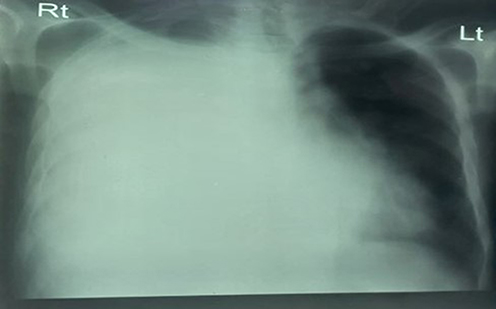 |
Figure 1 Right lung opacity with right Costo-phrenic angle distortion and air fluid level at the tip of the right lung (suggestive of empyema and lung abscess). |
The patient was diagnosed with perihepatic and perinephric abscess secondary to a possible ruptured appendicitis plus empyema; therefore, an emergency exploratory laparotomy was performed using a midline incision up to the midpoint between the xip sternum and umbilicus. Then the fascia was breached and a capsulated retroperitoneal fluctuating mass (containing the appendix (retrocecal) and massive pus were discovered. Appendectomy was done and the peritoneal cavity washed with three litera of warmed saline and the facia was closed after inserting a drain. Using the chest tube, 1 L pus mixed with blood was drained immediately and 100 mL of pus mixed with blood was drained per day for 3 consecutive days. There were multiple visible fragmented necrotized lung tissues inside the chest tube. Additionally, IV antibiotics (ceftriaxone and metronidazole) were commenced.
He stayed in the ICU for 1 week and in the surgical ward for 3 weeks. He was having intermittent fever, otherwise, his condition was normal. The patient attended appointmenta at the surgical referral clinic on different occasions over the following 2 months after discharge, and he was in good health. HIV test result and TB screening tests were negative, and his HA1C was 5.7%. Cytology samples taken from the subphrenic area and peritoneal wash showed the presence of sheets and sheets of neutrophils with a necrotic background.
Discussion
Acute appendicitis is the most common cause of emergency abdominal surgery in the pediatric population and requires prompt diagnosis and early treatment.10 Because of the atypical presentation of retrocecal appendicitis, its diagnosis could be missed or delayed, which may lead to a higher incidence of perforation and serious complications, as we saw in our case.7,10 The classic clinical manifestation of acute appendicitis is periumbilical pain localizing to the right iliac fossa with nausea and vomiting. Mild fever, leukocytosis, and right iliac fossa tenderness are usually present.11 A lung abscess is among the complications but it is very rare and only a few case reports have been published. Lung abscess symptoms are fever, cough, and sputum production over weeks.8,9 The presence of these symptoms and the chest tube drainage content (drainage of pus mixed with blood) and the occurrence of signs of emphysema after chest tube insertion indicate parenchymal damage.4,5
A classical cavity containing a gas-fluid level in an X-ray is a classical appearance of a lung abscess. Computed tomography is the gold standard diagnostic test for localization of intrapulmonary collections (differentiating lung abscess from empyema), as chest X-rays have poor sensitivity and specify and may mislead treatment. However, in such circumstances considering clinical symptom with chest X-ray findings would be important.11,12
In addition to the X-ray, which showed an air fluid level with massive opacity, this patient’s symptoms were not explained by empyema alone. The patient presented with a 4 weeks history of appendicitis symptoms and later 1 week symptoms of lung abscess. Furthermore, intraoperatively there was also pus mixed with lung tissue draining from the right crura of the diaphragm to the abdominal cavity, and postoperatively there was continuous bubbling at the abdominal surgical draining tube mixed with blood and damaged lung tissue, these findings strongly suggest the presence of communication between the lung and abdomen and visible parenchymal damage (lung abscess). Scientifically, after the development of appendicitis, perforation can occur after 48 hours in 80% of patients. Then abscess formation in the intraperitoneal cavity and the subphrenic can occurs within a week. If this abscess communicates with the lung, a lung abscess can develop 7–14 days after bacterial communication.4,5,10
In this patient’s case, there was a visible appendiceal abscess with a subphrenic extension. The commencement of a cough 3 weeks after the abdominal pain can also be the other strong justification. The cytology report (suppurative inflammation) and radiology findings consider the patient’s diagnosis. Surgical intervention is the treatment of choice for intraperitoneal, retroperitoneal, and large pulmonary abscesses.5,8,11 The patient outcomes and symptoms goalong with two previous similar case reports, where the patients were fully recovered with surgery and antibiotics like our patient.8,10
Conclusion
Appendicitis, specifically iretrocecal appendicitis, has subclinical symptoms and late presentation which could result in lung abscess as a complication may increase mortality. The level of suspicion of such a complication in lately presented patients should be raised and consideration given to the entity in the differential diagnosis. The early decision for surgery, commencement of broad-spectrum antibiotics and evidence of clinical diagnosis and auxiliary investigations has paramount importance in reducing post-operative hospital stay and patient costs, moreover, it decrease mortality by leading to early treatment.
Ethics Approval
Mizan-Tepi University’s College of Health Science granted ethical approval for this study (RN/00429/2012).
Informed Consent
Written informed consent was obtained from the patient for their anonymized information to be published in this article.
Acknowledgments
The Mizan-Tepi University, specifically the Department of surgery, staff members, and study participants are all sincerely thanked by the authors for their technical assistance and guidance.
Author Contributions
All authors made a significant contribution to the work reported, whether that is in the conception, study design, execution, acquisition of data, analysis and interpretation, or in all these areas; took part in drafting, revising, or critically reviewing the article; gave final approval of the version to be published; have agreed on the journal to which the article has been submitted; and agree to be accountable for all aspects of the work.
Disclosure
The authors report no conflicts of interest in this work.
References
1. Williams GR. Presidential Address: a history of appendicitis. With anecdotes illustrating its importance. Ann Surg. 1983;197:495. doi:10.1097/00000658-198305000-00001
2. Peksöz R, Albayrak Y, Atamanalp SS. Inflammatory parameters as predictive factors for complicated appendicitis: a retrospective cohort study”February 2022. Ann Med Surg. 2022;75(3):103391. doi:10.1016/j.amsu.2022.103391
3. Torres A, Menéndez R, Wunderink RG. Bacterial pneumonia and lung abscess. Murr Nadel’s Textbook Respirat Med. 2016:557–582.e22. PMCID: PMC7152161. doi:10.1016/B978-1-4557-3383-5.00033-6
4. Fitz RH. Perforating inflammation of the vermiform appendix with special reference to its early diagnosis and treatment. Am J Med Sci. 1886;92:321.
5. James T. The lung complications of appendicitis. JAMA. 1902;XXXIX(18):1117–1118. doi:10.1001/jama.1902.02480440037004
6. Mulholland MW, Lillemoe KD, Doherty GM, et al. Greenfield’s Surgery.
7. Dietrich A, Nicolas M, Iniesta J, Smith DE. Empyema and lung abscess as complication of a perforated appendicitis in a pregnant woman. Int J Surg Case Rep. 2012;3(12):622–624. doi:10.1016/j.ijscr.2012.08.015
8. Faizi FR, Farzam F. Perforated retrocecal appendicitis presenting with lung abscess-A case report. Radiol Case Rep. 2022;17(8):2754–2758. PMID: 35990571; PMCID: PMC9388878. doi:10.1016/j.radcr.2022.04.053
9. Landay MJ, Christensen EE, Bynum LJ, Goodman C. Anaerobic pleural and pulmonary infections. AJR Am J Roentgenol. 1980;134(2):233–240. PMID: 6766225. doi:10.2214/ajr.134.2.233
10. McKerrow WS, Thomson HJ. unusual complication of perforated appendix. Br Med J. 1982;284:1442. doi:10.1136/bmj.284.6327.1442
11. Saleem MM. Scrotal abscess as a complication of perforated appendicitis: a case report and review of the literature. Cases J. 2008;1:165. doi:10.1186/1757-1626-1-165
12. Kim S, Lim HK, Lee JY, et al. Ascending retrocecal appendicitis: clinical and computed tomographic findings. J Comp Ass Tomog. 2006;30(5):772–776. doi:10.1097/01.rct.0000228151.73528.8f
 © 2024 The Author(s). This work is published and licensed by Dove Medical Press Limited. The full terms of this license are available at https://www.dovepress.com/terms.php and incorporate the Creative Commons Attribution - Non Commercial (unported, v3.0) License.
By accessing the work you hereby accept the Terms. Non-commercial uses of the work are permitted without any further permission from Dove Medical Press Limited, provided the work is properly attributed. For permission for commercial use of this work, please see paragraphs 4.2 and 5 of our Terms.
© 2024 The Author(s). This work is published and licensed by Dove Medical Press Limited. The full terms of this license are available at https://www.dovepress.com/terms.php and incorporate the Creative Commons Attribution - Non Commercial (unported, v3.0) License.
By accessing the work you hereby accept the Terms. Non-commercial uses of the work are permitted without any further permission from Dove Medical Press Limited, provided the work is properly attributed. For permission for commercial use of this work, please see paragraphs 4.2 and 5 of our Terms.