")
Back to Journals » Veterinary Medicine: Research and Reports » Volume 14
Isolation and Molecular Detection of Mannheimia haemolytica and Pasteurella multocida from Clinically Pneumonic Pasteurellosis Cases of Bonga Sheep Breed and Their Antibiotic Susceptibility Tests in Selected Areas of Southwest Ethiopian Peoples Regional State, Ethiopia
Authors Alemu SA, Belachew YD, Tefera TA
Received 17 October 2023
Accepted for publication 19 December 2023
Published 27 December 2023 Volume 2023:14 Pages 233—244
DOI https://doi.org/10.2147/VMRR.S435932
Checked for plagiarism Yes
Review by Single anonymous peer review
Peer reviewer comments 2
Editor who approved publication: Professor Young Lyoo
Solomon Addisu Alemu,1 Yosef Deneke Belachew,2 Takele Abayneh Tefera3
1Bonga University, Department of Veterinary Medicine, Bonga, Ethiopia; 2Jimma University, School of Veterinary Medicine, Jimma, Ethiopia; 3National Veterinary Institute (NVI), Bishoftu, Ethiopia
Correspondence: Solomon Addisu Alemu, Department of Veterinary Medicine, Bonga University, P. O. Box= 334, Bonga, Southwest Ethiopian Regional State, Ethiopia, Email [email protected]
Background: Pneumonic pasteurellosis is a respiratory system disease of sheep caused by Mannheimia haemolytica, Pasteurella multocida, and Bibersteinia trehalosi responsible for the low productivity and economic loss resulting from death and treatment costs. This study was conducted to isolate and molecularly detect causative agents and antibiotic susceptibility tests from a nasal swab sample of the Bonga sheep breed that was suspected to have pneumonic pasteurellosis in selected areas of Southwest Ethiopian Peoples Regional State.
Methods: A cross-sectional study design was used along with purposive sampling of nasal swab samples from sheep that were brought to veterinary clinics during the study period. Bacterial isolation and phenotypic characterization were carried out using microbiological and biochemical tests that followed standard microbiological techniques. To molecularly confirm the isolates, PHSSA and KMT1, species-specific PCR primer genes were used. Using the disc diffusion method, molecularly confirmed isolates were subjected to an in vitro antibiotic susceptibility test.
Results: The 85 samples that were scrutinized had an overall isolation rate of 31.76%, whereas the isolates of Pasteurella multocida and Mannheimia haemolytica had species compositions of 40.7% and 59.25%, respectively. Overall, 12.5% of the Mannheimia haemolytica and 18.18% of the Pasteurella multocida species were verified from phenotypical isolates using the species-specific PCR primer genes PHSSA and KMT1, respectively. An in vitro antibiotic susceptibility test was carried out on all four PCR-confirmed isolates for seven commonly used antibiotics used to treat ovine pasteurellosis in the study area. It was found that both bacterial species were resistant to chloramphenicol and penicillin G.
Conclusion: Using phenotypic and molecular diagnostic techniques, the results of our current inquiry revealed that Pasteurella multocida and Mannheimia haemolytica are the causative agents of ovine pneumonic pasteurellosis in the study area.
Keywords: antibiotics, isolation, Mannheimia haemolytica, Pasteurella multocida, pneumonia, Bonga sheep breed
Introduction
Ethiopia has the largest livestock population in Africa that is distributed among diverse ecological conditions and production systems. Sheep production plays a significant role in the national economy and livelihood of smallholder farmers in Ethiopia.1,2 However, efficient utilization of this resource is limited by a combination of malnutrition, prevalent bacterial and parasitic diseases, management problems, poor genetic performance of indigenous breeds, marketing problems, social factors, and infrastructural constraints.3 Disease, particularly ovine pasteurellosis, is a major problem commonly encountered in sheep production as it affects groups or individuals of all age groups, causes great financial losses to sheep production,4 and is frequently diagnosed in veterinary clinics and abattoirs in Ethiopia.5
Mannheimia haemolytica, Pasteurella multocida, and Bibersteinia trehalosi are the three most commonly isolated bacterial agents in ovine pasteurellosis disease. This disease can affect all breeds of sheep and can be observed every season in the year.6 Several studies conducted in Ethiopia on pneumonic pasteurellosis in sheep have focused on the seroprevalence and bacteriological identification of these agents.7 The species of bacteria involved have been identified in sheep from different parts of Ethiopia.8 In Southwest Ethiopian Peoples’ Regional State, despite annual vaccination against ovine pneumonic pasteurellosis with a monovalent vaccine (P. multocida biotype A) in sheep, there are high rates of mortality and morbidity following respiratory diseases. A key problem in this study area is the absence of extensive studies on serotype identification and molecular characterization of P. multocida and M. haemolytica genes to produce multivalent preventive vaccines. This was accompanied by a complaint from field veterinarians and farmers in the area regarding the failure of vaccination against this disease and frequent occurrence of pneumonic pasteurellosis outbreaks in the Bonga sheep breed. This could be due to differences in the vaccine strains used and the serotypes of the organisms prevalent in the area. Based on the background information, there is a need for isolation and molecular detection of the causative agents of pneumonic pasteurellosis in the Bonga sheep breed and antibiotic susceptibility tests in selected areas of Southwest Ethiopian Peoples Regional State.
Materials and Methods
Description of the Study Area
We conducted the current research work around the Southwest Ethiopian People’s Regional State from October 2021 to August 2022. The Mizan-Aman town administration and Semen Bench district were selected from the Bench-Sheko zone, while the Chena district was selected from the Kaffa zone (Figure 1). Mizan-Aman town is located 561 km southwest of Addis Ababa, the capital city of the country, 214 km from Jimma town. The Semen Bench woreda and Chena districts are 25 km and 45 km northeast of Mizan-Aman town, respectively. The major economic activity of urban inhabitants is trading, whereas farming crops and livestock production are the major economic activities of the surrounding rural population.9
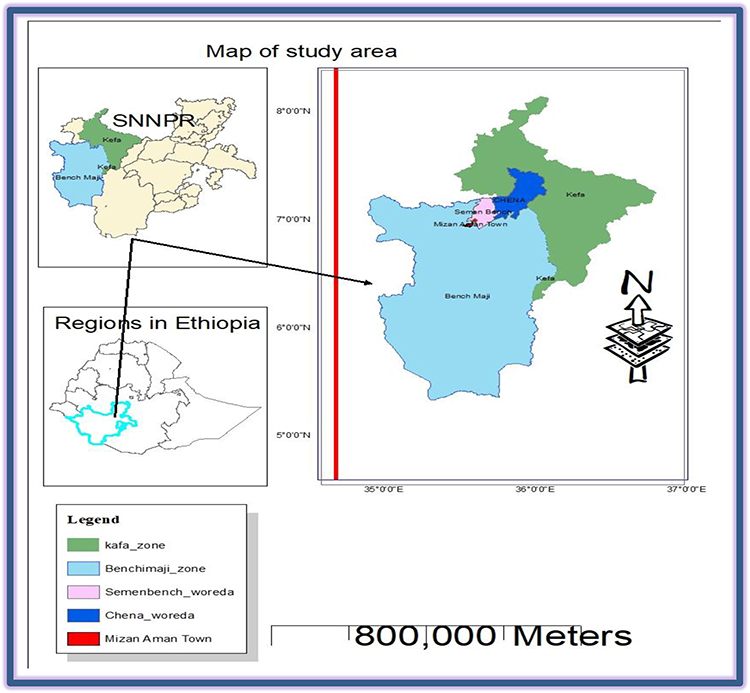 |
Figure 1 Map showing the location of the study area. |
Study Design and Study Populations
A cross-sectional study was conducted from October 2021 to August 2022 in selected veterinary clinics in the Southwest Ethiopian People’s Regional State. We selected veterinary clinics purposively based on availability of pneumonic clinical cases. The study population was a Bonga sheep breed of both sexes and all age groups with respiratory problems who were brought to veterinary clinics in the study areas during the study period. Sheep brought to veterinary clinics with respiratory problems were examined for symptoms of ovine pneumonic pasteurellosis. Based on clinical examinations, we included individuals clinically suffering from respiratory distress in the sampled populations. All information from the sampled animals was recorded on a recording format sheet developed for this purpose.
Sampling Method and Sample Size Determination
A non-probability purposive sampling technique was used, based on the presence of typical clinical signs of respiratory problems. The sample size in this study was determined on the basis of the availability of clinically pneumonic cases during the study period. The sample size in this study was determined based on the availability of clinically pneumonic sheep cases during the study period. Accordingly, 115 sheep were clinically examined at the veterinary clinics during the study period, and 85 sheep were found to be clinically pneumonic. Therefore, we have taken samples for bacteriological analysis from those 85 clinically pneumonic sheep.
Method of Nasal Swab Sample Collection
Clinical signs of pneumonic pasteurellosis were recorded for each Bonga sheep breed. We collected nasal swab samples from all pneumonic sheep found in veterinary clinics and suspected cases of ovine pneumonic pasteurellosis outbreaks. An assistant individually identified and restrained each pneumonic sheep, and they were kept fixed. After disinfection of the external part of the nose with 70% alcohol, a sterile cotton-tipped, 20–25-cm-long applicator stick moistened in Amies transport media was directed via the ventral nasal meatus to the nasopharynx, carefully inserted into the nostril and mucosa surface, and rolled gently. The collected samples were immediately transferred into sterile universal glass vials containing 3 mL of Amies transport medium, labeled, and stored in an ice box. All the collected samples were immediately transported to the microbiology laboratory of the Mizan-Aman regional veterinary diagnostic laboratory for bacteriological analysis.
Bacteriological Analysis
Microbiological isolation of M. haemolytica and P. multocida was performed at the Mizan-Aman regional veterinary diagnostic laboratory, using the standard bacteriological procedures recommended by Quinn et al10 and OIE.11 In the laboratory, aseptically collected nasal swab specimens were streaked on a blood agar base (HiMedia, India) supplemented with 5% sheep blood and immediately incubated under aerobic conditions at 37°C for 24–48 h. We carried out phenotypic detection of Pasteurella species based on colonial morphology, Gram staining, and biochemical characteristics, as described by Sneha et al.12 Culture-positive plates from blood agar and typical colonies were subjected to Gram staining to study the staining reactions and cellular morphology under a light microscope at 100× magnifications. Mixed gram-negative coccobacilli or short-rod bacteria were further subcultured on blood agar containing 5% sheep blood and MacConkey agar (HiMedia, India) plates for further analysis.
We characterized the growth of typical colonies on blood agar for hemolysis, the type of hemolysis, and the general appearance of the colonies (morphology, color, shape, size, consistency, and odor). We examined colonies on MacConkey agar in the presence or absence of growth, general appearance, and ability to ferment lactose, as described by Alemneh and Tewodros.2 Growth culture colony characteristics of round (smooth), greyish color, small-to-moderate size, and mucoid consistency, which were hemolytic or non-hemolytic on blood agar and MacConkey agar, were characterized. Narrow beta hemolysis on blood agar and growth on MacConkey agar (HiMedia, India) with lactose fermentation were classified as M. haemolytica. While those which were non-hemolytic on blood agar and did not grow on, MacConkey agar would group as P. multocida standard procedures described previously by Hawar et al13 and Abebe.8
Biochemical Tests
Pure cultures of a single colony type from both blood and MacConkey agar were transferred onto nutrient agar slants (HiMedia, India) for a series of primary and secondary biochemical tests, as described by Quinn et al.10 We conducted all activities, from primary to secondary biochemical tests, for the final identification of bacterial isolates at the species level. The primary biochemical tests conducted in this study were catalase, oxidase, and motility tests, to identify Pasteurella at the genus level. Bacterial isolates were identified using a variety of secondary biochemical tests and different sugar fermentation reactions. Secondary biochemical tests were performed using an indole test, and H2S production was tested in SIM medium, triple sugar iron (TSI) agar, and methyl red test (HiMedia, India). Sugar fermentation reactions (with glucose, lactose, sucrose, trehalose, and arabinose) were conducted according to procedures described by Quinn et al10 and Hawari et al.13
P. multocida on blood agar had non-hemolytic, round, smooth, or mucoid colonies; all isolates failed to grow on MacConkey but were able to produce indole. M. haemolytica forms a round, smooth, translucent, greyish with a β distinct zone of hemolysis on blood agar (HiMedia, India) and grows on MacConkey agar (HiMedia, India), but is unable to produce indole. In addition, M. haemolytica isolates were selected based on lactose and arabinose fermentation and a lack of fermenting Trehalosi sugars. We identified the isolates at the species level based on a series of tests carried out, from primary to secondary biochemical tests. The data were compared to confirm which species the isolates belonged to, as reported by Quinn et al.10
Bacterial Genomic DNA Extraction
The isolates of M. haemolytica and P. multocida were selected for molecular detection through genomic extraction of bacterial DNA using bacterial culture characteristics and biochemical tests. Bacterial cultures inoculated in nutrient broth with 30% glycerol from the study area were kept at ‒20°C and transported under cold chain to the National Veterinary Institute (NVI) in the molecular biology room for molecular detection. At NVI, a few colonies from phenotypically characterized pure cultures of M. haemolytica and P. multocida grown on nutrient slant agar for 24–48 h were transferred into 1.5 mL Eppendorf tubes, as described by Kumar et al.14 Bacterial genomic DNA was extracted using the Qiagen DNeasy Blood and Tissue Kit (Qiagen, German town, MD, USA) instructions.
PCR Master Mix Preparation for M. Haemolytica and P. Multocida
After the extraction of the target DNA, all P. multocida samples were subjected to PCR to detect specific genes. The PCR master mixes were prepared using a mixture of all components common to all reactions, except for template DNA. For molecular detection of P. multocida carried out with a PCR master mix of a 50 μL mixture containing master mix (Fermentas, Thermo Fisher Scientific, USA), 6 μL of 5 pmol of each primer (Eurofins MWG Operon, Germany), 0.25 μL of Taq DNA polymerase, Mgcl2, or PCR buffer, 3 μL of PCR nucleotides, 1 μL of 10 millimolar each base, and up to 28 μL of RNase free double distilled water were prepared according to company protocol. Finally, 5 μl of template DNA was added to the prepared PCR master mix to run the PCR protocol according to the manufacturer’s instructions.
Molecular detection of Pasteurella multocida was carried out using the PCR technique to amplify a specific fragment of the KMT1 gene sequence of Pasteurella multocida described previously by Townsend et al15 (Table 1). The forward primer sequence was 5’-ATCCGCTATTTACCCAGTGG-3’ and the reverse primer sequence was 5’-GCTGTAAACGAACTCGCCAC-3’, which amplified a segment of 460 base pairs. After master mix preparation was completed, optimized PCR conditions and amplification of the reaction mixtures were performed in a thermocycler PCR machine, according to the standard procedure (Table 2). A negative control consisting of all components of the reaction mixture, except the DNA template, was included in the PCR. The vaccine strain of P. multocida from NVI, Ethiopia was used as a positive control
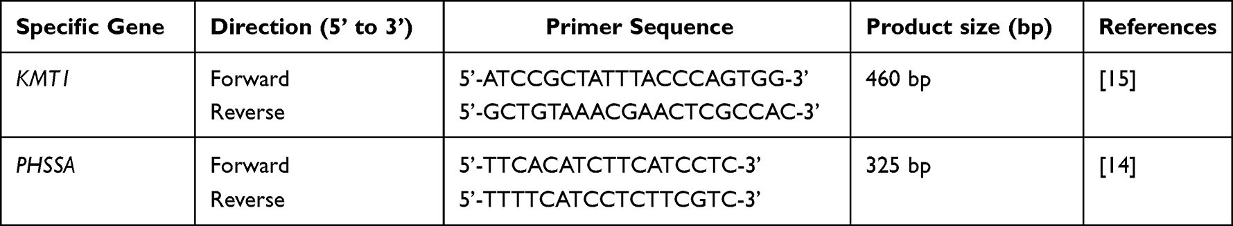 |
Table 1 Primer Pairs of KMT1 and PHSSA Genes Used for Detection of P. Multocida and M. Haemolytica in the Present Study, Respectively |
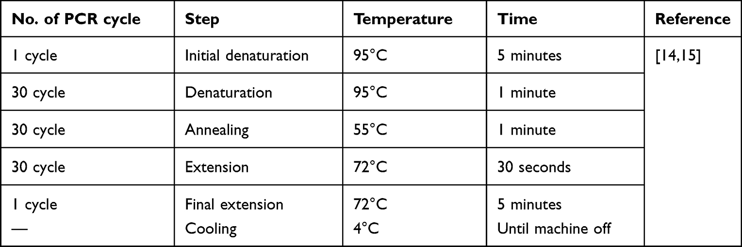 |
Table 2 PCR Protocol Condition Used for Amplification of M. Haemolytica and P. Multocida |
Similar to P. multocida, PCR master mix preparation for the detection of M. haemolytica isolates was confirmed by PCR for specific identification. PCR was carried out using primer pair sequence sets targeting PHSSA genes used in previous studies by Kumar et al.14 Forward primer sequence 5’- TTCACATCTTCATCCTC −3’ and the Reverse primer 5’- TTTTCATCCTCTTCGTC −3’ amplifying a segment of 325 base pairs (Table 1). The PCR master mix for M. haemolytica was carried out in a final volume of 25 μL of reaction mixture containing 2 μL of 5 pmol of each primer (Eurofins MWG Operon, Germany), 0.25 μL of Taq DNA polymerase, Mgcl2, or PCR buffer, 3 μL of PCR nucleotides, 1 μL of 10 millimolar each base, and up to 12 μL of RNase free double distilled water, prepared according to the manufacturer’s protocol. Finally, 5 μL of template DNA was added to the prepared PCR master mix to run the PCR protocol according to the manufacturer’s instructions. PCR amplification was performed using a thermocycler with thermal conditions (Table 2) to amplify PHSSA gene fragments. A negative control consisting of all components of the reaction mixture, except the DNA template and a positive control, was included in the PCR, using a template from the reference M. haemolytica isolate from the NVI culture collection.
Agarose Gel Electrophoresis Analysis
Agarose gel electrophoresis was used to separate the amplified PCR products at 120 V for 60 min in 2% agarose containing EtBr in 0.5 × Tris borate EDTA buffer using a marker 100 bp DNA ladder (Promega, Madison, Wisconsin, USA). The loading dye was used to load the PCR products into each well of a gel. Each PCR product (5 μL) was mixed with 6× loading buffer and loaded into a separate well of the pre-prepared gel, while we loaded 1 kb plus DNA molecular marker onto the first and last lanes and ran an electrophoresis apparatus (EC 2060, USA). Visualization of different band sizes of DNA bands was performed to analyze the PCR products under a UV transilluminator stained with gel and photographed in a gel documentation system.16
Antibiotic Susceptibility Tests
The antibiotic susceptibility evaluation in this study was used to determine which species of bacteria were resistant or susceptible to commonly used antibiotics in the study areas and to select the most appropriate antimicrobial agents for treatment against ovine pneumonic pasteurellosis. We selected commonly used antibiotics in the study area to treat ovine pneumonic pasteurellosis. Standard disc diffusion technique (Kirby–Bauer test performed the test) is based on the performance standards of the Clinical and Laboratory Standards Institute (CLSI).17 Each pure isolate was tested against seven antibiotics: Penicillin G (one unit), ampicillin (10 µg), gentamicin (10 µg), oxytetracycline (30 µg), chloramphenicol (30 µg), streptomycin (10 µg), and kanamycin (5 µg) (HiMedia, India). Escherichia coli ATCC 25922 strain, obtained from the Ethiopian Public Health Institute, was used for quality control. We base our interpretation of the results on the breakpoints provided by the CLSI guidelines (Table 3).
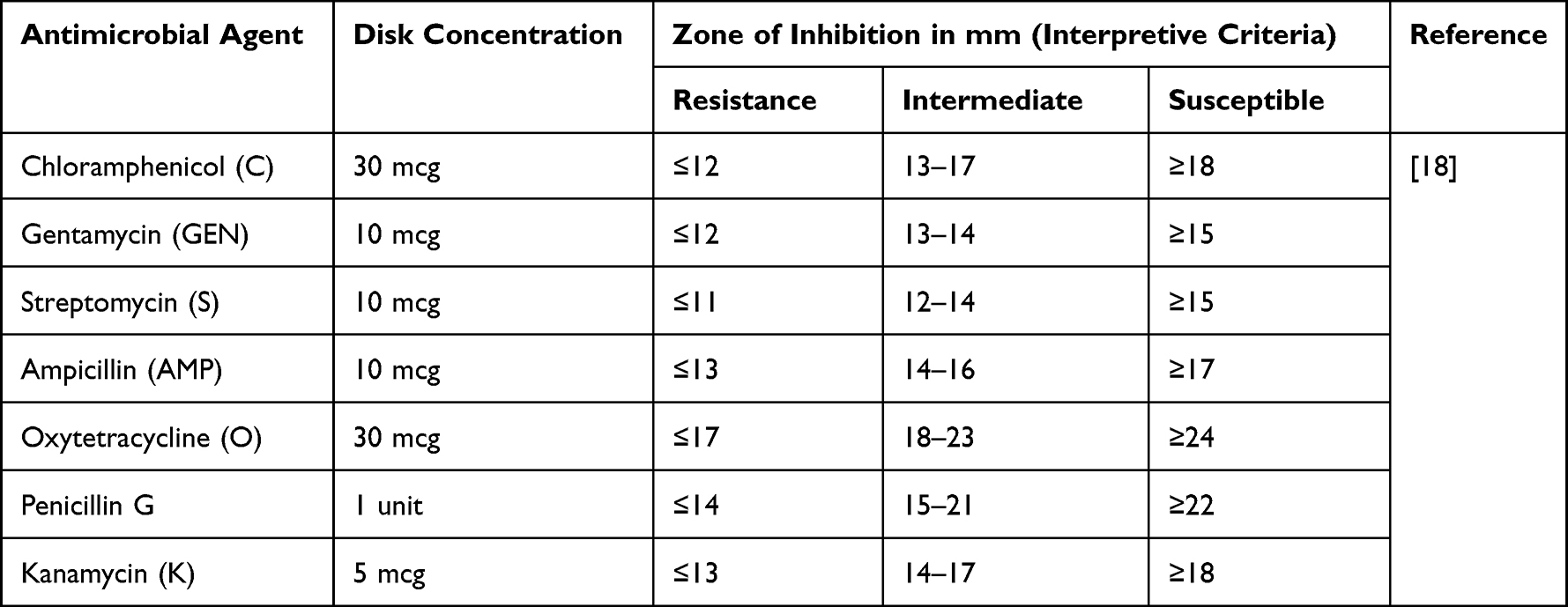 |
Table 3 Antibiotic Susceptibility Test Interpretive Criteria Used in This Study |
PCR confirmed that colonies from the pure culture were transferred into a test tube containing 5 mL TSB (HiMedia, India) and incubated at 37°C for 6 hr. The turbidity of the culture broth was adjusted using sterile saline solution or by the addition of more isolated colonies to obtain turbidity analogous to that of 0.5 McFarland standards (approximately 1×10⁸ CFU per mL). Mueller–Hinton agar plates (HiMedia) were prepared according to the manufacturer’s instructions. After the sterile cotton swab was immersed in the suspension and rotated against the side of the tube to remove excess fluid, it was spread uniformly on the surface of the Muller Hinton agar using a sterile cotton swab and allowed to stand for 3–5 min to observe any excess moisture from the medium before the antimicrobial disc was placed.18
Then, using sterile forceps, antibiotic discs were gently pressed on the plate to ensure complete contact with the agar surface and 4–5 discs were regularly placed in one plate, 3 cm apart and 1.5 cm from the edge antibiotic-impregnated paper discs. The plates were left for 30 min to diffuse the antibiotics into the disc, and the plates were inverted upside down and incubated at 35°C ± 2°C for 18–24 hr. Finally, each plate was examined. The diameter of the inhibition zone produced by the antimicrobial inhibition of bacterial growth was measured in millimeters using a digital caliper by laying it over the back of an inverted petri dish. The results were interpreted as susceptible when there was an absence of the growth of the tested bacteria around the antibiotic disc, intermediate when the tested bacterial growth was not full, and resistant when the presence of bacteria grew around the antibiotic disc according to the standardized table supplied by the manufacturer with the antibiotic disc and the Clinical and Laboratory Standards Institute.18
Data Management and Analysis
All data collected from field and laboratory experiments were coded, filtered, and recorded using Microsoft Excel. Descriptive analysis (percentage) was used to describe the isolation rate and the antibiotic susceptibility test results.
Results
Overall Isolation of Pasteurella Species
Eighty-five nasal swab samples were collected from clinically pneumonic cases of the Bonga sheep breed and processed using phenotypic methods (cultural characteristics and biochemical tests) for Pasteurella species, and 27 (31.76%) were found to be positive. The species composition of the isolated bacteria consisted of 16 (59.25%) and 11 (40.74%) isolates positive for M. haemolytica and P. multocida, respectively.
Biochemical test results revealed that all isolates of P. multocida were positive for indole, catalase, and oxidase; did not form hydrogen sulfide; and were negative for methyl red and motility tests. Upon biochemical testing, M. haemolytica isolates were found to be positive for catalase and oxidase, did not form hydrogen sulfide, and were negative for indole, methyl red, and motility. All the isolates successfully fermented glucose and fructose. M. haemolytica isolates fermented lactose but failed to ferment Trehalosi (Table 4).
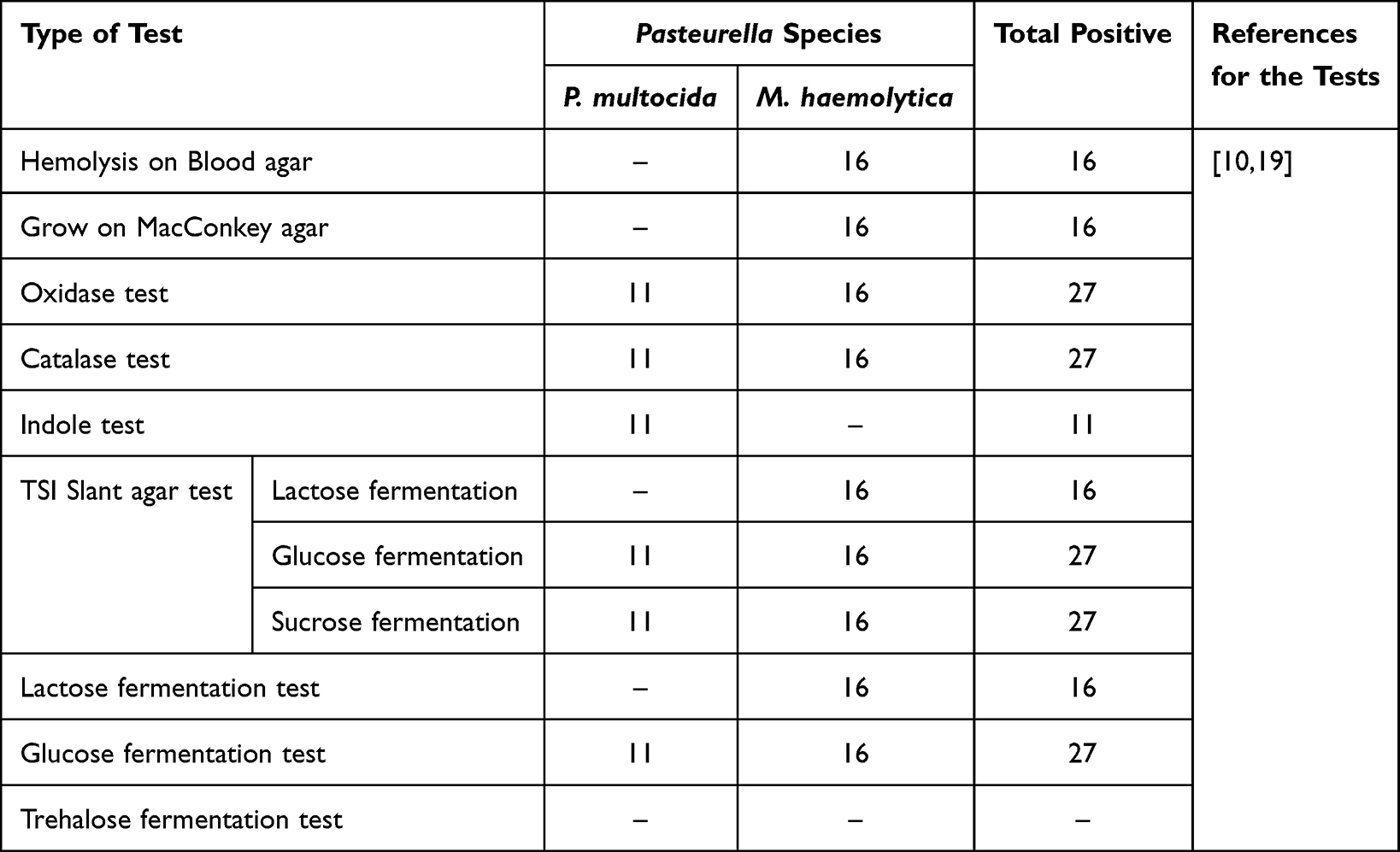 |
Table 4 Culture Characteristics and Biochemical Test Results for P. Multocida and M. Haemolytica Bacteria |
Molecular Detection of the Isolates
The phenotypic test results were confirmed by PCR using species-specific primers. In this study, extracted DNA from all 11 isolates of P. multocida was used to target the presence of species-specific PCR primers (Multocida toxin hydrolase gene) KMT1 genes as per the method described by Townsend et al.15 From the 11 isolates, two (on gel lanes 2 and 6) were detected using KMT1 gene amplified product size of 460 base pairs generated by electrophoresis (Figure 2A).
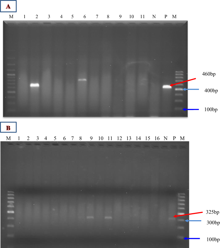 |
Figure 2 PCR product of PHSSA and KMT1 genes in 2% agarose gel electrophoresis. (A) Gel lane from left to right (1 to 11); gel lane 2 and 6 were positives for P. multocida of PCR product of KMT1 gene (460bp). (B) Gel lane number 1 to 16, gel lane 9 and 11 were positive for M. haemolytica in PCR product of PHSSA gene (325 bp). Abbreviations: N, negative control; P, positive control; M, DNA molecular marker ladder. |
Similarly, M. haemolytica isolates were identified by PCR amplification of PHSSA genes. Sixteen isolates identified as M. haemolytica by cultural and biochemical methods were subjected to species-specific targeting primer PCR (Pasteurella haemolytica species-specific antigen (PHSSA)) genes used in a previous study by Kumar et al14 were used in this study. We generated the desired amplification products lengths 325 base pairs upon electrophoresis (Figure 2B). The results revealed that only two isolates (in gel lanes 9 and 11) were confirmed to be M. haemolytica with PHSSA gene.
Antibiotic Susceptibility Tests
All pure isolates confirmed by PCR were subjected to in vitro antimicrobial susceptibility tests for seven commonly used antimicrobials in the study area (Table 5). The test results indicated 100% resistance to penicillin G and ampicillin in both P. multocida and M. haemolytica strains.
 |
Table 5 The Results of Antibiotic Susceptibility Tests of Pasteurella Species Isolated from Nasal Swabs of Clinically Pneumonic Sheep |
Discussion
Ovine pneumonic pasteurellosis is one of the most economically important infectious diseases of small ruminants with a high prevalence occurring throughout the world.20,21 Pasteurellosis is therefore a high-priority issue at the national and regional level due to the significant economic losses it causes through mortality, morbidity, and the high cost of treatment. In Ethiopia, pasteurellosis is a common respiratory infection and economically significant infectious diseases causing outbreaks of acute pneumonia in sheep/goats of all ages that may end with death of sheep and goats. Pasteurellosis is therefore a high-priority issue at the national and regional level due to the significant economic losses it causes through mortality, morbidity, and the high cost of treatment.22 The disease occurs in sheep due to complex factors that often interact to produce disease. Various conditions such as climate and weather change, transport stress, poorly ventilated housing and nutritional deficiencies are known to play a pre-disposing role as the animal’s immunity weakness. In such conditions, occurrence of normal flora in the upper respiratory tract and subsequent infection of the lungs is well documented.19
The main problem in the study area is absence of an extensive study on serotype identification and molecular characterization of P. multocida and M. haemolytica genes to produce multivalent preventive vaccine. Traditional therapy based on the extensive use of antibiotics, including mass medication of animals, has caused an increase in the incidence of multi-drug resistant, M. haemolytica strains in many parts of the world.23 The present finding of M. haemolytica and P. multocida bacterial isolation was based on phenotypic and molecular methods. The overall isolation rate of Pasteurella species using phenotypic methods was 27 (31.76%) from 85 nasal swab samples from clinically pneumonic cases of the Bonga sheep breed. The isolation rate is lower than the 34.2% reported by Legese et al24 in a selected part of central Ethiopia, 76.8% reported by Yemisrach et al25 in Wolaita Zone, SNNPR, and 40.7% reported by Tilaye et al26 in Bishoftu Town. This variation could be due to differences in agroecological conditions where sheep were raised, sample size differences, variation in serotype, and the presence of concurrent infections to suppress the immunity of sheep. However, the current findings were slightly higher than the 25% reported by Marru et al22 in Haramaya district, 21% that of Sadia27 in the East Shewa Zone of the Oromia Region, 3.38% Yami28 in Assosa and Bambasi District, Benishangul Gumuz Regional State, 22.15% Abebe8 in selected areas of Ethiopia, 11% reported by Deresse et al29 in Ethiopia, and 14.10% and 10.16% reported by Hussein et al30 and Mohamed et al,31 respectively. These differences may be associated with variations in husbandry practices, better animal health facilities, and sheep breeds.
All isolates belonging to P. multocida produced catalase-, oxidase-, and indole-positive acids by fermentation of glucose and sucrose and did not grow on MacConkey agar, whereas all isolates presumed to belong to M. haemolytica did not produce indole and grew on MacConkey agar. These results agree with the findings of Hawari et al13 and Tefere and Smola.32 In addition, M. haemolytica in the present study was cultured on blood agar as smooth round, white to gray colonies, and a small area of β-type hemolysis, which agreed with Hawari et al,13 whereas MacConkey agar appeared as small pink and pinpoint colonies, as shown by Alemneh and Tewodros.15 The biochemical reactions for the isolated M. haemolytica were negative for indole, and citrate, but positive for oxidase and catalase, in agreement with Quinn et al10 and Tefera and Smola.32
The presence of M. haemolytica and P. multocida was confirmed by species-specific PCR amplification using primers PHSSA and KMT1, respectively. The phenotypic isolates of P. multocida and M. haemolytica confirmed by PCR did not agree with the phenotypic method. The isolates confirmed by species-specific KMT1 for P. multocida resulted in 2/11 (18.2%) positivity, while 2/16 (12.5%) of all phenotypically detected M. haemolytica isolates were confirmed by species-specific PHSSA. The differences between phenotypic identification and PCR tool for the diagnosis of M. haemolytica and P. multocida may be related to several circulating serotypes of these bacteria, the high specificity and sensitivity of PCR as compared with the phenotypic method as stated by Kumar et al.14
In the present study, all P. multocida and M. haemolytica isolates confirmed by PCR were tested for their susceptibility to seven commonly used antibiotics used for the treatment of ovine pneumonic pasteurellosis in the study area. Antibiotic susceptibility tests were performed on the isolates using the Bauer-Kirby disc diffusion method. The results showed that antibiotic profiles of the two P. multocida isolates were 100% susceptible to Ampicillin, Gentamicin, and Oxytetracycline. This result is similar to the findings of the study conducted by Abebe8 in selected areas of Ethiopia and Marru22 in the Haramaya district. However, the antimicrobials Streptomycin and Kanamycin were (50%) susceptible and (50%) intermediate for P. multocida and Penicillin G and Chloramphenicol were resistant (Table 5). This finding agrees with those of Laxmi et al33 and Kalorey et al34 in India.
In addition, the antibiotic profiles of two M. haemolytica isolates confirmed by PCR revealed 100% susceptibility to ampicillin and gentamicin but resistance to penicillin G and chloramphenicol. However, the susceptibility to gentamicin is not similar to that reported by Marru et al.22 Penicillin G and Chloramphenicol are resistant against M. haemolytica and P. multocida isolates, which is similar to the findings of Marru et al22 in the Haramaya district, Eastern Hararghe. The development of resistance to antibiotics by all M. haemolytica and P. multocida isolates has been reported by Catry et al35 and Welsh et al.36 The antibiotic resistance observed in this study in Penicillin G and Chloramphenicol may be associated with improper use of antibiotics without prescription by qualified veterinarians, the widespread and erratic use of broad-spectrum antibiotics without proper isolation of the causative agents, and use without performing antibiotic susceptibility tests for treatment of the disease or from the presence of bacterial antibiotic resistance gene transfer from donor to recipient.
Conclusion and Recommendations
Our current study results show that P. multocida and M. haemolytica are causative agents of ovine pneumonic pasteurellosis, using phenotypic and molecular diagnostic techniques used in the study area. The presence of P. multocida and M. haemolytica was confirmed using species-specific PCR primers for KMT1 and PHSSA, respectively. This finding supports the need to develop a multivalent vaccine containing M. haemolytica. In Ethiopia, a monovalent vaccine (inactivated P. multocida biotype A) produced at the National Veterinary Institution is used to vaccinate against ovine pneumonic pasteurellosis. Antibiotic susceptibility tests showed that the most effective antimicrobial drug against PCR-confirmed isolates was ampicillin; oxytetracycline and gentamicin could be the antibiotic drugs of choice to treat ovine pneumonic pasteurellosis in the study area. However, both bacterial isolates were resistant to penicillin G and Chloramphenicol. Therefore, this study results helps to provide information for National Veterinary Institute to prepare effective multivalent/combined/vaccine that should contain M. haemolytica for prevention of ovine pasteurellosis in the study area. Also the finding of antibiotic susceptibility profile within the study area helps veterinary clinician workers of the study area get information on a better use of antibiotic for empirical therapy of ovine pasteurellosis. This can help veterinarians better select antibiotics for the treatment of pneumonic pasteurellosis in the study area. Hence, further investigations are needed to address the molecular characterization of virulent genes, and phylogenetic tree analysis and development of drug-resistance-encoding genes should be conducted for all molecularly detected Pasteurella species.
Data Sharing Statement
The data supporting the findings of this study are available from the corresponding author upon formal request.
Ethics Approval and Consent to Participate
Ethical approval for this study was obtained from Bonga University, Department of Veterinary Medicine animal research ethics committee with reference number REC-DVM/ERC/134/2021. Written informed consent was obtained for sample collection to keep the confidentiality of the owners at the time of sample collection. Sheep owners consented, and the benefits and outcomes of the research study briefed for the owners. The sample collection processes were performed safely, strictly protecting the welfare and wellbeing of the study animals during with relevant guidelines and regulations listed for ethics of animal research by the ethical committee of the university.
Acknowledgments
The authors would like to thank all staff members of Mizan Regional Veterinary Diagnostic Laboratory Workers, Southwest Ethiopian People’s Regional State district veterinary clinic workers, and National Veterinary Institute/NVI/molecular diagnostic room workers for their cooperation in different aspects of the study and unlimited assistance provided to us throughout the study period.
Author Contributions
All authors made a significant contribution to the work reported, whether in the conception, study design, execution, acquisition of data, analysis, and interpretation, or in all these areas, took part in drafting, revising, or critically reviewing the article; gave final approval of the version to be published; have agreed on the journal to which the article has been submitted; and agree to be accountable for all aspects of the work.
Funding
This research work was not funded by any sources or institutions.
Disclosure
The authors declare that they have no competing interests in this work.
References
1. Alemu and Merkel Sheep and Goat Production. Ethiopian sheep and goat productivity improvement Pro; 2008: 2–6.
2. Alemneh T, Tewodros AA. Pasteurellosis in small ruminants: biochemical isolation, characterization and prevalence determination in relation to associated risk factors in Fogera Woreda, North-West Ethiopia. Advan Biological Res. 2015;9:330–337.
3. Biffa D, Jobre Y, Chakka H. Ovine helminthosis, a major health constraint to productivity of sheep in Ethiopia. Animal Health Res Rev. 2017;7(1):107–118. doi:10.1017/S1466252307001132
4. Habashy HF, Fadel NG, El Shorbagy MM. Bacteriological and pathological studies on the causes of mortalities among sheep in sharkia-government farms. Egypt J Comparat Pathol Clin Path. 2009;22(1):130–146.
5. Tadele K, Fikadu Z. Review on the pneumonic pasteurellosis of cattle. Acad J Animal Dis. 2015;4:177–184.
6. Fulton R. Bovine Respiratory Disease Research (1983–2009). Animal Health Res Rev. 2009;10:131–139. doi:10.1017/S146625230999017X
7. Sisay T, Zerihun A. Diversity of M. haemolytica and P. trehalosi serotypes from apparently healthy sheep and in abattoir specimens in the high lands of Wollo, North Ethiopia. Veter Res Comm. 2003;27:3–14. doi:10.1023/A:1022088005887
8. Abebe Wurtu. Isolation and Identification of M. haemolytica, B. trehalosi and P. multocida from cattle and sheep from selected areas of Ethiopia. [Msc Thesis] Addis Ababa University College of Veterinary Medicine and Agriculture; 2018:34.
9. Zone B-S. Bench-Sheko Zone Livestock and Fishery development department socioeconomic information of livestock of Bench-Sheko zone. Bulletin No. 201. Mizan, SNNPRS, Ethiopia; 2019; 25–30.
10. Quinn M, Carter D, Lenard M. Pasteurella species and M. haemolytica. In: Veterinary Microbiology and Microbial Diseases.
11. OIE. Haemorrhagic septicaemia, Chapter 2.4.12. In: Terrestrial Manual. Paris, France: Office International Des Epizooties (OIE); 2012:739–750.
12. Sneha H, Daphal PPM, Mrunalini MP, Shelke PP, Sangle JD, Rahul P. Emergence of Virulent P. multocida and M. haemolytica in Sheep and Goats of Western Maharashtra, India. Internat J Curr Microbiol Applied Sci. 2018;7(9):1990–1998.
13. Hawari H, Hassawi DS, Sweiss M. Isolation and Identification of mannheimia haemolytica and pasteurella multocida in sheep and goats using biochemical tests and random amplified polymorphic DNA Analysis. J Biolog Sci. 2008;8:1251–1254. doi:10.3923/jbs.2008.1251.1254
14. Kumar J, Dixit SK, Kumar R. Rapid detection of M. haemolytica in lung tissues of sheep and from bacterial culture. Vet World. 2015;8:1073–1077. doi:10.14202/vetworld.2015.1073-1077
15. Townsend M, Frost J, Lee W, Papadimitriou M, Dawkins S. Development of PCR assays for species-and type-specific identification of Pasteurella multocida isolates. J Clin Microbiol. 1998;36(4):1096–1100. doi:10.1128/JCM.36.4.1096-1100.1998
16. Hegde A, Ballal M, Shenoy S. Detection of Pasteurella species by PCR. Indian J Med Micro. 2012;30:279–281. doi:10.4103/0255-0857.99485
17. Bauer AW, Kirby WM, Sherris JC, Turck M. Disc diffusion methods of antimicrobial sensitivity tests. J Clin Path. 1996;45:493. doi:10.1093/ajcp/45.4_ts.493
18. CLSI (Clinical and laboratory Standard Institute). Performance standards for clinical and laboratory standard institute (CLSI) performance for antimicrobial susceptibility testing; Twenty second informational supplement. CLSI Approv Stand Wayne. 2017;32:100–102.
19. Radostitis, Gay, Blood and Hinchcliff. Veterinary medicine. In: A Textbook of the Diseases of Cattle, Horses, Sheep, Pigs and Goats; Saunders Elsevier; 2007. 795–798.
20. Nicholas R. Respiratory Disease of Small Ruminants, Mycoplasma Diseases of Ruminants. CAB International; 2008:: 169–198.
21. Prabhakar P, Thangavelu A, Kirubaharan JJ, Chandran ND. Isolation and characterization of P. multocida isolates from small ruminants and avian origin. J Veterin Anim Sci. 2012;8(3):131–137.
22. Marru H, Takele A, Hassen A. A study on Ovine pneumonic pasteurellosis: isolation and identification of Pasteurellae and their antibiogram susceptibility pattern in Haramaya, Eastern Hararghe, Ethiopia. Biomed Cent Veterin Res. 2013;9:39.
23. Kehrenberg C, Schulze-Tanzil G, Martel JL, et al. Antimicrobial resistance in pasteurella and mannheimia: epidemiology and genetic basis. Vet Res Biomed Cent. 2001;32:323–339.
24. Abinet L, Abayneh T, Mamo G, et al. Molecular characterization of M. haemolytica isolates associated with pneumonic cases of sheep in selected areas of Central Ethiopia. Biomed Cent Micro. 2018;18:205–215.
25. Yonas Y, Goa A, Butako B. Study on the epidemiology and strain identification of small ruminant pneumonic pasteurellosis for vaccine production in selected districts of Wolaita Zone, SNNPR. Internat J Curr Res Biology Med. 2017;2(3):8–13.
26. Demissie T, Dawo F, Sisay T. Biochemical and antigenic characterization of mannheimia, pasteurella and mycoplasma species from naturally infected pneumonic sheep and goats, Bishoftu. Af J Bas Appl Sci. 2014;6:198–204.
27. Sadia H. Epidemiology of Ovine Pasteurollosis in Lume District, East Shewa Zone of Oromia Region, Ethiopia. [MSc Thesis] Addis Ababa University College of Veterinary Medicine and Agriculture, Department of Clinical Studies; 2017:4.
28. Yami B. Isolation, identification and antimicrobial susceptibility of P. multocida from cattle with hemorrhagic septicemia in Assosa and Bambasi districts, Benishangul Gumuz Regional state, Ethiopia. Inter J Animal Res. 2017;9:2301.
29. Deressa A, Lubke K, Tefera Z. Molecular detection of P. multocida and M. haemolytica in sheep respiratory infections in Ethiopia. J Appl Res Veterin Med. 2010;8:101–108.
30. Hussein. Kaoud AR, El-Dahshan MM, Zaki, Shimaa A, Nasir A. Occurrence of Mannheimia haemolytica and Pasteurella trehalosi Among Ruminants in Egypt. New York Science J. 2010;3(5):35–45.
31. Ragab MT, Hassan WH, Osman WA. Isolation, identification and antibiogram studies of P. multocida isolated from sheep and goats in siwa oasis. Global Vet. 2015;14:589–594.
32. Tefera G, Smola J. The utility of ENTERORapid 24 Kit for the identification of P. multocida and M. haemolytica. Veterinary Med. 2003;47:99–103. doi:10.17221/5811-VETMED
33. Laxmi N, Sarangi P, Gupta A, et al. Virulence gene profiling and antibiotic resistance pattern of Indian isolates of Pasteurella multocida of small ruminant origin. Compar Immunol Microbiol Infec Dis. 2015;38:33–39. doi:10.1016/j.cimid.2014.11.003
34. Kalorey DR, Yuvaraj SS, Vanjari PS, et al. PCR analysis of Pasteurella multocida isolates from an outbreak of Pasteurellosis in Indian sheep. Vet Res Comm. 2008;31:459–465.
35. Catry B, Decostere A, Schwarz S, Kehrenberg C, de Kruif A, Haesebrouck F. Detection of tetracycline-resistant and susceptible Pasteurellaceae in the nasopharynx of loose group-housed calves. Vet Res Commun. 2006;30:707–715. doi:10.1007/s11259-006-3347-8
36. Welsh D, Dye B, Payton E, Confer A. Isolation and antimicrobial susceptibilities of bacterial pathogens from bovine pneumonia. J Veterin Diagn Inve. 2004;16:426–431. doi:10.1177/104063870401600510
 © 2023 The Author(s). This work is published and licensed by Dove Medical Press Limited. The full terms of this license are available at https://www.dovepress.com/terms.php and incorporate the Creative Commons Attribution - Non Commercial (unported, v3.0) License.
By accessing the work you hereby accept the Terms. Non-commercial uses of the work are permitted without any further permission from Dove Medical Press Limited, provided the work is properly attributed. For permission for commercial use of this work, please see paragraphs 4.2 and 5 of our Terms.
© 2023 The Author(s). This work is published and licensed by Dove Medical Press Limited. The full terms of this license are available at https://www.dovepress.com/terms.php and incorporate the Creative Commons Attribution - Non Commercial (unported, v3.0) License.
By accessing the work you hereby accept the Terms. Non-commercial uses of the work are permitted without any further permission from Dove Medical Press Limited, provided the work is properly attributed. For permission for commercial use of this work, please see paragraphs 4.2 and 5 of our Terms.