")
Back to Journals » Cancer Management and Research » Volume 13
Investigation of Chromosome 1 Aberrations in the Lymphocytes of Prostate Cancer and Benign Prostatic Hyperplasia Patients by Fluorescence in situ Hybridization
Authors Miszczyk J , Przydacz M, Zembrzuski M, Chłosta PL
Received 21 November 2020
Accepted for publication 4 May 2021
Published 31 May 2021 Volume 2021:13 Pages 4291—4298
DOI https://doi.org/10.2147/CMAR.S293249
Checked for plagiarism Yes
Review by Single anonymous peer review
Peer reviewer comments 5
Editor who approved publication: Dr Ahmet Emre Eşkazan
Justyna Miszczyk,1 Mikołaj Przydacz,2 Michał Zembrzuski,2 Piotr L Chłosta2
1Department of Experimental Physics of Complex Systems, The H. Niewodniczański Institute of Nuclear Physics PAN, Krakow, Poland; 2Department of Urology, Jagiellonian University Medical College, Krakow, Poland
Correspondence: Justyna Miszczyk
Department of Experimental Physics of Complex Systems, The H. Niewodniczański Institute of Nuclear Physics PAN, Radzikowskiego 152 Street, Krakow, 31-342, Poland
Tel +48 126628409
Fax +48 126628458
Email [email protected]
Purpose: Radiotherapy is one of the most common treatments for prostate cancer. Finding a useful predictor of the therapeutic outcome is crucial as it increases the efficacy of treatment planning. This study investigated the individual susceptibility to radiation based on chromosome 1 aberration frequency measured by the FISH (fluorescence in situ hybridization) method.
Patients and Methods: Whole blood samples were collected from 27 prostate cancer (PCa) patients and 32 subjects with benign prostatic hyperplasia (BPH), who were considered as a control group. Samples were irradiated with 2 Gy of x-rays, cultured, harvested, and used in the FISH procedure.
Results: After irradiation, significantly higher levels of all studied chromosome 1 aberrations (except for deletions) in the group of PCa patients were revealed. Furthermore, in the lymphocytes of cancer patients, nearly five-fold higher frequencies of acentric fragments were observed compared to the BPH group. The highest individual radiosensitivities for all estimated biomarkers were seen in PCa patient cells who reported cancer incidence in the immediate family (CIF+).
Conclusion: The differences in chromosome 1 aberrations between PCa and BPH demonstrate that lymphocytes taken from patients with prostate cancer have higher radiosensitivity which might be related to hereditary or familiar inclinations. Therefore, this technique may find future application in searching biomarkers of the cellular radiotherapy response in prostate cancer patients.
Keywords: benign prostatic hyperplasia, chromosome 1, prostate cancer, radiotherapy planning, biomarkers, FISH method
Introduction
Cancers develop and progress because of genetic instability and the accumulation of mutations. A high frequency of chromosomal aberrations in human peripheral blood lymphocytes (HPBL) has been shown to be a risk factor for the cancer initiation process.1,2 The frequency of chromosome aberrations increases with radiation dose to the cells and serves as an indicator of the radiation dose received. In vitro irradiation experiments using HPBL can provide a dose-response relationship that can be used to estimate radiation dose to individuals based on the aberration frequency detected in their lymphocytes. Thus, chromosomal aberrations are considered to be valuable, internationally standardized, and validated biomarkers of biological effects after exogenous radiation exposures.2,3 Many cancer types are associated with specific chromosomal abnormalities.4 Among the various types of induced aberrations, dicentric and acentric fragments are eliminated from the peripheral blood during subsequent cell divisions (hence, they are called unstable aberrations). However, reciprocal translocations persist in peripheral blood lymphocytes for a long time,5 which is advantageous for using them as a biomarker in monitoring studies.1,2 Any exposure to low LET-radiation (Linear Energy Transfer, eg, X-rays) leads to a relatively homogeneous distribution of energy in the cell and initial DNA damage.6 Subsequent repair and post-repair mechanisms may lead to a selection of specific breakpoint locations along chromosomes. Due to impaired repair mechanisms, cells from subjects with increased or decreased radiosensitivity might express additional specific breakpoints.7
Prostate cancer (PCa) is the sixth most common cancer in the world, being the third most common male cancer,8 while benign prostatic hyperplasia (BPH) is among the most common age-related male disorders worldwide.9 The development of BPH, as well as normal prostate function, are known to be principally regulated by sex hormones and influenced by local inflammation.9,10 Recent data suggest that the action of androgens alone may not explain the hyperplastic development of the prostate gland, but that dietary and other lifestyle factors may also play an important role.9,11 Novel studies suggest that BPH might represent an element into the wide spectrum of disorders constituting the Metabolic Syndrome (MS).12
Cancer risk might be strongly influenced by hereditary and familial predisposition;13,14 however, social and environmental factors—particularly diet and lifestyle—also play an important role.14,15 The already known and common chromosomal abnormalities characteristic of prostate cancer are alterations in chromosomes 1, 7, 8, 10, 11, 16, 17, and X.16,17 Chromosome 1 is the largest, comprising 8.28% of the human male genome.16 Therefore, the aberrations in this chromosome are generally more frequently detected than those in other chromosomes. In addition, chromosome 1 undergoes frequent molecular changes in the case of prostate cancer, although many controversial findings have been reported; nevertheless, half of the most common prostate cancer susceptibility loci have been reported there.16,17
Radiotherapy—either alone or prescribed as an adjuvant therapy—is one of the standard methods for prostate cancer treatment.18 However, approximately 23–40% of prostate cancer patients still suffer from clinically relevant adverse acute gastrointestinal or genitourinary effects.18 Several technological advances over the last years have enhanced the precision of external beam radiotherapy delivery.19,20 Therefore, resulted in improved outcomes. Among them, intensity-modulated radiation therapy, stereotactic body, carbon ion, and proton therapy offer biological and physical advantages over conventional radiotherapy and demonstrated favorable outcomes.19,20 Establishing an appropriate method and biomarkers that will allow the reliable screening and selecting of individuals according to their level of radiosensitivity and genetic predisposition would help to better target the therapeutic procedure while avoiding the highest risk patients.
Many reports indicate that the FISH (fluorescence in situ hybridization) technique is a useful and effective approach for revealing structural rearrangements that are stable aberrations in individual chromosomes.1,5,21 Our previous results from classic cytogenetic studies revealed significantly higher numbers of unstable aberrations in all of the chromosomes of PCa patients compared to BPH subjects.22 It is thus essential to investigate whether particular chromosomes in the lymphocytes of PCa patients are more vulnerable to the induction of chromosomal aberrations as a result of therapeutic exposure. HPBLs are internationally used in biological dosimetry for radiation dose assessment.21,23 Lymphocytes circulate in the whole body and are all at the same stage of the cell cycle at the time of the radiation exposure as well as the time of the blood draw.23 They are at a significant advantage to other tissue cells. This study investigated different types of aberrations observed in chromosome 1 in non-irradiated and irradiated HPBLs from PCa and BPH patients, as well as the potential involvement of these abnormalities in hereditary or family predisposition to prostate cancer development.
Patients and Methods
Study Subjects
Whole blood samples were provided by the Department of Urology, Jagiellonian University Medical College. The investigated group consisted of 32 PCa patients and 27 BPH subjects, who were clinically considered as a healthy control group (PCa was excluded). The design of the study was described previously.22 Preliminary studies aimed to compare, on molecular and cellular levels, the response of HPBLs from PCa or BPH diseases to the X-rays. Briefly, every donor was asked to provide blood samples for classical chromosome aberration assessment (Dicentric assay), an alkaline DNA repair competence assay, and the FISH method. Information about each donor’s health, age, lifestyles, diets, habits, and cancer incidence in the immediate family (CIF) was collected via voluntary interviews. PCa appears earlier than BPH; the average age of the PCa patients was 62.1±5.5 years and that of the BPH group was 68.9±8.5 years. Patients from both groups had no known history of exposure to ionizing radiation, other than necessary for routine medical diagnosis. They no had a history of blood transfusion and a history of malignant tumors other than prostate cancer. At the moment of blood taking patients were before radiotherapy and chemotherapy treatment. For patients who reported a history of PC cancer in the immediate family, a number of affected relatives and degree of kinship were collected.
All procedures involving human participants were performed according to the ethical standards of the institutional and/or national research committee and according to the 1964 Helsinki declaration and its later amendments or comparable ethical standards. Consent was obtained by the study participants prior to study commencement. The Bioethics Committee of Jagiellonian University in Krakow approved the study methodology and the informed consent form used in this study (No. KBET/197/B/2012).
Sample Collection and X-Ray Treatment
Peripheral blood samples were collected into vacutainers containing lithium heparin by phlebotomy and then de-identified in the laboratory of The H. Niewodniczański Institute of Nuclear Physics Polish Academy of Sciences in Kraków, Poland (IFJ PAN). To evaluate individual susceptibility to radiation, one-half of the sample from each donor was irradiated with a 2 Gy X-ray dose using a Philips MCN 323 machine (250 kV, 10 mA). The dose rate was 1 Gy/min. The dose was chosen to simulate a typical for prostate cancer fraction dose used (1.8–2.0 Gy).24 The other half of the blood sample was not treated with X-rays to analyze the endogenous and exogenous chromosome 1 aberrations induced in vivo, eg, spontaneous aberrations.22,23 The X-ray irradiation procedures were previously described in detail.22 Immediately after the irradiation, the samples underwent cytogenetic culturing procedures.
Culture Conditions and FISH Procedure
Lymphocyte culturing was carried out according to a previously described standard methodology.1,17 After culturing, the lymphocytes were fixed according to a standard protocol.1,17 Metaphase spreads were dropped (1–2 drops) onto clean microscope slides, dried at room temperature, and stored at −20 °C before the FISH procedure. The slides with metaphase spreads underwent the FISH procedure using biotin-labeled whole chromosome probes for chromosome 1; the procedure was carried out according to the StarFish Cambio, Cambridge, United Kingdom painting protocol.
Microscopic and Statistical Analysis
Slides were examined at 1000x magnification with an epifluorescence microscope (Nikon Eclipse E400) equipped with a 100 W mercury lamp, an excitation filter of 515–560 nm, and a barrier filter of 590 nm for the visualization of DAPI and Cy-3 (red) signals. The aberrations of chromosome 1 were defined and measured according to guidelines and previously published data,1,17 as follows: translocation (T(1)) – a rearranged chromosome with a single centromere and at least two colors; acentric fragment (Face(1)) – a linear part of the painted chromosome without a centromere; deletion (Del(1)) – a visible deletion in one arm of the painted chromosome; insertion (Ins(1)) – an acentric chromosomal material from the painted chromosome inside a different chromosome. Representative examples of scored aberrations and normal chromosome 1 are presented in Figure 1.
 |
Figure 1 A representative example of a metaphase spread with normal chromosome 1 (A), translocation (B), acentric fragment (C), deletion (D), and insertion (E) detected by fluorescence in situ hybridization (FISH) using whole chromosome paints. |
Chromosomal aberrations in irradiated cells were counted in an average of 450 and 1768 metaphases for irradiated and not irradiated cells, respectively, and was dependent on the number of aberrations detected or acceptable metaphases available. On the basis of the measured aberrations, the following biomarkers were estimated: % AB.C(1), T(1)F, Face(1)F, Del(1)F, and Ins(1)F. The results were analyzed in STATISTICA 8.0. The standard deviation (S.D.) was calculated for all estimated biomarkers. The Mann–Whitney rank-sum test and Student’s t-test were used to compare the biomarkers of the two groups depending on whether the biomarkers were normally distributed, as verified by the Shapiro–Wilk test. P values of ≤0.05 were considered to be significant and are indicated in the tables.
Results and Discussion
Table 1 presents the estimated cytogenetic biomarker results of PCa patients compared with BPH subjects based on cellular responses to an X-ray dose of 2 Gy. These were measured with the FISH technique for whole chromosome 1. To facilitate literature comparisons, the results were standardized to a dose of 1 Gy per 100 metaphases.
 |
Table 1 The Mean Cellular Biomarkers Evaluated for PCa and BPH Patient Cells Irradiated with X-Rays |
After irradiation, the percentage of cells with aberrant chromosome 1 and the frequencies of translocations, acentric fragments, and insertions were significantly higher in the PCa patients than in the BPH patients. Acentric fragments were almost five-fold more frequent in the cells of PCa patients after radiation than in those of BPH donors. A similar trend was observed for the number of insertions; however, their incidence was lower than acentric fragments. No significant difference between the PCa and BPH subjects was found regarding deletions. These findings indicated that chromosome 1 in the PCa patients was more vulnerable to genotoxic action than the same chromosome in the BPH subjects. The presented data are in line with previous results of the classical cytogenetic assay and alkaline version of DNA repair competence assay that were conducted in the same patients.22 The results showed that PCa patients had a higher level of DNA damage in lymphocytes than BPH subjects that were not repaired during post-radiation exposure incubation. A similar study design has been presented by Hille et al,25 were found that spontaneous yields of acentric chromosome fragments and simple exchanges were significantly increased in lymphocytes of prostate cancer patients before the onset of therapy, indicating chromosomal instability in these patients. Simple exchanges were evaluated in chromosomes 2 and 4.25 To evaluate FISH as a method for predicting radiosensitivity, Beaton-Green et al,26 examined the incidence of translocations in HPBLs, after exposure to in vitro radiation, suggesting that the FISH technique should be considered as a potential predictor of radiosensitivity. Additionally, in their next work in vitro irradiated blood samples from prostate cancer patients showed statistically significant differences between the radiosensitive PCa and control cohorts for the mean number of dicentric chromosomes per cell and the mean number of excess fragments per cell.27 In the pilot work of En-Zein et al,28 the authors evaluated chromosomal instability of prostate cancer using fluorescence in situ hybridization assay using two probes targeting specific regions on the X chromosome and chromosome 1. Results showed a significantly higher mean level of chromosome 1 spontaneous breaks, without specification what exactly the type of aberration was studied.
To investigate the individual radiosensitivity and the impact of genetic and familial predisposition, we performed an in vivo analysis of aberrations in non-irradiated lymphocytes. We analyzed whether the observed increased or decreased frequency of aberrations in response to irradiation was due to a higher background frequency (influence of endogenous and exogenous factors or familiar predisposition) or due to enhanced radiosensitivity of the prostate cancer lymphocytes. For this sub-analysis, we included only donors who had a positive family history of prostate cancer. In the PCa group, nine of the patients reported cancer incidence in their immediate family (CIF+ with no. of codes: 5, 6, 9, 36, 49, 52, 55, 58, 60), and 10 patients reported no family history of cancer (CIF-). In the control BPH group, 10 patients were CIF+ and 10 were CIF-. In this sub-analysis, groups were smallest than in groups when cells were irradiated with X-rays because for a few donors data about their family history were limited or the proliferation index was too low for obtaining enough metaphases suitable in the hybridization process. The results of the analyzed aberration frequency of chromosome 1 in non-irradiated lymphocytes from the two patient groups are presented in Table 2.
 |
Table 2 The Mean Frequency of Endogenous and Exogenous Chromosome 1 Aberrations Found in vivo, Evaluated in Non-Irradiated Cells from PCa and BPH Patients |
An analysis of chromosome 1 aberrations in lymphocytes before irradiation (in vivo) showed significantly higher levels of all studied endogenous and exogenous chromosome 1 aberrations, except for translocations (Table 2) in cells from PCa patients. These results are similar to those of Hille et al25 and El-Zein et al28 who also examined spontaneous chromosome instability in PCa patients and healthy controls indicating that spontaneous chromosome instability could be a risk factor for prostate.
Finally, individual radiosensitivity was estimated. For each patient, the frequency of aberrations at the in vivo level was subtracted from the frequency of aberrations evaluated for the same donor in cells irradiated with X-rays. Table 3 shows a comparison between the cellular radiosensitivity presented as different estimated biomarkers in cells of PCa and BPH patients after stratification by CIF.
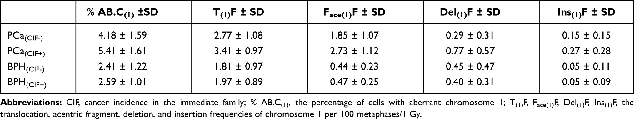 |
Table 3 Comparison of Cellular Radiosensitivity of Chromosome 1 Aberrations Obtained for PCa and BPH Patients, After Stratification by Cancer in the Immediate Family |
We observed the highest values of cellular radiosensitivity for all estimated biomarkers in cells from CIF+ PCa patients compared with the other subgroups (PCa(CIF-), BPH(CIF+), and BPH(CIF-)). The largest difference in radiosensitivity was observed for acentric fragments between the PCa(CIF+) and BPH(CIF+) groups (nearly six-fold higher for PCa(CIF+)). Additionally, in the PCa patient group, a significant, prominent difference was observed in the frequency of deletions (p<0.05) between subgroups of patients with or without cancer in the immediate family.
This result might be related to the report that the deletion of region 1p36 (CAPB) is most commonly observed as a terminal syndrome in men.29 Prostate cancer susceptibility loci in chromosome 1 have also been reported at the region 1q24-25 (denoted as the HPC1 gene locus) and at 1q42-43 (PCAP).29,30 Therefore, further research is needed on the suggested types of aberrations using larger patient groups, stratification by cancer incidence in the immediate family, and probes specific to the 1q24-25, 1q42.2–43, and 1p36 regions. Individual variability also necessitates deeper molecular insight and further studies in terms of smoking, diet, and other lifestyle factors. These studies would benefit from the use of molecular markers specific to early and late events in cancer progression, which are critical for these types of diseases.
Recent advances in chromosome staining using the FISH technique facilitate the fast and reliable measurement of simple translocations, which are an excellent biomarker for retrospective biological dosimetry of absorbed dose.31 Our results show that the frequency of acentric fragments detected in response to X-ray exposure can be proposed as an efficient predictor of susceptibility to radiation treatment in PCa patients. Figure 2 presents individual radiosensitivity (RS) as the frequency of acentric fragments (Face(1)F) of each PCa patient as evaluated as the dispersion from the average RS of the whole PCa group subtracted by the value obtained for the BPH group in the peripheral blood lymphocytes. The x-axis representing the mean RS value (2.05). The lines above and below the x-axis showed on the histogram indicating the standard deviation (±SD=1.08) of the mean RS value. The patient identification number is typed on a single bar.
 |
Figure 2 The individual radiosensitivity (RS) presented as the frequency of acentric fragments (Face(1)F) of each PCa patient. The RS value is calculated for each donor. The central cross-line (the x-axis) represents the mean RS value and how the RS value varies among the donors. The lines (above and below the x-axis) showed on the histogram are the standard deviation (±SD) of the mean RS value. The patient identification number is typed on a single bar. |
After subtraction of spontaneous chromosome 1 aberrations, the FISH results revealed differences in susceptibility to radiation, expressed as significantly increased or decreased frequencies of acentric fragments for chromosome 1 in the group of cancer patients compared to the BPH stage, which suggests that this technique and biomarkers in future might be proposed as screening and selecting of individuals to estimate individual susceptibility to radiation according to their level of radiosensitivity and genetic predisposition. Similar trends were observed in our previously published data.22,32 In literature, studies analyzing chromosomal aberrations have shown great promise to predict cancer risk and individual sensitivity.2,33 Analyzing the frequency of chromosomal aberrations in lymphocytes after in vitro irradiation have shown great promise to predict late sequelae due to cancer treatment.34 This suggests that cancer patients are predisposed to radiosensitivity compared to the general population. Radiotherapy inflicts acute and chronic toxicities to the normal tissue surrounding the tumor which is represented by HPBLs.35 Since HPBLs traffic throughout the body, which include irradiation field, could potentially be used to interrogate radiation injury to normal tissue during irradiation of tumors.36 Accordingly with data, following a standard regime of thirty fractions of 2Gy, 98.8% of the blood pool has been exposed to more than 0.5 Gy.35
The observed differences between PCa and BPH patients might be due to different factors. Our previous study indicated that lymphocytes from PCa patients have a higher level of DNA damage that was not repaired during post-challenging exposure incubation. Furthermore, DNA damage was less efficiently repaired than BPH subjects. Another open issue is the role of genotoxic and carcinogenic agents experienced by study subjects at the time of FISH testing on the level of spontaneous aberration frequency and radiosensitivity. We can not exclude more complex models or even the simultaneous occurrence of multiple events and their interaction during radiation therapy. The hypothesis is still under investigation because the effect might be influenced by the initial amount of DNA damage, physical activity, smoking status, etc. Notable, despite technological advances in chromosome identification, the mechanisms behind the origination and transmission of chromosomal aberrations after irradiation remain unclear.37 Many research groups suggest that any double-strand breaks (dsb) can participate in aberration formation, others postulate that only clustered can be involved.37 Understanding the relationship of aberrations with other endpoints, such as pathological grade, Gleason score, and conversion to malignancy, is one of the challenging tasks in biological dosimetry and radiotherapy.
Compared to classic cytogenetics, the method applied in this study could be a useful and more rapid method to identify overly sensitive or resistant patients. The FISH method is clinically applied to detect genetic abnormalities or the identification of novel oncogenes in cancer patients. However, before this method can be applied as a reliable and sensitive biomarker of individual predisposition to treatment, further effort is needed to examine whether the observed variation in patient radiosensitivity is associated with health recovery outcomes or with molecular and medical observations. Our studies are performed under ex vivo irradiation conditions, it is speculative at this time that this technique can be translated to clinical use for radiation assessment. The experimental or clinical implementation of the FISH technique is time-consuming and costliness. Additionally, all steps require experienced personnel. The lower sensitivity might happen due to a technical failure in the probes hybridization process with chromosome 1. In our studies in total, 26,554 irradiated cells and 68,945 not-irradiated were examined. However, future studies on bigger PCa group together with correlation studies with pathological grading, staging, and Gleason score are planned. Valuable will be studies comparing the incidence of chromosome aberrations before and after receiving radiotherapy. Further effort is needed to examine if the observed variation in radiosensitivity of PCa donors in the induced frequency of aberrations ich chromosome 1 could be associated with other molecular and medical observations, or with a health recovery outcome before it will be applied as a reliable and sensitive biomarker of the individual predisposition to the treatment.
Conclusions
Our preliminary investigation found statistical differences in the radiosensitivity of lymphocytes of PCa patients and BPH subjects. Among the various aberrations that can be analyzed using the FISH technique, acentric fragments of chromosome 1 appeared to be specifically detected in response to radiation treatment for prostate cancer. Thus, acentric fragments can be proposed as a biomarker of radiosensitivity for future detailed studies. This report is a part of cytogenetic and molecular research reflecting individual differences in the HPBLs response which aimed to find processes underlying the observed effects. Understanding these processes might bring fundamental insights to optimize radiotherapy and better exploit the influence of chromosomal instability.
Acknowledgments
We would like to thank the Department and Clinic of Urology, Collegium Medicum, The Jagiellonian University, Kraków, Poland for supplying us with the samples, and Z. Drąg, Ph.D. for performing interviews and coding questionnaires. Sincere gratitude to mentor, Professor Antonina Cebulska-Wasilewska for the research supporting, crucially valuable discussions, motivation, and suggestions are highly appreciated. In particular, the authors gratefully acknowledge the donors for providing blood samples, technical staff (mainly Joanna Wiltowska) for FISH slides preparing, irradiation, dosimetry, and Kamila Rawojć Ph.D. for editorial support.
Funding
Work partly supported by grants KBN 6P05CO5421 and MNiSW 2520/B/P01/2010/39.
Disclosure
The authors declare no conflicts of interest in this work.
References
1. Beskid O, Binkova B, Dusek Z, et al. Chromosomal aberrations by fluorescence in situ hybridization (FISH) - Biomarker of exposure to carcinogenic PAHs. Mutat Res. 2007;690:62–70. doi:10.1016/j.mrfmmm.2007.02.023
2. Bonassi S. Chromosomal aberration in peripheral blood lymphocytes of healthy subjects and risk of cancer. In: Vijayalaxmi OG, editor. Chromosomal Alterations. Berlin, Heidelberg: Springer; 2007.
3. Cebulska-Wasilewska A. Response to challenging dose of X-rays as a predictive assay for molecular epidemiology. Mutat Res. 2003;544:289–297. doi:10.1016/j.mrrev.2003.07.003
4. Grade M, Difilippantonio MJ, Camps J. Patterns of chromosomal aberrations in solid tumors. Recent Results Cancer Res. 2015;200:115–142.
5. Boei JJ, Vermeulen S, Natarajan AT. Detection of chromosomal aberrations by fluorescence in situ hybridization in the first three post-irradiation divisions of human lymphocytes. Mutat Res. 1996;349:127–135. doi:10.1016/0027-5107(95)00171-9
6. Rana S, Kumar R, Sultana S, Sharma RK. Radiation-induced biomarkers for the detection and assessment of absorbed radiation doses. J Pharm Bioallied Sci. 2010;2(3):189–196. doi:10.4103/0975-7406.68500
7. Mladenov E, Magin S, Soni A, Iliakis G. DNA double-strand break repair as determinant of cellular radiosensitivity to killing and target in radiation therapy. Front Oncol. 2013;3:113. doi:10.3389/fonc.2013.00113
8. Grönberg H. Prostate cancer epidemiology. Lancet. 2003;361:859–864. doi:10.1016/S0140-6736(03)12713-4
9. Chughtai B, Forde JC, Thomas DDM, et al. Benign prostatic hyperplasia. Nature Rev. 2016;16032.
10. Mirone V, Fusco F, Verze P, et al. Androgens and benign prostatic hyperplasia. Eur Urol Supp. 2006;5:410–417. doi:10.1016/j.eursup.2006.02.004
11. Rakel D. Chapter 60 - Benign prostatic hyperplasia. In: Integrative Medicine. Elsevier,
12. Gallardo FF, Quintar AA. The pathological growth of the prostate gland in atherogenic contexts. Exp Gerontol. 2021;148:111304. doi:10.1016/j.exger.2021.111304
13. Singh R, Eeles RA, Durocher F, et al. High risk genes predisposing to prostate cancer development – do they exist? Prostate Cancer Prostatic Dis. 2000;3:241–247. doi:10.1038/sj.pcan.4500478
14. Frank C, Sundquist J, Hemminki A, Hemminki K. Familial associations between prostate cancer and other cancers. Eur Urol. 2017;71:162–165. doi:10.1016/j.eururo.2016.07.031
15. Ballon-Landa E, Parsons JK. Nutrition, physical activity, and lifestyle factors in prostate cancer prevention. Curr Opin Urol. 2018;28:55–61. doi:10.1097/MOU.0000000000000460
16. Saramäki OR, Visakorpi T. Chromosomal aberrations in prostate cancer. Front Biosci. 2018;12:3287–3301. doi:10.2741/2312
17. Smith JR, Freije D, Carpten JD, et al. Major susceptibility locus for prostate cancer on chromosome 1 suggested by a genome-wide search. Science. 1996;274:1371–1374. doi:10.1126/science.274.5291.1371
18. Robinson D, Garmo H, Lissbrant IF, et al. Prostate cancer death after radiotherapy or radical prostatectomy: a nationwide population-based observational study. Eur Urol. 2018;73:502–511. doi:10.1016/j.eururo.2017.11.039
19. Podder TK, Fredman ET, Ellis RJ. Advances in radiotherapy for prostate cancer treatment. Adv Exp Med Biol. 2018;1096:31–47.
20. Takakusagi Y, Hiroyuki Katoh H, Kano K. Preliminary result of carbon-ion radiotherapy using the spot scanning method for prostate cancer. Radiat Oncol. 2020;15:127. doi:10.1186/s13014-020-01575-7
21. Cytogenetic dosimetry: applications in preparedness for and response to radiation emergencies, IAEA report. Vienna; 2011.
22. Cebulska-Wasilewska A, Miszczyk J, Balegenowa N, et al. Studies of the susceptibility to radiation of prostate cancer or BPH patients and healthy donors. In: Cebulska-Wasilewska A, Osipov AN, Darroudi F, editors. Rapid Diagnosis in Populations at Risk from Radiation and Chemicals. Vol. 73. IOS Press Amsterdam; 2010:211–220. ISBN 978-1-60750-644-7.
23. Prasanna PGS, Hamel CJC, Escalada ND, et al. Biological dosimetry using human interphase peripheral blood lymphocytes. Mil Med. 2002;167:10–12. doi:10.1093/milmed/167.suppl_1.10
24. Fowler JF. The radiobiology of prostate cancer including new aspects of fractionated radiotherapy. Acta Oncol. 2005;44(3):265–276. doi:10.1080/02841860410002824
25. Hille A, Hofman-Hüther H, Kühnle E, et al. Spontaneous and radiation-induced chromosomal instability and persistence of chromosome aberrations after radiotherapy in lymphocytes from prostate cancer patients. Radiat Environ Biophys. 2010;49:27–37. doi:10.1007/s00411-009-0244-x
26. Beaton LA, Marro L, Samiee S, et al. Investigating chromosome damage using fluorescent in situ hybridization to identify biomarkers of radiosensitivity in prostate cancer patients. Int J Radiat Biol. 2013;89:1087–1093. doi:10.3109/09553002.2013.825060
27. Beaton LA, Ferrarotto C, Marro L, et al. Chromosome damage and cell proliferation rates in vitro irradiated whole blood as markers of late radiation toxicity after the radiation therapy to the prostate. Int J Radiat Oncol Biol Phys. 2013;85:1346–1352. doi:10.1016/j.ijrobp.2012.09.026
28. El-Zein R, Gu Y, Sierra MS, et al. Chromosomal instability in peripheral blood lymphocytes and risk of prostate cancer. Cancer Epidemiol Biomarkers Prev. 2005;14:748–752. doi:10.1158/1055-9965.EPI-04-0236
29. Xu J, Zheng SL, Chang B, et al. Linkage of prostate cancer susceptibility loci to chromosome 1. Hum Genet. 2001;189:335–345. doi:10.1007/s004390100488
30. Berthon P, Valeri A, Cohen-Akenine A, et al. Predisposing gene for early-onset prostate cancer, localized on chromosome 1q42.2-43. Am J Hum Genet. 1998:1416–1424.
31. Miszczyk J, Cebulska-Wasilewska A. Retrospective biological dosimetry of the absorbed dose – training on the estimation of the radiation dose of a person presumably exposed to X-ray radiation by FISH. In: Cebulska-Wasilewska A, Osipov AN, Darroudi F, editors. Rapid Diagnosis in Populations at Risk from Radiation and Chemicals. Vol. 73. IOS Press Amsterdam. 2010;211–220. ISBN 978-1-60750-644-7.
32. Miszczyk J, Cebulska-Wasilewska A, Glazar B, et al. Comparison between susceptibilities to radiation treatment of lymphocytes from prostate cancer (PC) with BPH patients. Eur Urol Suppl. 2012;11(4):126–127. doi:10.1016/S1569-9056(13)60184-0
33. Vodenkova S, Polivkova Z, Musak L, et al. Structural chromosomal aberrations as potential risk markers in incident cancer patients. Mutagenesis. 2015;30:557–563. doi:10.1093/mutage/gev018
34. Distel LVR, Neubauer S, Keller U, et al. Individual differences in chromosomal aberrations after in vitro irradiation of cells from healthy individuals, cancer and cancer susceptibility syndrome patients. Radiat Oncol. 2006;81:257–263. doi:10.1016/j.radonc.2006.10.012
35. Wilson JD, Hammond EM, Higgins GS, Petersson K. Ultra-high dose rate (FLASH) radiotherapy: silver bullet or fool’s gold? Front Oncol. 2020;9:1563. doi:10.3389/fonc.2019.01563
36. Miszczyk J, Rawojć K, Panek A, et al. Do protons and X-rays induce cell-killing in human peripheral blood lymphocytes by different mechanisms? Clin Transl Radiat Oncol. 2018;31(9):23–29. doi:10.1016/j.ctro.2018.01.004
37. Kaddour A, Colicchio B, Buron D, et al. Transmission of induced chromosomal aberrations through successive mitotic divisions in human lymphocytes after in vitro and in vivo radiation. Sci Rep. 2017;7(1):3291. doi:10.1038/s41598-017-03198-7
 © 2021 The Author(s). This work is published and licensed by Dove Medical Press Limited. The full terms of this license are available at https://www.dovepress.com/terms.php and incorporate the Creative Commons Attribution - Non Commercial (unported, v3.0) License.
By accessing the work you hereby accept the Terms. Non-commercial uses of the work are permitted without any further permission from Dove Medical Press Limited, provided the work is properly attributed. For permission for commercial use of this work, please see paragraphs 4.2 and 5 of our Terms.
© 2021 The Author(s). This work is published and licensed by Dove Medical Press Limited. The full terms of this license are available at https://www.dovepress.com/terms.php and incorporate the Creative Commons Attribution - Non Commercial (unported, v3.0) License.
By accessing the work you hereby accept the Terms. Non-commercial uses of the work are permitted without any further permission from Dove Medical Press Limited, provided the work is properly attributed. For permission for commercial use of this work, please see paragraphs 4.2 and 5 of our Terms.