")
Back to Journals » Veterinary Medicine: Research and Reports » Volume 5
Influence of feeding on serum canine pancreatic lipase immunoreactivity concentrations
Authors Steiner J, Ruaux C, Williams D
Received 17 June 2014
Accepted for publication 28 August 2014
Published 17 October 2014 Volume 2014:5 Pages 139—142
DOI https://doi.org/10.2147/VMRR.S69576
Checked for plagiarism Yes
Review by Single anonymous peer review
Peer reviewer comments 2
Editor who approved publication: Dr Jeffrey MB Musser
Jörg M Steiner, Craig G Ruaux, David A Williams
Gastrointestinal Laboratory, College of Veterinary Medicine and Biomedical Sciences, Texas A&M University, College Station, TX, USA
Abstract: Measurement of serum concentration of pancreatic lipase immunoreactivity (PLI) has been shown to be highly specific for exocrine pancreatic function and sensitive for the diagnosis of canine pancreatitis. Currently, it is recommended that food be withheld for at least 12 hours before collecting a blood sample for analysis from dogs. However, it is unknown whether feeding has any influence on serum canine PLI concentration. Thus, the goal of this study was to evaluate the influence of feeding on serum canine PLI concentrations in healthy dogs. Food was withheld from eight healthy adult Beagle dogs for at least 17 hours and a baseline serum sample (0 minutes) was collected. Dogs were fed and serum samples were collected at 15, 30, 45, 60, 75, 90, 105, 150, 180, 210, 240, 300, 360, 420, and 480 minutes. There was no significant difference in serum canine PLI concentrations at any time after feeding (P=0.131). We conclude that feeding has no significant influence on serum canine PLI concentrations.
Keywords: dog, pancreatic function, pancreatitis, biomarker, diagnostic test
Introduction
The main function of the gastrointestinal tract is to digest macromolecular nutrients and absorb their breakdown products. Digestion is accomplished by synthesis and secretion of digestive enzymes, mainly by the exocrine pancreas.1 A small portion of digestive enzymes secreted reaches the vascular space.2 There are different potential mechanisms by which digestive enzymes may reach the vascular space. Digestive enzymes may be secreted into the gastrointestinal lumen exclusively, but may then be partially reabsorbed. Also, digestive enzymes may leak into the vascular space. Normal cells could leak a small amount of digestive enzymes into the vascular space or senescent cells undergoing apoptosis could release their content of digestive enzymes. Finally, digestive enzymes could be actively secreted into the vascular space. Depending on the mechanism by which digestive enzymes reach the vascular space, this mechanism may or may not be affected by food intake.
Serum concentrations of digestive enzymes have been used as minimally invasive markers for gastrointestinal disorders in the dog. For example, serum trypsin-like immunoreactivity (TLI) concentration has been shown to be highly sensitive and specific for canine exocrine pancreatic insufficiency.3 Similarly, the measurement of serum pancreatic lipase immunoreactivity (PLI) has been shown to be highly specific for the exocrine pancreas4,5 and highly sensitive for canine pancreatitis,6,7 with specificities reported to be as high as 97.5%4 and sensitivities in most studies ranging between 70% and 95%, depending on the study population.6,7 It has been shown that feeding leads to a significant increase in serum TLI concentration.3,8 While this increase was significant in both dogs and cats it did not appear to be clinically important in clinically healthy cats.8 These studies have led to the recommendation that serum samples for evaluation of TLI be collected after withholding food for at least 12 hours in both dogs and cats. More recently, assays for the measurement of canine pancreatic lipase (by canine pancreatic lipase immunoreactivity [cPLI]) in serum have been developed and analytically validated.9,10 These assays have been shown to be highly specific for exocrine pancreatic function and highly sensitive for a diagnosis of pancreatitis.4,6 Based on the findings for serum TLI, it is currently recommended to withhold food for at least 12 hours before collecting a serum sample for measurement of cPLI. However, it is unknown whether serum cPLI is affected by feeding. Therefore, the goal of this study was to evaluate the influence of feeding on serum cPLI concentrations in healthy dogs.
Materials and methods
Eight healthy adult Beagle dogs belonging to a research colony were enrolled in this study. The study protocol was reviewed and approved by the Animal Care and Use Committee responsible for the research colony. None of the dogs showed any abnormalities on physical examination, complete blood count, or serum chemistry profile. An indwelling jugular catheter was placed, and food was withheld from the dogs for at least 17 hours. A baseline serum sample (0 minutes) was collected and dogs were fed the daily ration (based on the calculation of the maintenance energy requirement for the current body weight of each dog; however, the exact amount of food given was not recorded) of a commercial dry canine maintenance diet. Further serum samples were collected at 15, 30, 45, 60, 75, 90, 105, 150, 180, 210, 240, 300, 360, 420, and 480 minutes after feeding. Serum samples were frozen at −80°C, shipped on dry ice, and stored frozen at −80°C until analysis.
Serum cPLI was measured using an in-house sandwich enzyme-linked immunosorbent assay (ELISA) that had been validated previously.9 Briefly, 96-well flat-bottom ELISA plates were coated with affinity purified anti-canine pancreatic lipase antibodies. Nonspecific binding sites of the plate were blocked by incubation with a commercially available blocking solution. Standards, controls, and samples were diluted 1 in 200 in phosphate-buffered saline with 1% bovine serum albumin and 0.05% Tween (buffer A) and were loaded in duplicates. Then the plate was incubated and afterwards washed. Biotinylated anti-cPL antibodies were added to the plate and, after incubation and washing, horseradish peroxidase-labeled streptavidin was added. After another wash cycle, the plate was developed for 15 minutes using a TMB (3,3′,5,5′-tetramethylbenzidine dihydrochloride) substrate solution; the color reaction was stopped using 4 M acetic acid and 0.5 M sulfuric acid, and the plates were read at 450 nm. Standard curves were calculated using a four-parameter curve fit.
Datasets for all time points were evaluated for normality using the D’Agostino-Pearson omnibus normality test. Serum cPLI concentrations at different time points after feeding were compared with baseline concentrations using repeated-measures, one-way analysis of variance, followed by a Dunnett’s multiple comparison test (GraphPad Prism 3.0). Statistical significance was assigned for values of P<0.05.
Results
Serum cPLI concentrations at baseline ranged from 1.8 to 73.7 μg/L, with a mean ± standard deviation of 28.9±28.6 μg/L (Table 1 and Figure 1). Datasets for all time points passed normality testing. There was no significant difference in serum cPLI concentrations at any time after feeding when compared with baseline concentrations (P=0.131). However, there were fluctuations in serum cPLI concentrations over time. Two of the dogs had baseline serum cPLI concentrations in the lower range of the reference interval and one dog even below the lower limit of the reference interval. Serum cPLI concentrations decreased below the lower limit of the reference interval for one of the two dogs with a baseline in the lower range of the reference interval. None of the serum cPLI concentrations measured at any time point were increased over the upper limit of the reference interval for serum PLI concentration in dogs (2.2–102.1 μg/L).
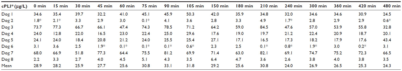 | Table 1 Serum cPLI concentrations after feeding |
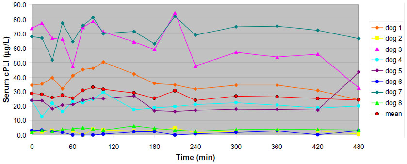 | Figure 1 Serum cPLI concentrations in eight healthy Beagle dogs after feeding. |
Discussion
There was no statistically significant difference in serum cPLI concentrations at any time point after feeding when compared with baseline concentrations. There was, however, some variability in serum cPLI concentration between different measurements in the same dog, but no general pattern could be observed. Thus, it can be concluded that, at least in clinically healthy dogs, the measurement of PLI concentrations in serum is not affected by feeding. A previous study evaluated the effect of various diets on serum PLI concentrations, and dogs fed a diet with a crude fat content of 5% or 16%, dogs fed a diet with a crude fat content of 16% and a pancreatic enzyme supplement, and dogs fed a diet with a crude fat content of 5% and a pancreatic enzyme supplement and medium chain triglyceride oil did not show a significant difference in serum cPLI concentrations.11 These findings would agree with the findings of the current study.11
Withholding food in patients prior to sample collection for cPLI analysis may be less important than for measurement of other marker substances. However, it must be stressed that the dogs used in the current as well as the previous study were clinically healthy, and dogs evaluated for a potential diagnosis of pancreatitis may show a different response.11 Further research would be required to evaluate the effect of feeding on serum cPLI concentrations in dogs with pancreatitis or suspected pancreatitis. Feeding may result in lipemia in some individuals, which may in turn have an effect on the performance of the cPLI assay. However, the commercially available assay for measurement of cPLI (Spec cPL®, Idexx Laboratories, Westbrook, ME, USA) has been shown to be unaffected by even severe lipemia.12 Although there are differences between the ELISA for measurement of cPLI used here and the Spec cPL ELISA, they are overall very similar in that they are sandwich ELISAs, so it would appear unlikely that the cPLI ELISA utilized here would be affected by lipemia. Also, none of the dogs evaluated in this study developed gross lipemia, although this may be different for dogs evaluated for possible pancreatitis.
None of these eight healthy dogs had serum cPLI concentrations above the upper limit of the reference interval at any time point. The highest serum cPLI concentration in any dog and at any time point was 84.5 μg/L, which was well below the upper limit of the reference interval of 102.1 μg/L. Thus it would appear that stimulation of pancreatic enzyme secretion (ie, through feeding) is not sufficient to increase serum concentrations of cPLI, which in turn would suggest that reabsorption of lipase from the intestinal lumen is not a likely origin of pancreatic lipase in the serum of dogs. It also would suggest that if secretion of pancreatic lipase directly into the vascular space were the mechanism by which pancreatic lipase reaches the vascular space, stimulation of secretion onto the luminal side of acinar cells does not mirror stimulation of secretion onto the vascular side of acinar cells. However, further studies are required to definitively determine the origin of pancreatic lipase in the serum in dogs.
Conclusion
In conclusion, in healthy dogs, feeding causes no significant changes in serum cPLI concentrations. However, it remains to be determined if the same is true in dogs with pancreatitis.
Disclosure
JMS serves as the director of the Gastrointestinal Laboratory at Texas A&M University. DAW and JMS are paid consultants for Idexx Laboratories. CGR has no conflicts of interest in this work. Both the Gastrointestinal Laboratory and Idexx Laboratories offer measurement of serum cPLI concentration on a fee-for-service basis. This material was presented in part at the 2002 Forum of the American College of Veterinary Internal Medicine.
References
Rinderknecht H. Pancreatic secretory enzymes. In: Go VLW, DiMagno EP, Gardner JD, Lebenthal E, Reber HA, Scheele GA, editors. The Pancreas: Biology, Pathobiology and Disease. 2nd ed. New York, NY, USA: Raven Press; 1993. | |
Steiner JM. Exocrine pancreas. In: Steiner JM, editor. Small Animal Gastroenterology. 1st ed. Hannover, Germany: Schlütersche-Verlagsgesellschaft mbH; 2008. | |
Williams DA, Batt RM. Sensitivity and specificity of radioimmunoassay of serum trypsin-like immunoreactivity for the diagnosis of canine exocrine pancreatic insufficiency. J Am Vet Med Assoc. 1988;192:195–201. | |
Neilson-Carley SC, Robertson JE, Newman SJ, et al. Specificity of a canine pancreas-specific lipase assay for diagnosing pancreatitis in dogs without clinical or histologic evidence of the disease. Am J Vet Res. 2011;72(3):302–307. | |
Steiner JM, Berridge BR, Wojcieszyn J, Williams DA. Cellular immunolocalization of gastric and pancreatic lipase in various tissues obtained from dogs. Am J Vet Res. 2002;63(5):722–727. | |
McCord K, Morley PS, Armstrong J, et al. A multi-institutional study evaluating the diagnostic utility of the Spec cPL and SNAP(R) cPL in clinical acute pancreatitis in 84 dogs. J Vet Intern Med. 2012;26(4):888–896. | |
Trivedi S, Marks SL, Kass PH, et al. Sensitivity and specificity of canine pancreas-specific lipase (cPL) and other markers for pancreatitis in 70 dogs with and without histopathologic evidence of pancreatitis. J Vet Intern Med. 2011;25(6):1241–1247. | |
Steiner JM, Williams DA. Influence of feeding on serum feline trypsin-like immunoreactivity. Am J Vet Res. 1999;60(7):895–897. | |
Steiner JM, Teague SR, Williams DA. Development and analytic validation of an enzyme-linked immunosorbent assay for the measurement of canine pancreatic lipase immunoreactivity in serum. Can J Vet Res. 2003;67(3):175–182. | |
Steiner JM, Williams DA. Development and validation of a radioimmunoassay for the measurement of canine pancreatic lipase immunoreactivity in serum of dogs. Am J Vet Res. 2003;64(10):1237–1241. | |
James FE, Mansfield CS, Steiner JM, Williams DA, Robertson ID. Pancreatic response in healthy dogs fed diets of various fat compositions. Am J Vet Res. 2009;70(5):614–618. | |
Huth SP, Relford R, Steiner JM, Strong-Townsend MI, Williams DA. Analytical validation of an ELISA for measurement of canine pancreas-specific lipase. Vet Clin Pathol. 2010;39(3):346–353. |
 © 2014 The Author(s). This work is published and licensed by Dove Medical Press Limited. The full terms of this license are available at https://www.dovepress.com/terms.php and incorporate the Creative Commons Attribution - Non Commercial (unported, v3.0) License.
By accessing the work you hereby accept the Terms. Non-commercial uses of the work are permitted without any further permission from Dove Medical Press Limited, provided the work is properly attributed. For permission for commercial use of this work, please see paragraphs 4.2 and 5 of our Terms.
© 2014 The Author(s). This work is published and licensed by Dove Medical Press Limited. The full terms of this license are available at https://www.dovepress.com/terms.php and incorporate the Creative Commons Attribution - Non Commercial (unported, v3.0) License.
By accessing the work you hereby accept the Terms. Non-commercial uses of the work are permitted without any further permission from Dove Medical Press Limited, provided the work is properly attributed. For permission for commercial use of this work, please see paragraphs 4.2 and 5 of our Terms.