")
Back to Journals » Research and Reports in Urology » Volume 15
Experience in Retrocaval Ureter at Saint Paul’s Hospital Millennium Medical College: A Case Series
Authors Mideksa AG , Huluka TY, Dino MS, Ahmed MM
Received 2 August 2023
Accepted for publication 19 September 2023
Published 22 September 2023 Volume 2023:15 Pages 431—436
DOI https://doi.org/10.2147/RRU.S419718
Checked for plagiarism Yes
Review by Single anonymous peer review
Peer reviewer comments 4
Editor who approved publication: Dr Panagiotis J Vlachostergios
Adugna Getachew Mideksa, Tolesa Yadeta Huluka, Masresha Solomon Dino, Mensur Mohammed Ahmed
Department of Surgery Urology Division, Saint Paul’s Hospital Millennium Medical College, Addis Ababa, Ethiopia
Correspondence: Adugna Getachew Mideksa; Mensur Mohammed Ahmed, Email [email protected]; [email protected]
Abstract: Retrocaval ureter is a rare congenital vascular anomaly described as the passage of the ureter behind the inferior vena cava (IVC) and then turning around the IVC to attain the final lateral position. The condition is usually associated with obstruction in the ipsilateral kidney, causing different degrees of hydronephrosis and complications associated with urinary stasis, such as stone formation. Imaging has a crucial role in the diagnosis and management of retrocaval ureter. CT urography may be the procedure of choice to confirm the diagnosis and avoid retrograde ureteropyelography. Indications for treatment include flank pain, recurrent infection, hydronephrosis, and stone formation due to obstruction. Surgical management is standard and can be done through either an open, laparoscopic, or robotic approach. In this case series, we are going to see two cases of retrocaval ureter in a 56-year-old male and a 14-year-old male child who presented with a right flank of less than a couple of months duration. The first case has an associated horseshoe kidney and a solitary secondary stone. Both cases were surgically managed with open ureteral division, relocation, and ureteroureterostomy. Both have uneventful post-operative follow-ups.
Keywords: retrocaval ureter, circumcaval ureter, pre-ureteral vena cava, horseshoe kidney
Introduction
Retrocaval ureter (also called circumcaval ureter) is a rare congenital anomaly in which the ureter passes posterior to the inferior vena cava (IVC). 1 In fact, it is a vascular anomaly rather than a ureteral abnormality. Based on autopsy results, it is said to occur in 1 in 1500 lives, with an overall incidence of about 0.006%–0.17%.2
Abnormal persistence of the right subcardinal vein anterior to the ureter during embryogenesis of the IVC is said to be the reason for the retrocaval ureter. The developing right ureter passes behind and medial to the IVC over the pedicle of the L3-L4 vertebrae, then exits between the IVC and aorta, returning to its normal position.3
It is usually asymptomatic but becomes symptomatic in the third or fourth decades of life as a result of kinking of the ureter posterior to the IVC. Most patients present with right-flank pain, recurrent urinary tract infection (UTI), renal stones, and hydronephrosis.2
Clinical Presentation
Case 1
A 56-year-old male patient who is newly diagnosed with hypertension on amlodipine 10 mg po per day for the past month presented with 10-day history of right-side flank pain that was intermittent and stabbing in nature, exacerbated by exercise, and prolonged standing. Despite this, he denies any history of decreased urine output, reddish discoloration of urine, or leg swelling. His BP was 130/90, and other vital signs were in the normal range. He was investigated with urine analysis that showed protein +1, serum creatine 0.95 mg/dl, and urea of 35.0 mg/dl.
He was later investigated with abdominal ultrasound, which showed a large left kidney cortical renal cyst and mild-to-moderate right-side proximal hydroureteronephrosis, an otherwise unremarkable ultrasound. He was then investigated with CT-Urography, which showed a horseshoe kidney with the isthmus fusing between the abdominal aorta and IVC, a left-side renal cortical simple cyst, and a right-side moderate hydroureteronephrosis, with the dilated proximal ureter running posterior to the IVC and containing a stone measuring 0.9 cm with a HU of 800, and the ureter progressed distally behind the IVC (Figure 1A).
 |
Figure 1 (A) CT-scan image – axial, coronal and sagittal sections (C stands for left side renal cyst, U is dilated right proximal ureter located lateral and behind the IVC, A is aorta, s stands for the stone in the right proximal dilated ureter and I is inferior vena cava). (B) Intraoperative image of retrocaval ureter. |
He was then taken to the operation room for the above assessment, and the ureter approached through the right 12th rib subcostal flank incision. Upon developing the retroperitoneal plane, the ureter is seen curving behind the IVC, as shown in Figure 1B After careful dissection of the ureter, it was divided distal to the curvature and mobilised anterior to the IVC. Intraoperatively, the stone was found to have migrated to the renal pelvis, and flexible ureterorenoscopy was used to remove the stone with a Dormia stone basket. Ureteroureterostomy was completed with a tension-free end-to-end and stented anastomosis. A drainage tube was kept for a day. The patient had a smooth hospital course and was discharged on the third day. A double J stent was removed after 4 weeks post-surgery, with no hydronephrosis seen on the follow-up ultrasound.
Case 2
A 14-year-old male child who is a seventh-grade student presented with a history of right-side flank pain of 2-month duration. He has no history of urine colour or amount change nor a history of fever, chills, or rigor. He had a follow-up at a different governmental general hospital with the diagnosis of right moderate hydronephrosis from the age of one. The physical examination was unremarkable. Abdominal ultrasound was done, which showed the right kidney measures 10.3 cm by 4.4 cm with a cortical thickness of 0.87 cm and a moderate degree of hydronephrosis with proximal ureteric dilatation, and serum creatine was 0.6 mg/dl.
CT-urography was done, showing contrast uptake by both kidneys, and contrast excretion from the left ureter was noted. The delayed image was only taken until 15 min, which did not show contrast excretion to the collecting system (Figure 2A); hence, we did a retrograde pyelography, which shows the characteristic S-shaped ureter (Figure 2B).
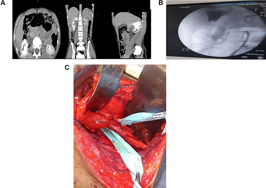 |
Figure 2 (A) Abdominopelvic CT-scan – axial, sagittal and coronal section. (B) Retrograde pyelography showing the S-shaped retrocaval ureter. (C) Intraoperative image of retrocaval ureter. |
Surgical intervention was decided with the above diagnosis, and the ureter was approached through a 12th rib subcostal flank incision. The gerota's fascia was opened from the caudal region to find a retrocaval ureter, which has an aperistaltic segment running behind the IVC (Figure 2C).
The ureter was carefully dissected and transected caudally, and a tension-free, water-tight, and stented ureterouretrostomy was done. There was no drain output after 24 hr, hence the drain and transurethral catheter were removed. The patient was kept in the ward for 2 days and discharged with a smooth post-op course. Stent removal was done after 4 weeks, and the patient had no sign of hydronephrosis on subsequent ultrasound scans.
Discussion
Urologists frequently refer to the preureteral vena cava as the circumcaval or retrocaval ureter, words that are physically accurate but confusing in terms of development.4 The phrase pre-ureteral vena cava emphasises that altered vascular development, not ureteral development, is what causes the circumcaval ureter. The more appropriate phrase is this.
The retrocaval ureter is a rare congenital anomaly that results from the persistence of the posterior cardinal vein during embryonal development of the IVC.4 Based on radiologic appearance, it can be classified as Type 1, the more common one and similar to our cases, which present with sickle-shaped severe or moderate hydronephrosis with extreme medial deviation of the middle ureteral segment, usually medial to the pedicle or across the midline at the L3 level. Radiographically, it has an “S” or “fish hook” deformity at the level of obstruction. The second form of the retrocaval ureter is characterised by mild hydronephrosis with a less medial deviation of the ureter. The obstruction is due to the compression of the IVC on the ureter, which will lead to a sickle-shaped ureter at the level of obstruction.5,6 The retrocaval ureter (RCU) is typically located on the right side of the body. However, there are some rare conditions where it may be found on the left side, like in cases of situs inversus, duplication of the IVC, or persistent left subcardinal vein.7 Majorities of patients with type 2 retrocaval ureter are asymptomatic in their early lives and may seek help because of right-flank pain with urinary tract infections and haematuria in the third or the fourth decade.8 Our index cases presented with a short duration of right-side flank pain.
Anomalies related to RCU can occur in up to 21% of the patients. Cardiovascular abnormalities, genitourinary anomalies (such as horseshoe kidney, missing or ectopic opposite kidney, and agenesis of vasor uterus), Turner syndrome, and imperforated anus are among these congenital anomalies.9 In our first case, a horseshoe kidney was present along with an unusual isthmus that passed in front of the aorta and behind the IVC (Figure 1A). Therefore, all patients with RCU should be screened for these disorders.
Stone in the retrocaval ureteral loop is exceedingly rare and has only been reported in a few case reports.10 In our first case, there was a 0.9-cm measuring stone within the proximal ureter, which was removed with a dormia stone basket using flexible ureterorenoscopy through the proximal ureterostomy.
Ultrasonography and CT or MRI have been useful in defining vascular malformation. Abdominal sonography can demonstrate the presence of hydronephrosis, but it is poor at delineating the ureter. With an intravenous urography study, one can easily see hydroureteronephrosis with upward curving and abrupt tapering of the ureter; the rest of the middle and distal thirds of the ureter might not be visible because of obstruction. A spiral CT scan delineates both the ureter and IVC, is considered the investigation of choice, and avoids retrograde ureteropyelography.1 Anatomically obstructed anomalies can be differentiated from functional obstructions using nuclear renal furosemide scanning.11 Compared to CT and retrograde pyelography, MRI can show the path of a preureteral vena cava and may be a more accurate and minimally intrusive imaging technique.12 Sometimes the diagnosis may not be straightforward from imaging and one case reported in which the obstruction from a pre-ureteral cava was thought to be a ureteropelvic junction obstruction managed with subsequent pyeloplasty that failed.13
Some of the indications for surgical correction of the retrocaval ureter are the presence of complications like obstructive uropathy, leading to nephropathy, urolithiasis, hematuria, and urinary tract infections.8 Surgical correction involves ureteral division, relocation, and ureteroureteral or ureteropelvic reanastomosis. The retrocaval segment, which may be aperistaltic, is often excised or bypassed. The blood flow to the ureter from the renal artery and aorta superiorly and the iliac vessels inferiorly must be taken into consideration. The surgical approach could be open, laparoscopic, or robotic based on the surgeon’s experience and surgical setup.14 Laparoscopic and robotic reconstruction of the ureter in a preureteral vena cava via both the transperitoneal and retroperitoneal approaches in children has been described. Even though open and laparoscopic approaches are the most commonly used methods of repair, robot-assisted surgery has been seen as the latest advancement for surgical correction of retrocaval ureter.15
Conclusion
The diagnosis of retrocaval ureter, a rare congenital malformation, is crucial due to its potential clinical implications. A high index of suspicion is necessary, as delayed diagnosis can lead to complications and kidney function loss. Imaging should be used to assess complications and associated genitourinary anomalies before surgical intervention. Our case series highlights the importance of early diagnosis using advanced imaging techniques like CT urography and retrograde pyelography. The first case involved a horseshoe kidney with an unusual isthmus location and a secondary stone. Both conditions were diagnosed preoperatively, and the stone was managed with flexible ureterorenoscopy. Surgical management is recommended for symptomatic patients and those showing progressive renal derangement. This case series contributes to the growing body of knowledge on this rare condition and serves as a valuable resource for clinicians and urologists worldwide.
Consent for Publication
A written informed consent has been obtained from the patient to have the case details and any accompanying images published. No institutional review board approval was required to publish the case details.
Acknowledgment
We would like to acknowledge Abebe Ketema Aderu, who was involved in patient case management.
Author Contributions
All authors made a significant contribution to the work reported, whether that is in the conception, study design, execution, acquisition of data, analysis and interpretation, or in all these areas; took part in drafting, revising, or critically reviewing the article; gave final approval of the version to be published; have agreed on the journal to which the article has been submitted; and agree to be accountable for all aspects of the work.
Funding
There was no funding for this report.
Disclosure
The authors report no conflicts of interest in this work.
References
1. Kumar Jangid M, Ahmad R, Jangid P, Pandre SK, Kaur G. Retrocaval ureter with proximal hydroureteronephrosis presenting as flank pain in a child: a rare case report with review of literature; 2019. Available from: https://www.researchgate.net/publication/344332447.
2. Ahmed M, Alhassan A, Sadiq MA, Lawal AT, Bello A, Maitama HY. Variable presentation of retrocaval ureter: report of four cases and review of literature. Niger Postgrad Med J. 2017;24(2):126–129. doi:10.4103/npmj.npmj_48_17
3. Agarwal S, Goel S, Sankhwar S, Garg G. Circumcaval ureter/retrocaval ureter. BMJ Case Rep. 2018;2018. doi:10.1136/bcr-2018-225449
4. Lesma A, Bocciardi A, Rigatti P. Circumcaval ureter: embryology. Eur Urol Suppl. 2006;5(5):444–448. doi:10.1016/j.eursup.2006.02.008
5. El-Assmy A, ElShal AM, Mekkawy R, El-Kappany H, Ibrahiem EHI. Erectile and ejaculatory functions changes following bipolar versus monopolar transurethral resection of the prostate: a prospective randomized study. Int Urol Nephrol. 2018;50(9):1569–1576. doi:10.1007/s11255-018-1950-6
6. Maestroni U, Dinale F, Ziglioli F, Bocchialini T, Guarino GG, Campobasso D. Robotic transposition and ureteroureterostomy of type 1 retrocaval ureter. J Surg Case Reports. 2023;2023(6). doi:10.1093/jscr/rjad352
7. Gupta R, Kesar A, Mahajan A, Mehta A, Masood S. Transperitoneal laparoscopic ureteropyeloplasty of retrocaval ureter: single surgeon experience and review of literature. Asian J Endosc Surg. 2022;15(1):90–96. doi:10.1111/ases.12970
8. Liu E, Sun X, Guo H, Li F, Liu S, Wang K. Retroperitoneoscopic ureteroplasty for retrocaval ureter: report of nine cases and literature review. Scandinavian Journal of Urology. 2016;1805. doi:10.1080/21681805.2016.1177589
9. Perimenis P, Gyftopoulos K, Athanasopoulos A, Pastromas V, Barbalias G. Retrocaval ureter and associated abnormalities. Int Urol Nephrol. 2002;33:19–22. doi:10.1023/A:1014436432109
10. Al-hajjaj M, Aljool AA. Urology case reports retrocaval ureter with ureteral calculus: case report. Urol Case Reports. 2020;33:101288. doi:10.1016/j.eucr.2020.101288
11. Pienkny AJ, Herts B, Streem SB. Contemporary diagnosis of retrocaval ureter. J Endourol. 1999;13(10):721–722. doi:10.1089/end.1999.13.721
12. Uthappa MC, Anthony D, Allen C. Case report: retrocaval ureter: MR appearances. Br J Radiol. 2002;75(890):177–179. doi:10.1259/bjr.75.890.750177
13. Junejo N, Vallasciani S, Peters C, et al. High retrocaval ureter: an unexpected intraoperative finding during robotic redo pyeloplasty. Urol Case Rpt. 2018;20:19–21.
14. Atawurah H, Opoku P, Maison M, Asante-asamani A, Owusu-ansah M. Case report retrocaval ureter: report of two cases. Case Reports in Urology. 2019;2019:10–13. doi:10.1155/2019/2815748
15. Prakash R, Monika K, Handu AT, Naredi BK, Menon P, Rao KLN. Retrocaval ureter with stone in the retrocaval segment of the ureter. Pediatric Surgery International. 2010;26:863–865. doi:10.1007/s00383-010-2607-4
 © 2023 The Author(s). This work is published and licensed by Dove Medical Press Limited. The full terms of this license are available at https://www.dovepress.com/terms.php and incorporate the Creative Commons Attribution - Non Commercial (unported, v3.0) License.
By accessing the work you hereby accept the Terms. Non-commercial uses of the work are permitted without any further permission from Dove Medical Press Limited, provided the work is properly attributed. For permission for commercial use of this work, please see paragraphs 4.2 and 5 of our Terms.
© 2023 The Author(s). This work is published and licensed by Dove Medical Press Limited. The full terms of this license are available at https://www.dovepress.com/terms.php and incorporate the Creative Commons Attribution - Non Commercial (unported, v3.0) License.
By accessing the work you hereby accept the Terms. Non-commercial uses of the work are permitted without any further permission from Dove Medical Press Limited, provided the work is properly attributed. For permission for commercial use of this work, please see paragraphs 4.2 and 5 of our Terms.