")
Back to Journals » Clinical Ophthalmology » Volume 15
Evaluation of Efficacy and Lateral Gaze Incomitance in Symmetrical and Asymmetrical Surgery for Concomitant Esotropia and Exotropia
Authors Ismail AA, Abdelkader MF , Mohamed AA, Abdelaziz ST
Received 30 June 2021
Accepted for publication 13 August 2021
Published 26 August 2021 Volume 2021:15 Pages 3613—3621
DOI https://doi.org/10.2147/OPTH.S326659
Checked for plagiarism Yes
Review by Single anonymous peer review
Peer reviewer comments 4
Editor who approved publication: Dr Scott Fraser
Ahmed Alaadin Ismail, Mohamed Farouk Abdelkader, Asmaa Anwar Mohamed, Sahar Torky Abdelaziz
Ophthalmology Department, Faculty of Medicine, Minia University, Minia, Egypt
Correspondence: Sahar Torky Abdelaziz Email [email protected]
Purpose: Evaluation of asymmetrical and symmetrical horizontal strabismus surgery as regards efficacy and postoperative lateral gaze incomitance.
Patients and Methods: This prospective comparative interventional case series study included 40 patients of age more than 3 years with alternating horizontal strabismus. Patients were divided according to the type of horizontal deviation into esotropia and exotropia groups, which were further subdivided into asymmetrical and symmetrical subgroups in each type. The surgery was defined as symmetrical procedure if the surgery was done on the same muscle in the two eyes and it was defined as asymmetric in recess-resect procedures in one eye, and three horizontal muscles surgery. Preoperative and postoperative measurements were done in primary position, right, and left gaze using alternate prism cover test and hand-held orthopedic goniometer.
Results: At the end of follow-up period, a statistically insignificant difference (P value = 0.8057) was present in the success rate between the asymmetrical and symmetrical subgroups of esotropia (90 and 86.67%, respectively) and it was 100% in both exotropia subgroups. Lateral gaze incomitance results were different between esotropia and exotropia subgroups. In esotropia, a statistically significant difference was reported at 1 month (0.009), which became insignificant at 6 months (0.077) and 12 months (0.077) between asymmetrical and symmetrical subgroups. In exotropia subgroups, there was a statistically insignificant difference in lateral gaze incomitance at 1, 6, and 12 months (P = 1).
Conclusion: Asymmetrical procedure had the same efficacy and success rate of symmetrical procedure for the correction of concomitant horizontal deviations in primary gaze without persistent postoperative lateral gaze incomitance, especially in exotropia.
Clinical Trial Registration: NCT04199286.
Keywords: asymmetrical, symmetrical, esotropia, exotropia, horizontal strabismus, lateral incomitance
Introduction
Establishment of a specific diagnosis and treatment goals is very important in forming a treatment plan before proceeding to strabismus surgery.1
Horizontal strabismus surgery is either a symmetric procedure if the surgery is carried out on the same rectus muscle in the two eyes or an asymmetric procedure in recess-resect procedures in one eye, or three horizontal rectus muscles surgery.2
Lateral incomitance was defined as difference between angle of deviation in primary position and angle of deviation in lateral gaze.3 It is considered significant if the difference is 5 to 10 prism diopters or there is a reduction of 20% or more in lateral gaze from the primary position.4–6
Graeber and Hunter reported 11.8% postoperative gaze incomitance after asymmetrical surgery in patients with comitant strabismus.2
The aim of the current study is to prospectively evaluate efficacy as well as the incidence of postoperative lateral incomitance following asymmetrical and symmetrical horizontal rectus muscle surgeries in patients with alternating comitant esotropia and exotropia. This can help in decision-making of the surgical plan with consideration of postoperative primary and lateral gaze results.
Patients and Methods
This was a prospective interventional randomized comparative case series study that was carried out in the Ophthalmology Department, Minia University Hospital, Egypt in the period from December 2019 to May 2021.
This study was carried out in accordance with the ethical standards of the ethics committee of the Faculty of Medicine, Minia University and with the 1964 Helsinki declaration and its later amendments or comparable ethical standards.
Informed written consent was obtained from all patients or their legal guardians after it was determined that they met the inclusion criteria.
Forty patients with alternating horizontal strabismus seeking surgery were recruited from the outpatient clinic of the ophthalmology department of Minia University Hospital.
Patients more than 3 years old with alternating esotropia or exotropia, no previous muscle surgery, and no preoperative lateral incomitance were included in the study.
We excluded patients with amblyopia, paralytic, restrictive or unilateral strabismus, and those with cerebral palsy, mental retardation, and media opacity. Also, patients who needed 4- horizontal muscle surgery were excluded.
All patients were subjected to proper history taking, general examination, assessment of visual acuity whenever possible depending on the age and patient’s cooperation using Snellen’s acuity chart or Kay picture test crowded log MAR book (Kay Pictures, Tring, UK), anterior segment examination, pupillary reaction, cycloplegic refraction, and fundus examination.
Orthoptic examination included cover-uncover, alternate cover, and alternate prism cover using accommodative target in near and far with and without glasses if present.
Examination of the ocular motility in six cardinal directions was performed to exclude the restrictive and paralytic strabismus, and to detect any muscular over-action or under-action.
The difference between distant and near angle of deviation measurements were ≤ 5 prism diopters (PD) and the target angle for surgery was the mean between them if there was any difference.
Lateral gaze measurements were done at 30 degrees from the primary position using a hand-held orthopedic goniometer while asking the patient to maintain fixation on the distant target at 6 meters (Figure 1).
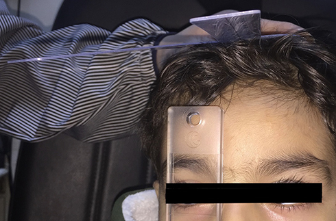 |
Figure 1 Measurement of right gaze deviation at 30 degree using hand-held orthopedic goniometer. |
Patients with inferior oblique overaction were measured with the same method after simultaneous neutralization of the vertical deviation using vertical base-up or base-down prisms according to the examined eye.
Preoperative lateral gaze measurements were done twice at different times by 2 different authors before surgery.
Change in lateral comitance was detected by difference in right and left gaze measurements and it was considered significant if the difference was more than 10 PD.
Simple random sampling was used where patient names were drawn from a pool where every patient had an even probability of being chosen for either asymmetrical or symmetrical procedure.
Surgical Procedure
All surgical procedures were performed under general anesthesia by the same surgeon (STA) using standard surgical tables for correction of esotropia and exotropia.7
Symmetrical surgery included bilateral medial rectus recession in esotropic patients or bilateral lateral rectus recession in exotropic patients.
Asymmetrical surgery was defined as recess-resect muscle surgery in the same eye or 3 muscle surgery (recess-resect in one eye and one muscle recession in the other eye).
Three-muscle surgery was done if the angle of deviation was more than 50 PD in exotropia and 60 PD in esotropia.7
Follow Up
All patients were examined postoperatively every week for the first month and then every 3 months until the end of follow up.
Patients were examined for evaluation of the angle of deviation in primary position and lateral (right and left) gazes using hand-held orthopedic goniometer, and for any postoperative complications.
Postoperative alignment less or equal to 10 PD was considered a successful result, while under-correction and overcorrection were considered with postoperative angles more than 10 PD.
Statistical Method
The collected data were coded, tabulated, and statistically analyzed using Statistical Package for Social Sciences program (SPSS) (software version 25; SPSS Inc., IBM Corp., NY, USA, 2017).
Descriptive statistics were done for parametric quantitative data by mean ± standard deviation and for non-parametric quantitative data by median and interquartile range (IQR), while they were done for categorical data by number and percentage.
Distribution of the data was done by Shapiro–Wilk test.
Analyses were done for non-parametric quantitative data using Mann–Whitney test between the 2 groups and Wilcoxon signed rank test between different times within the same group.
Analyses were done for qualitative data using Chi Square test (expected number per cell > 5) and Fisher’s exact test (expected number per cell < 5).
The level of significance was taken at P value < 0.05.
Results
Forty patients were enrolled in our study, with their demographic data presented in Table 1.
 |
Table 1 Demographic Data of the Study Patients |
Six esotropic patients (24%) were partially accommodative and 19 patients (76%) were non-accommodative. Three patients with esotropia (12%) had inferior oblique over-action ranged between +3 to +4 without any alphabetical pattern detected in up or down gaze, and they underwent inferior oblique myectomy with the horizontal procedure.
All exotropic patients had constant alternating exotropia except only one patient who had basic intermittent exotropia (Table 1).
All our patients had normal accommodation convergence/accommodation (AC/A) ratio.
Fundus examination was normal in all patients.
Patients were divided according to the type of deviation into esotropia and exotropia surgery groups.
Twenty-five patients who had esotropia surgery were subdivided into 2 subgroups:
Group I: included 10 patients who had asymmetrical procedure for esotropia.
Group II: included 15 patients who had symmetrical procedure for esotropia.
Fifteen patients who had exotropia surgery also were subdivided into 2 subgroups:
Group III: included 10 patients who had asymmetrical procedure for exotropia.
Group IV: included 5 patients who had symmetrical procedure for exotropia.
In esotropic patients who underwent asymmetrical surgery, the mean recession of medial rectus was 6.36±1.02 mm (range: 5–7 mm) while the mean resection of the lateral rectus was 8.6±1.64 mm (range: 6–10 mm). In exotropic patients, the mean recession of lateral rectus was 10±0 mm and the mean resection of medial rectus was 7±0 mm.
In esotropic patients who underwent symmetrical surgery, the mean recession of medial rectus was 6.83±0.04 mm (range: 6–7 mm) while in patients with exotropia, the mean of lateral rectus recession was 8.4±1.26 mm (range: 7–10 mm).
In partially accommodative esotropia patients (3 in each esotropia subgroup), the target angle for surgery was the residual angle with glasses (range: 30–50 PD). The angle of deviation without glass was higher (range: 65–70 PD).
In asymmetrical esotropia patients, the ratio between right/left eyes was 3/6 and one patient had surgery in 3 muscles (2 muscles in the right eye and the third in the left eye).
In asymmetrical exotropia procedure, 6 patients had their surgery in the right eye and the remaining 4 patients had surgery in 3 muscles (2 muscles in the right eye and the third in the left eye).
Mean postoperative follow-up was 16.4±2.2 months (range: 12–18 months).
As Regards Esotropia Subgroups I and II
Mean preoperative angle of deviation in group II (62±8.4) was higher than group I (49.5±11.7) which was statistically significant (P=0.011).
There was a statistically insignificant difference in the success rate (P value = 0.8057) between asymmetrical (subgroup I) and symmetrical (subgroup II) procedures (90% and 86.67%, respectively) at the end of follow up period.
As regards postoperative alignment in primary position, there was a statistically insignificant difference between the 2 subgroups after 1 month (P = 0.479), 6 months (P = 0.111), and 12 months (P = 0.065) (Table 2).
 |
Table 2 Primary Angle of Deviation in Esotropia Subgroup I and II Before and After Surgery |
One patient (10%) in asymmetrical esotropia subgroup had consecutive exotropia 30 PD and her parents refused additional surgery. Two patients (13.33%) in symmetrical esotropia subgroup had residual deviation. One patient had residual esotropia 20 PD with +2 refraction and he was satisfied with his correction while the other patient had residual esotropia 45 PD and her spherical equivalent was −10.5D in right eye and −11.5D in left eye (esotropia with myopia) and she refused additional surgery for residual deviation.
The mean of postoperative gaze difference in the asymmetrical subgroup was 1.8±3.7 compared with 0±0 in the symmetrical subgroup at 1 month which was a statistically significant difference (P = 0.009) which became insignificant at 6 and 12 months (P = 0.077) (Table 3).
 |
Table 3 Gaze Difference in Esotropia Subgroup I and II Before and After Surgery |
As Regards Exotropia Subgroups III and VI
Mean preoperative angle of deviation in group III (64±10.2) was higher than group IV (38±7.6) which was statistically significant (P=0.002).
Success rate was 100% in all exotropia patients either in group III or VI.
Mean of postoperative alignment in primary position was 1.9±0.3 in group III and 1.4±0.5 in group IV after 1 month with a statistically significant difference (P = 0.046) which became insignificant at 6 and 12 months (P = 0.708 and 0.690, respectively) (Table 4).
 |
Table 4 Primary Angle of Deviation in Exotropia Subgroup III and IV Before and After Surgery |
The mean of postoperative lateral gaze difference in group III and VI was 0±0 which was statistically insignificant at 1, 6, and 12 months (P =1) (Table 5).
 |
Table 5 Gaze Difference in Exotropia Subgroup III and IV Before and After Surgery |
Postoperative ductions and versions were within normal range.
None of our patients had postoperative diplopia in primary or side gaze as they were young with subsequent suppression.
There were no serious complications encountered in this study apart from under-correction and overcorrection.
Discussion
This study was designed mainly to detect the effect of asymmetrical surgery on lateral gaze and if there was a significant lateral gaze incomitance or not.
There is controversy about the postoperative lateral gaze incomitance induced by asymmetrical surgeries and its effect on the side gaze. Wright pointed out that the recess-resect procedure in treatment of exotropia can produce lateral incomitance with significant esotropia on the operated side with subsequent diplopia in lateral gaze that may persist for a long period.8 On the other hand, Kushner reported that lateral incomitance had little significance in the recess-resect procedure.9
Lateral gaze incomitance can produce distressing double vision in side gazes. Many authors have used unilateral muscle surgery to correct comitant esotropia or exotropia as regards the efficacy, but few considered lateral gaze incomitance or even measure it by mean of prism diopter.
Pollard and Manley reported that 20% of patients who underwent unilateral medial rectus muscle recession for small angle esotropia developed lateral incomitance in the direction of gaze toward the operated muscle.10 Similar lateral incomitance toward the operated muscle was observed by others but it was not measured in prism diopters.11–14
Others have reported no or mild abduction limitation after unilateral lateral rectus muscle recessions and it was reduced with time.13,15,16
There were few studies discussing postoperative lateral gaze incomitance in both symmetrical and asymmetrical surgeries.2 This study was conducted to detect the effectiveness of asymmetrical horizontal strabismus surgery and if it produced lateral gaze incomitance comparing it with symmetrical surgery.
As Regards the Postoperative Alignment and Efficacy
In the current study, asymmetrical surgery had an almost similar success rate to symmetrical surgery (90 and 86.67%, respectively) in esotropia patients. This was similar to the results of Polling et al. as they detected no difference between bilateral medial rectus recession and recess-resect in postoperative alignment in infantile esotropia.17
In contrast to our results, Kim and Choi in their study found that bilateral medial rectus recession had a higher significant success (80.70% compared with 56.52% in recess-resect) and a lower re-operation rate than recess-resect procedure.18
There were several studies about the efficacy of symmetrical and asymmetrical surgeries in exotropia especially in the intermittent type because of variable and unstable postoperative alignment.19–22
Successful alignment was achieved in all exotropia patients in our study in either asymmetrical or symmetrical subgroups even with the large angle of deviation in the asymmetrical subgroup (mean 64±10.2).
Kim et al. reported that successful alignment was similar between symmetrical and asymmetrical groups (P = 0.070) in patients with an exotropia of 40 PD but in patients with an exotropia ≥ 45 PD, asymmetrical group had successful alignment three-fold more than the symmetrical group at the end of the follow-up period (P = 0.006).19
Choi et al. reported different results from our study as they had higher success rates after bilateral lateral rectus recession compared with unilateral recess/resect procedure at the end of postoperative follow-up.20
Jeoung et al. reported that unilateral recess/resect procedure resulted in a better outcome than bilateral lateral rectus recession in exotropes with a dominant eye.21
Lee et al. found that the success rate in the bilateral lateral rectus recession group was double the success rate in the unilateral lateral rectus recession-medial rectus plication group (P = 0.021) in patients with basic intermittent exotropia.22
As Regards Postoperative Lateral Incomitance
Significant postoperative lateral incomitance was observed towards the opposite gaze of the operated eye in asymmetrical esotropia subgroup in 1 esotropic patient (out of 10) which decreased with follow-up time.
Lateral incomitance was not reported in the asymmetrical or symmetrical subgroups of exotropia patients.
So, even the lateral gaze incomitance occurred after asymmetrical surgery, it did not persist and became less distressing with time.
Although Cifuentes et al. reported a small percentage of postoperative induced lateral incomitance similar to our study, it was in exotropic patients not in esotropic (one patient out of 26). Their success rate was 100% in esotropia patients and slightly less in the exotropia group (84 and 82% at near and distance, respectively).23
These results were different from Deacon et al. who detected significant and persistent lateral incomitance in their case series study with greater effect in gaze towards the operated eye and 3 of their patients had gaze diplopia toward the operated eye. Deacon et al. explained this by a high dose of surgery needed to correct large deviations in these patients. This different result could be explained by the different surgeons who performed the surgical techniques, and they did not compare it with symmetrical procedure.24
Graeber and Hunter also detected that asymmetric surgery produced lateral incomitance. 12% of their comitant strabismus patients developed postoperative gaze incomitance with about 90% of them having had asymmetrical surgery. About one-third of their patients complained of lateral gaze intolerable results as they were adults. Their surgical techniques were performed by different surgeons using different surgical techniques. Also, there was a larger change in comitance noted in their patients who had surgery on the oblique muscles and the vertical rectus muscles at the same time of horizontal surgery and they explained this finding by the tertiary actions of these muscles which was different from our cases that had inferior oblique myectomy with horizontal muscle surgery.2
Yoon and Kim reported significant postoperative lateral incomitance in the patients who had unilateral lateral rectus recession in comparison to the patients who had symmetrical bilateral lateral rectus recession (P = 0.006).25
Few studies discussed the lateral gaze incomitance after horizontal strabismus surgery and only Cifuentes et al.23 had similar results to this study as regards the low incidence of lateral incomitance but in a different type of horizontal strabismus. Differences in the current study from the others may be explained by average amounts of surgical muscle dosage and all the surgical procedures were done by the same surgeon following the same surgical technique in all study groups either recession or resection.
There were some limitations in our study; first the sample size, so a larger sample size is recommended to confirm our results, and second the need for a longer period of follow up.
Finally, surgeons should assess the preoperative deviation in right and left gazes and consider the results not only in the primary position but also in these gazes before deciding to do symmetrical or asymmetrical procedures.
Conclusion
Asymmetrical horizontal strabismus surgery is an effective and alternative procedure to symmetrical surgery in both esotropia and exotropia patients without persistent postoperative lateral gaze incomitance or limitation.
Data Sharing Statement
Data are available upon request.
Ethics
Approval was obtained from the Research Ethics Committee of Faculty of Medicine, Minia University (NO: 69-7/2018). All procedures performed in studies involving human participants were in accordance with the ethical standards of the institutional and/or national research committee and with the 1964 Helsinki Declaration and its later amendments or comparable ethical standards.
Informed Consent
Informed written consent was obtained from all patients or their legal guardians included in the study.
Acknowledgment
This paper was uploaded to the Minia University, University repository as a Master thesis in 2019.26
Author Contributions
All authors made a significant contribution to the work reported, whether that is in the conception, study design, execution, acquisition of data, analysis and interpretation, or in all these areas; took part in drafting, revising or critically reviewing the article; gave final approval of the version to be published; have agreed on the journal to which the article has been submitted; and agreed to be accountable for all aspects of the work.
Funding
No financial support was received for this submission.
Disclosure
The authors reported no conflicts of interest in this work.
References
1. Wright KW, Strube YNJ. Principles of strabismus surgery. In: Color Atlas of Strabismus Surgery (Strategies and Techniques). Farzavandi S, editor. Los Angeles, USA: Springer; 2015:7–12.
2. Graeber CP, Hunter DG. Changes in lateral comitance after asymmetric horizontal strabismus surgery. JAMA Ophthalmol. 2015;133(11):1241–1246.
3. Moore S. The prognostic value of lateral gaze measurements in intermittent exotropia. Am Orthopt J. 1969;19(1):69–71.
4. Parks MM. Concomitant exodeviations. In: Duane TD, editor. Clinical Ophthalmology, Ocular Motility and Strabismus. Hagerstown, MD: Harper & Row; 1975:113.
5. Von Noorden GK. Exodeviations. In: Binocular Vision and Ocular Motility: Theory and Management of Strabismus.
6. Burke MJ. Intermittent exotropia. Int Ophthalmol Clin. 1985;25:53.
7. Coats DK, Olitsky SE. Surgical decision making. In: Strabismus Surgery and Its Complications. Philipp M, Himberger M, editor. Berlin Heidelberg: Springer-Verlag; 2007:37–39.
8. Wright KW. Exotropia. In: Wright KW, Spiegel PH 2nd, editors. Pediatric Ophthalmology and Strabismus. New York: Springer-Verlag; 2003:224–231.
9. Kushner BJ. Exotropic deviations: a functional classification and approach to treatment. Am Orthopt J. 1988;38:81–93.
10. Pollard ZF, Manley D. Unilateral medial rectus recession for small-angle esotropia. Arch Ophthalmol. 1976;94(5):780–781.
11. Stack RR, Burley CD, Bedggood A, Elder MJ. Unilateral versus bilateral medial rectus recession. J AAPOS. 2003;7(4):263–267.
12. Weakley DR
13. Feretis D, Mela E, Vasilopoulos G. Excessive single lateral rectus muscle recession in the treatment of intermittent exotropia. J Pediatr Ophthalmol Strabismus. 1990;27(6):315–316.
14. Menon V, Singla MA, Saxena R, Phulijele S. Comparative study of unilateral and bi-lateral surgery in moderate exotropia. J Pediatr Ophthalmol Strabismus. 2010;47(5):288–291.
15. Dadeya S, Kamlesh MS. Long-term results of unilateral rectus recession in intermittent exotropia. J Pediatr Ophthalmol Strabismus. 2003;40:283–287.
16. Deutsch JA, Nelson LB, Sheppard RW, Burke MJ. Unilateral lateral rectus recession for the treatment of exotropia. Ann Ophthalmol. 1992;24:111–113.
17. Polling JR, Eijkemans MJ, Esser J, et al. A randomised comparison of bilateral recession versus unilateral recession–resection as surgery for infantile esotropia. Br J Ophthalmol. 2009;93(7):954–957.
18. Kim E, Choi DG. Comparison of surgical outcomes between bilateral medial rectus recession and unilateral recess-resect for infantile esotropia. Ophthalmic Epidemiol. 2019;26(2):102–108.
19. Kim KE, Yang HK, Hwang JM. Comparison of long-term surgical outcomes of 2-muscle surgery in children with large angle exotropia: bilateral versus unilateral. Am J Ophthalmol. 2014;157(6):1214–1220.
20. Choi J, Chang JW, Kim SJ, Yu YS. The long-term survival analysis of bilateral lateral rectus recession versus unilateral recession-resection for intermittent exotropia. Am J Ophthalmol. 2012;153(2):343–351.
21. Jeoung JW, Lee MJ, Hwang JM. Bilateral lateral rectus recession versus unilateral recess-resect procedure for exotropia with a dominant eye. Am J Ophthalmol. 2006;141(4):683–688.
22. Lee HJ, Kim SJ, Yu YS. Long-term outcomes of bilateral lateral rectus recession versus unilateral lateral rectus recession-medial rectus plication in children with basic type intermittent exotropia. Eye (Lond). 2019;33(9):1402–1410.
23. Cifuentes DL, Pineles SL, Demer JL, Velez FG. Surgical success and lateral incomitance following three-muscle surgery for large-angle horizontal strabismus. JAAPOS. 2018;22(1):17–21.
24. Deacon BS, Fray KJ, Grigorian AP, et al. Unilateral strabismus surgery in patients with exotropia results in postoperative lateral incomitance. J AAPOS. 2014;18(6):572–575.
25. Yoon CH, Kim SJ. Lateral incomitancy and surgical results in intermittent exotropia. Br J Ophthalmol. 2014;98:1404–1408.
26. Ismail AA, Abdelkader MF, Mohamed AA, Abdelaziz ST Assessment of Symmetrical and Asymmetrical Horizontal Strabismus Surgery: Efficacy and Lateral Incomitance, 2019 [dissertation]. Faculty of Medicine- Minia University; 2019.
 © 2021 The Author(s). This work is published and licensed by Dove Medical Press Limited. The full terms of this license are available at https://www.dovepress.com/terms.php and incorporate the Creative Commons Attribution - Non Commercial (unported, v3.0) License.
By accessing the work you hereby accept the Terms. Non-commercial uses of the work are permitted without any further permission from Dove Medical Press Limited, provided the work is properly attributed. For permission for commercial use of this work, please see paragraphs 4.2 and 5 of our Terms.
© 2021 The Author(s). This work is published and licensed by Dove Medical Press Limited. The full terms of this license are available at https://www.dovepress.com/terms.php and incorporate the Creative Commons Attribution - Non Commercial (unported, v3.0) License.
By accessing the work you hereby accept the Terms. Non-commercial uses of the work are permitted without any further permission from Dove Medical Press Limited, provided the work is properly attributed. For permission for commercial use of this work, please see paragraphs 4.2 and 5 of our Terms.