")
Back to Journals » Veterinary Medicine: Research and Reports » Volume 14
Eukaryotic Infections in Dairy Calves: Impacts, Diagnosis, and Strategies for Prevention and Control
Authors Robi DT , Mossie T, Temteme S
Received 27 September 2023
Accepted for publication 27 November 2023
Published 1 December 2023 Volume 2023:14 Pages 195—208
DOI https://doi.org/10.2147/VMRR.S442374
Checked for plagiarism Yes
Review by Single anonymous peer review
Peer reviewer comments 2
Editor who approved publication: Professor Young Lyoo
Dereje Tulu Robi,1 Tesfa Mossie,2 Shiferaw Temteme1
1Ethiopian Institute of Agricultural Research, Tepi Agricultural Research Center, Tepi, Ethiopia; 2Ethiopian Institute of Agriculture Research, Jimma Agriculture Research Center, Jimma, Ethiopia
Correspondence: Dereje Tulu Robi, Email [email protected]
Abstract: Eukaryotic infections are common among dairy calves and can have significant impacts on their health and growth rates. Fungal infections caused by Aspergillus fumigatus, Trichophyton verrucosum, and Candida albicans can cause respiratory diseases, dermatophytosis, and diarrhea, respectively. Protozoan parasites, including Cryptosporidium parvum, Giardia duodenalis, and Eimeria spp., are also common in dairy calves. C. parvum is highly contagious and can cause severe diarrhea and dehydration, while Giardia duodenalis can lead to poor growth and is transmissible to humans through contaminated food or water. Eimeria spp. can cause coccidiosis and lead to reduced growth rates, poor feed conversion, and death. The common helminthic infections in dairy calves include Ostertagia ostertagi, Cooperia spp., Fasciola hepatica, and Strongyloides papillosus. These parasitic infections significantly impact calf health, growth, and dairy industry productivity. Diagnosis of these infections can be made through fecal samples using microscopy or molecular methods. However, diagnosis of the infections can be challenging and requires a combination of clinical signs and laboratory tests such as culture and PCR. Preventing and controlling eukaryotic infections in dairy calves requires several measures. Good hygiene and sanitation practices, proper management strategies, and timely treatment of affected animals are important. It is also necessary to avoid overcrowding and consider vaccination against ringworm. Further research is needed to better understand the epidemiology and characterization of eukaryotic infections in dairy calves, which will help in the development of more effective prevention and control strategies. In general, good hygiene practices, appropriate management strategies, and timely treatment of affected animals are crucial in preventing and controlling the infections, ensuring the health and well-being of dairy calves.
Keywords: protozoa, fungus, helminths, diagnosis methods, prevention strategies, dairy calves
Introduction
Dairy farming is an important sector of agriculture industry, providing a significant source of food and income worldwide. However, infectious diseases pose a major threat to the health and productivity of dairy calves.1,2 Eukaryotic infections caused by protozoan parasites, helminths, and fungi are common problems among dairy calves. The term “eukaryotic infections” encompasses a wide array of pathogens that belong to the domain Eukarya, including protozoa, fungi, and helminths.3 Unlike bacteria or viruses, these microorganisms possess complex cellular structures with membrane-bound organelles, reflecting a higher level of biological organization.4 These pathogens can affect different parts of the body, including the digestive system, respiratory system, and urogenital system. Therefore, it is crucial to understand the nature of infections and their management strategies to ensure the welfare of dairy calves and the profitability of dairy farming.5,6
Protozoan and fungal infections are significant concerns in the health of dairy calves, representing major eukaryotic health problems. These infections can lead to substantial economic losses attributed to decreased milk production, treatment costs, and mortality.3,7 Moreover, some of these infections can also be transmitted to humans, posing a public health concern.8 In addition to protozoa and fungi, helminthic infections, such as roundworms and tapeworms, have also been identified as critical contributors to the health burden of dairy calves.9 These parasites can lead to gastrointestinal disturbances, decreased nutrient absorption, and anemia, thereby impeding calf growth and performance.10,11 Several studies have reported the prevalence of the infections in dairy calves worldwide. A study conducted by12 and3 in Ethiopia reported a prevalence of 20.1% for coccidiosis and 13.8% for cryptosporidiosis, respectively, in dairy calves. Similarly, a study by13 in Bangladesh reported a prevalence of 55.6% for cryptosporidiosis in dairy calves.
The diagnosis of eukaryotic infections in dairy calves poses unique challenges due to the diversity of pathogens and their varied clinical presentations. Traditional diagnostic methods, such as microscopic examination of fecal samples and other, are often labor-intensive and lack sensitivity and specificity.4 However, advancements in molecular techniques, such as polymerase chain reaction (PCR) and next-generation sequencing (NGS), have revolutionized the identification and characterization of eukaryotic pathogens. These methods enable precise detection, differentiation, and quantification of various pathogens, enhancing our ability to diagnose infections accurately.14,15
Effective management of eukaryotic infections in dairy calves involves a combination of preventive measures, early diagnosis, and appropriate treatment.16,17 Preventive measures include maintaining good hygiene and sanitation practices, such as using clean and dry bedding, and implementing biosecurity measures. Early diagnosis can be achieved through regular monitoring and diagnostic testing, while appropriate treatment may involve the use of antiparasitic drugs or antifungal agents.18 Therefore, the aim of this review is to provide an overview of the common eukaryotic infections affecting dairy calves, their effects on the animals, and the current strategies used for their management. We also highlight recent advancements in the diagnosis and treatment of the infections, as well as areas for future research. Understanding these pathogens and employing management strategies is essential to prevent and control the diseases in dairy calves.
Eukaryotic Infections in Dairy Calves
Fungal Infections
Aspergillus fumigatus (A. fumigatus) is a ubiquitous fungus found in soil, decaying vegetation, and other organic matter.19 It is also known to colonize the respiratory tract of animals, including dairy calves. Infection with A. fumigatus in dairy calves can result in a variety of clinical signs, including coughing, fever, and decreased appetite. In severe cases, it can lead to pneumonia, respiratory distress, and death.5
The transmission of A. fumigatus to dairy calves occurs through contaminated feed or bedding material, and environmental conditions such as high humidity and poor ventilation can facilitate its growth and spread.20 Calves that are immune compromised or stressed due to factors such as transport or weaning may be more susceptible to infection. In dairy calves, A. fumigatus can cause a range of respiratory diseases, including pneumonia, bronchopneumonia, and aspergillosis. Clinical signs of Aspergillus-related respiratory disease in calves include coughing, labored breathing, and fever.21 In severe cases, the disease can progress rapidly and lead to death.
A study conducted by22 investigated the prevalence of A. fumigatus in the lungs of dairy calves with respiratory distress. The study found that the infection was the most frequently isolated fungus from the lungs of affected calves. Moreover, the study identified an association between the presence of A. fumigatus and the severity of respiratory distress in the calves. Another study by23 investigated the pathogenicity of A. fumigatus in dairy calves. The study found that A. fumigatus was capable of causing severe lung lesions and mortality in experimentally infected calves. The study also identified several virulence factors that were involved in its pathogenicity, including the production of toxins and proteases.
Diagnosis of A. fumigatus infection in dairy calves can be challenging as clinical signs may be non-specific and other pathogens may also cause similar symptoms. However, a combination of clinical signs, radiographic findings, and laboratory tests such as culture and PCR help to confirm the presence of A. fumigatus.24 Treatment of infection in dairy calves typically involves the use of antifungal medications such as itraconazole or voriconazole (Table 1). However, prevention and control measures such as improving ventilation, reducing humidity, timely treatment of affected animals and avoiding contaminated feed and bedding material can reduce the risk of infections in dairy calves.21,25,26
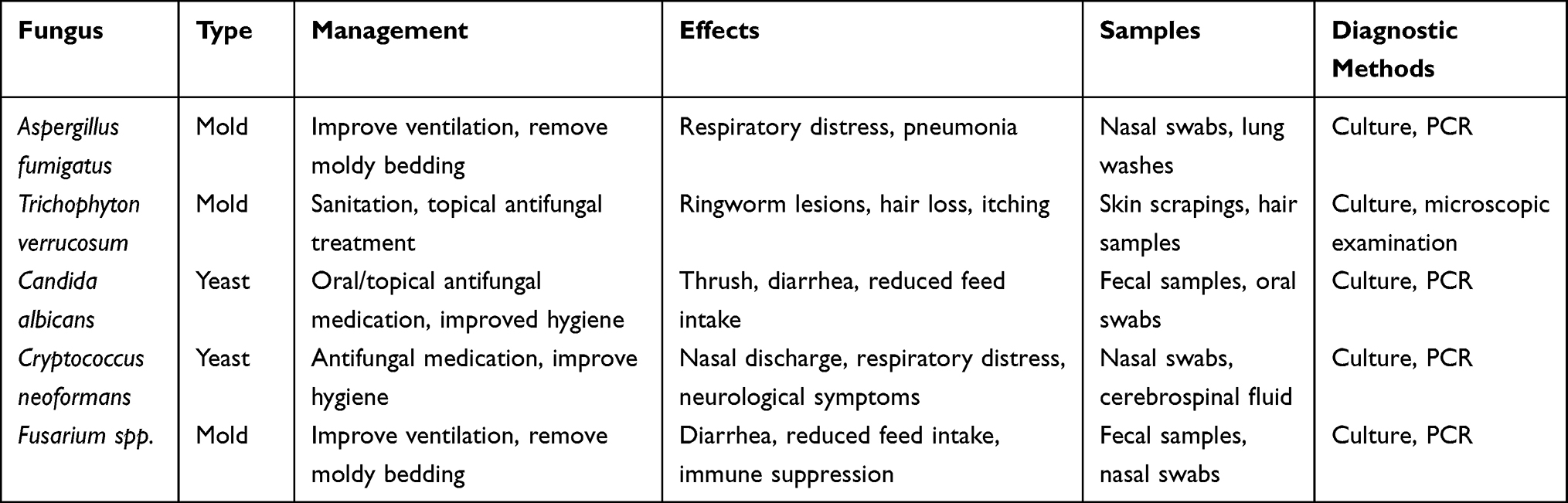 |
Table 1 Fungal Types and Their Management, Effects, Diagnostic Samples, and Methods |
Trichophyton verrucosum (T. verrucosum) is a dermatophyte fungus that commonly infects dairy calves, causing a skin disease known as bovine dermatophytosis or ringworm. This fungal infection is highly contagious and can spread rapidly between calves, leading to significant economic losses in the dairy industry. It is a zoonotic pathogen that can also infect humans who come in contact with infected animals or contaminated surfaces.27–29 T. verrucosum is primarily found in humid and cold environments, making it more prevalent in northern regions. Calves between 1–6 months of age are most susceptible to infection, as their immune systems are not fully developed. The fungus can be transmitted through direct contact with infected animals or through contaminated environments, such as bedding or equipment.29–31
The clinical signs of bovine dermatophytosis include circular patches of hair loss, scab formation, and crusty lesions on the skin of affected calves. The lesions occur on any part of the body but are most commonly seen on the head, neck, and shoulders. Infected calves may also experience itching and discomfort, leading to reduced feed intake and weight loss.32,33 According to a study by34 and,35 T. verrucosum was identified as the most common dermatophyte isolated from dairy calves with ringworm infections in Brazil and Italy, respectively. The study also found that the prevalence of ringworm infection was higher in calves that were not treated with fungicides and in farms with poor hygienic practices. This suggests that proper management practices and regular treatment with fungicides can help prevent and control T. verrucosum infections in dairy calves.
The diagnosis of bovine dermatophytosis is usually based on clinical signs and confirmed by fungal culture or microscopic examination of skin scrapings.31 Diagnosis of the disease in dairy calves can be done through a combination of clinical signs, fungal culture, and microscopic examination.34 Fungal culture can confirm the presence of etiological agent of the disease, while microscopic examination can identify the fungal structure and aid in species identification.27,29
Treatment options for the disease include topical antifungal agents such as miconazole or clotrimazole, as well as other systemic antifungal drugs. Implementing good hygienic practices and avoiding overcrowding of calves are the key prevention measures of the disease.36,37 Vaccination against ringworm is also available in some countries and can be a useful preventive measure.28 Prevention of T. verrucosum infection in dairy calves is essential to control the spread of the disease (Table 1).
Candida albicans (C. albicans) is a fungal pathogen that can cause infections in various animal species, including dairy calves. In dairy farming, C. albicans infections in calves have been associated with a range of clinical signs, including diarrhea, reduced appetite, and poor growth rates.38,39 C. albicans is a type of yeast that commonly colonizes the mucosal surfaces of mammals, including dairy calves. In healthy animals, C. albicans is typically present in low numbers and does not cause disease. However, in certain circumstances, when the animal’s immune system is compromised, C. albicans can cause a range of infections, including oral thrush, skin infections, and systemic infections.40,41 The pathogenesis of the C. albicans infection in dairy calves involves colonization of gastrointestinal tracts particularly the rumen and abomasum even if not fully understood. Then it causes inflammation and tissue damage. In some cases, it can also spread to other organs, such as the liver and lungs, causing systemic infections in dairy calves.38
Several risk factors have been identified for C. albicans infections in dairy calves, including poor hygiene, inadequate nutrition, and stress.38 Furthermore, the infections can be more common in calves that are housed in crowded or unsanitary conditions. Diagnosis of the infection in dairy calves can be challenging, as it requires the identification of the organism in clinical samples such as feces, oral swabs, or blood cultures (Table 1). Diagnosis can be done through culture and PCR testing.42 Treatment of C. albicans infections in calves typically involves the use of antifungal drugs, such as fluconazole and nystatin.42 Prevention of the infections in dairy calves involves good management practices, providing adequate nutrition and hydration, and minimizing stress.43,44
Cryptococcus neoformans (C. neoformans) is a fungal pathogen that can cause serious disease in both humans and animals. C. neoformans is a yeast-like fungus that is found worldwide in soil, bird droppings, and other organic matter.45,46 This pathogen can cause disease in immune compromised hosts. In dairy calves, the fungus can cause cryptococcosis, which is characterized by respiratory symptoms, neurological problems, and other clinical signs.47 The exact mode of transmission of C. neoformans in dairy calves is not fully understood, but it is thought to occur via inhalation of fungal spores from contaminated feed or bedding. Furthermore, immunocompromised calves are at higher risk of developing cryptococcosis, as their weakened immune systems make them more susceptible to infection.48,49
Clinical signs of cryptococcosis in dairy calves can vary, but commonly include respiratory symptoms such as coughing and dyspnea, as well as neurological symptoms like ataxia and seizures.50 In some cases, the disease can also cause skin lesions and ophthalmic problems. Diagnosis of cryptococcosis in dairy calves is typically made through a combination of clinical signs, laboratory testing, and imaging studies.47 The diagnosis of the disease in dairy calves is challenging since clinical signs are often non-specific and overlap with other diseases. Therefore, A combination of clinical examination, laboratory tests and histopathological analysis are required for definitive diagnosis.45
Treatment of C. neoformans in dairy calves usually involves a combination of antifungal drugs and supportive care. Fluconazole is the most commonly used antifungal medication, and has been shown to be effective in treating cryptococcosis in calves.18 However, successful treatment also depends on early diagnosis and prompt initiation of therapy. Prevention of C. neoformans in dairy calves involves maintaining good hygiene practices, including regular cleaning and disinfection of feed and bedding areas (Table 1). Furthermore, it is important to monitor calf health and implement appropriate measures to manage immunocompromised individuals.17,26
Fusarium is of filamentous fungi commonly found in soil and plant debris. The genus Fusarium fungus contains economically important species that cause a wide range of health problems in calves, including respiratory and gastrointestinal disease.51 The fungus has been identified as major cause of mycotoxicosis in dairy calves which leads to reduce growth rate, feed intake and diarrhea. In severe cases, mycotoxicosis can even lead to death.52 The mycotoxins produced by Fusarium are trichothecenes, zearalenone, and fumonisins. These toxins can contaminate feed and forage, leading to ingestion by dairy calves.53,54 The toxins damage the intestinal lining leading to inflammation and reduced nutrient absorption following ingestion of toxins. Furthermore, the toxins disrupt the immune system, leaving the calf vulnerable to other infections.55,56
One of the most common Fusarium species in dairy calves is Fusarium verticillioides. This fungus produces fumonisin mycotoxins, which can cause a range of health problems, including neurological disorders, liver and kidney damage, and reduced growth rates.57 Another Fusarium species that can infect dairy calves is Fusarium graminearum, which produces deoxynivalenol (DON) mycotoxins. It is a potent inhibitor of protein synthesis and can cause feed refusal, vomiting, and diarrhea in dairy calves. DON can cause vomiting, diarrhea, and reduced feed intake in calves.58,59 In addition to causing mycotoxicosis, Fusarium spp. can also cause systemic infections in dairy calves. Fusarium solani is known to cause pneumonia in calves. Fusarium oxysporum can cause disseminated infections in calves with compromised immune systems.55
Preventing Fusarium infection in dairy calves can be challenging, as the fungi are ubiquitous in the environment.60 However, several strategies can be employed to reduce the risk of infection. These include ensuring proper ventilation and hygiene in calf housing facilities, using clean and high-quality feed, and avoiding the use of contaminated bedding (Table 1). Moreover, proper storage and handling of feed can help to minimize mycotoxin exposure.38 To prevent Fusarium-related mycotoxicosis in dairy calves, it is important to implement measures such as proper feed storage and handling, routine mycotoxin testing of feed, and the use of mycotoxin binders in feed. These strategies can help reduce the risk of Fusarium contamination and minimize the negative impact on calf health.13
Protozoa Infections
Protozoa are single-celled organisms that cause various health problems in humans and animals. Cryptosporidium parvum, Giardia duodenalis, and Eimeria species are major types of protozoa parasites (Table 2).
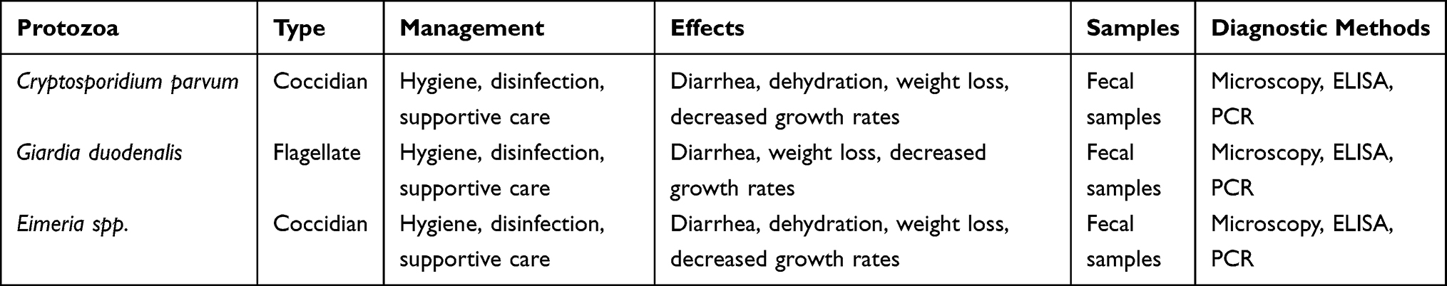 |
Table 2 Protozoa Types and Their Management, Effects, Diagnostic Samples, and Methods |
Cryptosporidium parvum (C. parvum) is a protozoan parasite that commonly infects dairy calves, causing diarrhea and other gastrointestinal symptoms.61 The C. parvum is highly contagious and transmitted through direct contact with infected animals, as well as through contaminated water and feed.62,63 Several studies have investigated the prevalence and impact of C. parvum in dairy calves. The prevalence of C. parvum was range from 6.3% to 39.7%64 with infection rates highest in calves between one and three weeks of age. Another study by65 found that C. parvum infection in dairy calves was associated with reduced weight gain and increased mortality.
C. parvum is ubiquitous in the environment and can survive for long periods in moist and cool conditions. The main source of infection for dairy calves is contaminated water, feed, or bedding material.66 The parasite has a complex life cycle, with both asexual and sexual stages occurring in the host’s intestinal tract. Infected cows can shed millions of oocysts (the infective stage of the parasite) in their feces, which can contaminate the environment and spread the infection to susceptible calves.63 Once ingested, C. parvum oocysts release sporozoites that invade the intestinal epithelium, causing damage and inflammation. This leads to malabsorption, maldigestion, and osmotic diarrhea, which can result in severe dehydration and electrolyte imbalances.67,68 The severity of the disease depends on various factors, such as the calf’s age, immune status, nutritional status, and concurrent infections.69
Clinical signs of C. parvum infection in dairy calves include watery diarrhea, dehydration, weight loss, and lethargy. In severe cases, infection can result in death.63 The diagnosis of cryptosporidiosis in dairy calves can be challenging, as the clinical signs can be similar to those of other gastrointestinal diseases. However, several diagnostic methods are available, including fecal examination, antigen detection assays, and molecular techniques, as described in a review by70 and.68
The treatment of the infection in dairy calves involves rehydration, electrolyte therapy, and supportive care. Antimicrobial agents, such as halofuginone and nitazoxanide, are also effective in reducing the severity and duration of diarrhea.71 However, the emergence of drug-resistant strains of C. parvum has limited the efficacy of the drugs in some cases. Prevention and control of the infection in dairy calves involve proper hygiene and sanitation, good management practices, and vaccination.72,73 Vaccination against C. parvum can reduce the severity and incidence of the disease and has been shown to be effective in field trials.74,75
Giardia duodenalis (G. duodenalis), also known as Giardia intestinalis or Giardia lamblia, is a protozoan parasite that infects the small intestine of various animals, including dairy calves. It is a significant cause of diarrhea and poor growth in young calves, leading to economic losses in the dairy industry.76 In addition to causing disease in dairy calves, G. duodenalis can also be transmitted to humans through contaminated food or water. This zoonotic potential highlights the importance of effective control measures to prevent transmission of the parasite between animals and humans.77
The transmission of G. duodenalis in dairy calves occurs through the ingestion of cysts, which are the parasite’s infective stage, shed in the feces of infected animals. The transmission of G. duodenalis in dairy calves occurs through the ingestion of cysts, which are the parasite’s infective stage, and are shed in the feces of infected animals. The cysts can survive for prolonged periods in the environment, particularly in damp and cool conditions, making it challenging to control the spread of infection.78 Once ingested, the cysts release trophozoites, the active form of the parasite, which attach to the intestinal wall, causing damage to the intestinal villi and reducing nutrient absorption. This results in diarrhea, weight loss, and decreased feed efficiency, ultimately leading to decreased milk production and potential mortality in severe cases.76 The infection can also cause damage to the intestinal lining, leading to malabsorption of nutrients and impaired growth in dairy calve.77
Several diagnostic methods are available to detect G. duodenalis infection in dairy calves, including fecal flotation, ELISA, and PCR. Diagnosis of Giardia infections in dairy calves can be challenging, as the parasite is not always detectable in fecal samples and may require multiple sampling and testing methods.79 According to a study by,80 microscopy and immunological assays are the most commonly used diagnostic methods, although PCR-based techniques are becoming increasingly popular due to their high sensitivity and specificity in calve.
Treatment of the infections in dairy calves typically involves the use of antiparasitic drugs such as metronidazole or fenbendazole, although resistance to these drugs has been reported in some regions (Siwila, 2017). Prevention of the infections in dairy calves requires good hygiene practices, such as regular cleaning and disinfection of feeding and watering equipment, as well as minimizing contact with contaminated environments and infected animals.77,81
The genus Eimeria is protozoan parasites that can cause coccidiosis in dairy calves. Coccidiosis is a common disease in young calves that can lead to reduced growth rates, poor feed conversion, and even death in severe cases.82,83 Eimeria are ubiquitous in the environment, and infection typically occurs through ingestion of oocysts shed in the feces of infected animals.84
There are several species of Eimeria that can infect dairy calves, including Eimeria (E. bovis), Eimeria (E. zuernii), Eimeria (E. auburnensis), and Eimeria) E. ellipsoidalis).13 These parasites can be found in the environment and are typically transmitted through fecal-oral contamination. Once ingested, Eimeria oocysts release sporozoites that invade the cells lining the intestinal wall in dairy calves. The parasites then reproduce asexually, causing damage to the intestinal lining and leading to diarrhea, dehydration, and weight loss.85 Clinical signs of coccidiosis in dairy calves include diarrhea, anorexia, lethargy, and dehydration. Each species has a unique pathogenesis and clinical presentation.86 E. bovis, for example, is associated with severe diarrhea and weight loss, while E. zuernii causes less severe clinical signs but can lead to more chronic infections.87 Eimeria spp infections are particularly common in young calves, as their immune systems are not yet fully developed, making them more susceptible to infection.13
Diagnosis of Eimeria spp infection in dairy calves can be challenging, as the clinical signs are nonspecific and can be caused by other gastrointestinal pathogens. However, fecal flotation and microscopic examination can reveal the presence of oocysts, which are the infective stage of the parasite. It is important to note that not all infected calves will shed oocysts in their feces, so a negative fecal test does not necessarily rule out infection. Diagnosis is typically made through more advanced diagnostic techniques such as PCR and ELISA.23
Preventing and controlling Eimeria spp infections in dairy calves is critical to maintaining calf health and reducing economic losses. Management practices, such as proper sanitation and hygiene, can help reduce the risk of Eimeria infection. Moreover, anticoccidial drugs can be used to treat and prevent Eimeria infections in dairy calves. However, the overuse of these drugs can lead to the development of drug-resistant strains of Eimeria, making it important to use them judiciously.59 Additionally, some producers may choose to use medicated feed or oral medications to help control the parasites.13
Helminthic Infections
Helminthic infections are a prevalent health concern in dairy calves, impacting growth, productivity, and overall herd management (Table 3). These infections are primarily caused by parasitic worms, including nematodes, trematodes and cestodes.88 Ostertagiasis is a gastrointestinal parasitic infection that predominantly affects young ruminants, including dairy calves. Ostertagiasis is caused by the nematode Ostertagia ostertagi, commonly affecting dairy calves.89 This disease poses significant challenges to the dairy industry due to its impact on calf health, growth, and overall productivity.90 Understanding the epidemiology, clinical signs, diagnosis, and management of Ostertagiasis is crucial for effective control and prevention.91
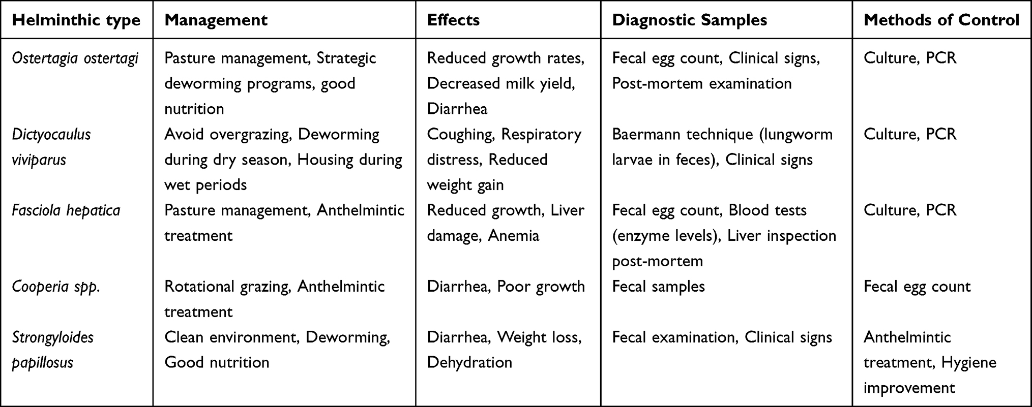 |
Table 3 Common Helminthic and Their Management, Effects, Diagnostic Samples, and Methods |
The disease is prevalent in temperate regions with humid climates, where the larvae of O. ostertagi can survive on pasture. Calves grazing on contaminated pastures are at high risk of infection.92 Calves raised in confinement systems may experience lower exposure compared to those on pasture. High stocking densities and poor manure management contribute to increased contamination of grazing areas.93 Ostertagia nematodes have a complex life cycle involving both direct and indirect transmission.94 Cattle become infected by ingesting infective larvae from contaminated pastures. Once ingested, these larvae penetrate the gastric glands, leading to the development of L4 larvae, which emerge into the abomasum, causing tissue damage and clinical symptoms.95 Clinical manifestations of Ostertagiasis in dairy calves include poor growth rates, weight loss, diarrhea, and suboptimal feed conversion efficiency. These signs are primarily attributed to the damage caused by the parasite’s larval migration and the subsequent inflammatory response in the abomasal lining.96
Accurate diagnosis of Ostertagiasis involves a combination of clinical signs, fecal examination, and laboratory techniques. Fecal egg count (FEC) is a commonly used diagnostic method to quantify parasite burden.97 Furthermore, the use of serological assays and polymerase chain reaction (PCR) techniques can enhance diagnostic accuracy.82,95,98 The management and control of the disease require a multi-faceted approach. Implementing strategic deworming protocols based on calf age, grazing history, and FEC results can help reduce parasite load.91 Pasture management practices such as rotational grazing, maintaining hygiene, and minimizing overcrowding can also mitigate the risk of infection. Furthermore, promoting calf immunity through proper nutrition and vaccination can aid in reducing the impact of the disease.99,100
The genus Cooperia are a group of small intestinal nematodes belonging to the family Trichostrongylidae. They are recognized as major contributors to the gastrointestinal parasite burden in dairy calves.101 The detrimental effects of Cooperia infections on calf health and productivity have made them a subject of intense research and management efforts.102 The infection has a direct lifecycle involving both free-living and parasitic stages. The infective third-stage larvae (L3) are ingested by calves while grazing, eventually migrating to the abomasum and small intestine.95 The larvae develop into adults that attach to the intestinal mucosa, leading to nutrient depletion, inflammation, and potential tissue damage. These physiological disruptions contribute to diarrhea, weight loss, and decreased growth rates in infected calves.103
Clinical signs of Cooperia infections vary in severity and can include diarrhea, dehydration, anemia, and poor body condition. Diagnostic methods encompass fecal egg counts (FEC), which quantify the parasite burden, as well as fecal culture and larval differentiation techniques to identify the specific Cooperia spp.97 Molecular methods, such as polymerase chain reaction (PCR), aid in species identification and differentiation.104,105 Cooperia infections are influenced by various factors, including geographical location, climate, management practices, and host immunity.106 Calves in group housing systems are particularly susceptible due to higher exposure levels. Environmental conditions, such as humidity and temperature, also play a crucial role in larval survival and infectivity. Accurate diagnosis is essential for effective management.107
Anthelmintic resistance has become a concern in Cooperia spp control. Rotation of different anthelmintic classes and the use of combination therapies are recommended to mitigate resistance development.105,108 Strategic deworming protocols, based on calf age, infection risk, and anthelmintic efficacy, are crucial for successful treatment. Integrated parasite management (IPM) strategies are essential to control Cooperia spp infections. These strategies involve a combination of measures, including strategic deworming, pasture management, nutrition optimization, and genetic selection for resistance.109,110 However, indiscriminate use of anthelmintics may lead to the development of drug-resistant strains, underscoring the need for judicious and targeted treatment.105,111 Recent research has shed light on various aspects of Cooperia spp infections, including host immunity, genetic resistance, and alternative control methods such as biological agents and plant-based treatments.112 Developing a deeper understanding of the host-parasite interaction and identifying novel control strategies are crucial for sustainable management of the infections in dairy calves.109
Dictyocaulus viviparus (D. viviparous), commonly known as the lungworm, is a parasitic nematode that affects cattle, particularly dairy calves. Lungworm infections impose substantial economic burdens on dairy producers due to reduced calf growth rates, increased veterinary costs, and decreased milk production in affected animals. Implementing effective control measures can mitigate these losses and improve overall herd health.113
The life cycle of D. viviparus involves both direct and indirect transmission.114 The primary host is the bovine, where the adult lungworms reside in the bronchi and bronchioles of the lungs. Female lungworms produce eggs that are coughed up by the host and excreted in the feces. These eggs hatch into first-stage larvae (L1) within the feces and develop into infective third-stage larvae (L3) over a period of several days.115,116 These L3 larvae of are then ingested by grazing calves during feeding, completing the indirect life cycle. Upon ingestion, L3 larvae penetrate the intestinal wall and migrate through the bloodstream to the lungs. This migration can cause a localized inflammatory response, leading to coughing, respiratory distress, and reduced feed intake. The presence of adult lungworms in the bronchi can further exacerbate the clinical signs and cause chronic respiratory issues.114
Lungworm infections commonly occur in grazing systems where calves are exposed to contaminated pastures. The infective third-stage larvae (L3) are ingested during grazing and migrate to the lungs, causing damage to the respiratory tract.82 Climate, management techniques, and herd immunity factors all influence the seasonal and geographical prevalence of D. viviparus infections. The infection in dairy calves can have various detrimental effects, including reduced weight gain and poor feed conversion efficiency.117 Calves with compromised lung function are also more susceptible to secondary infections, such as bacterial pneumonia, which can significantly impact calf mortality rates. Moreover, respiratory issues in infected calves can result in long-term lung damage, affecting overall health and productivity even after the infection is cleared.114
Accurate diagnosis of D. viviparus infection involves analyzing fecal samples for the presence of lungworm eggs. Techniques like the Baermann technique or fecal flotation are commonly used to detect these eggs. Early diagnosis is crucial for effective management and intervention.98,118 Maintaining clean and hygienic living conditions, rotational grazing to reduce exposure to contaminated pastures, and strategic deworming protocols are prevention and control strategies for lungworm infestation in dairy calves. These protocols may involve the use of anthelmintic drugs administered at appropriate intervals to target both adult lungworms and larvae.109 Anthelmintic medications may be used in these procedures at appropriate intervals to target both adult lungworms and larvae.
Fasciola hepatica (F. hepatica) is a digenetic trematode that infests the liver of numerous mammalian species, including dairy calves. Fasciola hepatica infections impose substantial economic losses on dairy calf producers due to decreased growth rates, impaired feed efficiency, veterinary expenses, and potential mortality. Furthermore, the infections may lead to trade restrictions for livestock and animal products.119,120
The complex life cycle of F. hepatica starts with the release of eggs through bovine feces. Upon reaching aquatic environments, miracidia hatch from eggs and infect specific freshwater snail species. Within the snail, miracidia undergo a series of developmental stages, eventually leading to the emergence of cercariae. Cercariae are released into water bodies and can directly infect cattle by penetrating their skin or being ingested with contaminated herbage.121 F. hepatica larvae migrate through liver tissue, causing inflammation, fibrosis, and tissue damage. This can lead to clinical signs such as anorexia, decreased milk production, and weight loss. Moreover, migrating larvae can cause mechanical damage to bile ducts, obstructing bile flow and inducing cholangitis. Severe infections may result in hepatic necrosis, impaired liver function, and even death.122
The distribution of F. hepatica is influenced by climatic conditions, grazing practices, and management strategies. The infection prevalence varies seasonally and geographically, with regions characterized by high humidity and abundant vegetation favoring transmission.121 Accurate diagnosis of F. hepatica infection is crucial for effective management. Techniques include fecal examination for eggs using sedimentation or flotation methods, and serological tests like ELISA for detection of specific antibodies. Advanced imaging techniques like ultrasound can aid in identifying liver damage caused by migrating larvae.123–125
Anthelmintic drugs, such as triclabendazole, are the primary approach to treating F. hepatica infections. However, drug resistance has been reported, requiring monitoring and judicious use of the drugs. Integrated control strategies involve pasture management, grazing rotation, and strategic deworming to reduce transmission risk.119,126 Preventing the infection involves minimizing exposure to contaminated water and pasture. Proper snail control measures, such as habitat modification and molluscicides, can reduce snail populations.127 Vaccination against F. hepatica has also shown promise in some studies, offering a novel approach to disease prevention.128
Strongyloides papillosus (S. papillosus) is a nematode parasite that primarily affects young ruminants, especially dairy calves. S. papillosus infections contribute to significant economic losses in the dairy industry. Reduced growth rates, increased veterinary costs, and decreased milk production all contribute to the financial burden on producers.129
The life cycle of S. papillosus involves direct transmission through the fecal-oral route. Infective third-stage larvae (L3) develop in the environment from eggs passed in the feces of infected animals.130 The infective L3 larvae can penetrate the skin of calves, leading to internal migration through the body of host. This unique feature sets the infection apart from other gastrointestinal parasites, contributing to its persistence and ability to cause chronic infections.131
Strongyloides papillosus infection can result in a range of clinical symptoms in dairy calves. These include diarrhea, weight loss, decreased feed efficiency, and reduced growth rates.132 Furthermore, the migration of larvae in the body of host can cause tissue damage, leading to inflammation and secondary infections. The severity of clinical signs can vary based on factors such as age of the calf, nutritional status, and overall health.129 Several factors contribute to the prevalence of S. papillosus infection in dairy calves, including management practices, environmental conditions, and host susceptibility.132
Accurate diagnosis of S. papillosus infection is crucial for effective management. Various diagnostic methods are available, including fecal egg counts, larval culture, and serological tests.98 Differentiating S. papillosus eggs from those of other gastrointestinal parasites can be challenging due to their morphological similarity. Molecular techniques, such as polymerase chain reaction (PCR), offer promising avenues for improving diagnostic accuracy.
Implementing effective management strategies is essential to mitigate the impact of the infections in dairy calves. These strategies encompass both preventive and therapeutic measures. Pasture management, rotational grazing, and maintaining proper hygiene in calf housing facilities can help reduce environmental contamination and larval exposure.109,129 Anthelmintic treatment, based on veterinary recommendations and targeted at the appropriate stage of the parasite’s life cycle, remains a cornerstone of control efforts.100
Conclusion
Fungal, protozoal, and helminthic infections pose significant health risks for dairy calves. Among the most common pathogens affecting these animals are Aspergillus fumigatus, Trichophyton verrucosum, Candida albicans, Cryptosporidium parvum, Giardia duodenalis, and Eimeria spp. Common helminthic infections in dairy calves, caused by parasitic worms including Ostertagia ostertagi, Cooperia spp., Fasciola hepatica, and Strongyloides papillosus, present significant challenges to the dairy industry. They can cause respiratory diseases, dermatophytosis, diarrhea, weight loss, and decreased growth rates, leading to economic losses and the potential transmission to humans. Preventive measures such as good hygiene and sanitation practices, timely treatment of affected animals, and proper ventilation can help reduce the risk of these infections. Vaccination against ringworm and C. parvum is also available and can be useful in preventing these infections. Early and accurate diagnosis of the infections is critical to initiate appropriate treatment with antifungal medications and antiparasitic drugs, as well as provide supportive care and electrolyte therapy. Implementing effective control measures on dairy farms is crucial in preventing the transmission of eukaryotic infections. This not only safeguards the health and welfare of both animals and humans but also minimizes economic losses in the dairy industry. Moreover, further research is needed to better comprehend the epidemiology and characterization of eukaryotic infections in dairy calves.
Acknowledgments
I want to convey our genuine appreciation to the Ethiopian Institute of Agricultural Research (EIAR) for their invaluable aid in this review. Their proficiency and aid have played a crucial role in upholding the precision and excellence of my work. Moreover, I wish to express my sincere gratitude to all the people and entities who have contributed to this review. Their assistance, whether in providing data, perspectives, or comments, has been vital in helping me meet my review objectives.
Disclosure
The authors report no conflicts of interest in this work.
References
1. Maunsell F, Donovan GA. Biosecurity and risk management for dairy replacements. Vet Clin N Am. 2008;24:155–190. doi:10.1016/j.cvfa.2007.10.007
2. Grout L, Baker MG, French N, Hales S. A review of potential public health impacts associated with the global dairy sector. GeoHealth. 2020;4. doi:10.1029/2019GH000213
3. Ebiyo A, Haile G. Prevalence and factors associated with cryptosporidium infection in calves in and around Nekemte Town, East Wollega Zone of Ethiopia. Vet Med Int. 2022;2022:1–7. doi:10.1155/2022/1468242
4. Cho YI, Yoon KJ. An overview of calf diarrhea - infectious etiology, diagnosis, and intervention. J Vet Sci. 2014;15:1–17. doi:10.4142/jvs.2014.15.1.1
5. Radostitis OM. A Text Book of Disease of Cattle, Horses, Sheep, Pigs and Goats. Harcount Publisher Ltd.; 2007.
6. MacGregor P, Nene V, Nisbet ERR, Knoll LJ. Tackling protozoan parasites of cattle in sub-Saharan Africa. PLoS Pathog. 2021;17:e1009955. doi:10.1371/journal.ppat.1009955
7. Volpato A, Tonin AA, Machado G, et al. Gastrointestinal protozoa in dairy calves: identification of risk factors for infection. Rev MVZ Cordoba. 2017;22:5910–5924.
8. Kifleyohannes T, Nødtvedt A, Debenham JJ, Terefe G, Robertson LJ. Cryptosporidium and Giardia In Livestock In Tigray, Northern Ethiopia and associated risk factors for infection: a cross-sectional study. Front Vet Sci. 2022;8. doi:10.3389/fvets.2021.825940
9. van Seventer JM, Hochberg NS. Principles of infectious diseases: transmission, diagnosis, prevention, and control. Intern Encycl Public Health. 2016;22–39. doi:10.1016/B978-0-12-803678-5.00516-6
10. Osorio JS. Gut health, stress, and immunity in neonatal dairy calves: the host side of host-pathogen interactions. J Anim Sci Biotechnol. 2020;11. doi:10.1186/s40104-020-00509-3
11. Budny-Walczak A, Śpitalniak-Bajerska K, Szołtysik M, Pogoda-Sewerniak K, Kupczyński R. Effects of iron supplementation on metabolism in calves receiving whole milk. Animals. 2023;13:477. doi:10.3390/ani13030477
12. Tamrat H, Mekonnen N, Ferede Y, Cassini R, Belayneh N. Epidemiological study on calf diarrhea and coccidiosis in dairy farms in Bahir Dar, North West Ethiopia. Ir Vet J. 2020;73. doi:10.1186/s13620-020-00168-w
13. Chandra Deb L, Ahmed SSU, Baidhya CC, et al. Prevalence of Eimeria spp. with associated risk factors in dairy calves in Sylhet, Bangladesh. Vet Med Sci. 2022;8:1250–1257. doi:10.1002/vms3.776
14. Nowrousian M. Next-generation sequencing techniques for eukaryotic microorganisms: sequencing-based solutions to biological problems. Eukaryotic Cell. 2010;9:1300–1310. doi:10.1128/EC.00123-10
15. Gupta N, Verma VK. Next-generation sequencing and its application: empowering in public health beyond reality. Microb Technol Welf Soc. 2019;313–341. doi:10.1007/978-981-13-8844-6_15
16. Constable PD. Treatment of Calf Diarrhea: antimicrobial and Ancillary Treatments. Vet Clin N Am. 2009;25:101–120. doi:10.1016/j.cvfa.2008.10.012
17. McGuirk SM. Disease Management of Dairy Calves and Heifers. Vet Clin N Am. 2008;24:139–153. doi:10.1016/j.cvfa.2007.10.003
18. Garvey M, Meade E, Rowan NJ. Effectiveness of front line and emerging fungal disease prevention and control interventions and opportunities to address appropriate eco-sustainable solutions. Sci Total Environ. 2022;851:158284. doi:10.1016/j.scitotenv.2022.158284
19. Latgé J-P. Aspergillus fumigatus and Aspergillosis. Clin Microbiol Rev. 1999;12:310–350. doi:10.1128/CMR.12.2.310
20. Headley SA, Müller MC, de Oliveira TES, et al. Diphtheric aspergillosis tracheitis with gastrointestinal dissemination secondary to viral infections in a dairy calf. Microb Pathog. 2020;149. doi:10.1016/j.micpath.2020.104497
21. Callan RJ, Garry FB. Biosecurity and bovine respiratory disease. Vet Clin Food Anim. 2002;18:57–77. doi:10.1016/S0749-0720(02)00004-X
22. Pusz W, Pląskowska E, Weber R, Kita W. Assessing the abundance of airborne fungi in a dairy cattle barn. Pol J Environ Stud. 2015;24:241–248. doi:10.15244/pjoes/29201
23. Pellegrino M, Alonso V, Vissio C, et al. Gliotoxinogenic Aspergillus fumigatus in the dairy herd environment. Mycotoxin Res. 2013;29:71–78. doi:10.1007/s12550-013-0162-2
24. Elad D, Segal E. Diagnostic aspects of veterinary and human aspergillosis. Front Microbiol. 2018;9. doi:10.3389/fmicb.2018.01303
25. Seyedmousavi S, Guillot J, Arné P, et al. Aspergillus and aspergilloses in wild and domestic animals: a global health concern with parallels to human disease. Med Mycol. 2015;53:765–797. doi:10.1093/mmy/myv067
26. Gorden PJ, Plummer P. Control, management, and prevention of bovine respiratory disease in dairy calves and cows. Vet Clin N Am. 2010;26:243–259. doi:10.1016/j.cvfa.2010.03.004
27. Papini R, Nardoni S, Fanelli A, Mancianti F. High infection rate of Trichophyton verrucosum in calves from Central Italy. Zoonoses Public Health. 2009;56:59–64. doi:10.1111/j.1863-2378.2008.01157.x
28. Tartor YH, El-Neshwy WM, Merwad AMA, et al. Ringworm in calves: risk factors, improved molecular diagnosis, and therapeutic efficacy of an Aloe vera gel extract. BMC Vet Res. 2020;16. doi:10.1186/s12917-020-02616-9
29. Guo Y, Ge S, Luo H, et al. Occurrence of Trichophyton verrucosum in cattle in the Ningxia Hui autonomous region, China. BMC Vet Res. 2020;16. doi:10.1186/s12917-020-02403-6
30. Hameed K, Riaz Ch F, Nawaz MA, et al. Trichophyton verrucosum infection in livestock in the Chitral district of Pakistan. J Infect Dev Ctries. 2017;11:326–333. doi:10.3855/jidc.7925
31. Dalis JS, Kazeem HM, Kwaga JKP, Kwanashie CN. Prevalence and distribution of dermatophytosis lesions on cattle in Plateau State, Nigeria. Vet World. 2019;12:1484–1490. doi:10.14202/vetworld.2019.1484-1490
32. Swai ES, Sanka PN. Bovine dermatophytosis caused by trichophyton verrucosum: a case report. Vet World. 2012;5:297–300. doi:10.5455/vetworld.2012.297-300
33. Bianchi MV, Silveira S, Mósena ACS, et al. Pathological and virological features of skin lesions caused by BVDV in cattle. Braz J Microbiol. 2019;50:271–277. doi:10.1007/s42770-018-0019-0
34. Spanamberg A, Ravazzolo AP, Araujo R, Franceschi N, Ferreiro L. Bovine ringworm - Detection of Trichophyton verrucosum by SYBR-Green real-time PCR. Med Mycol Case Rep. 2023;39:34–37. doi:10.1016/j.mmcr.2023.01.002
35. Agnetti F, Righi C, Scoccia E, et al. Trichophyton verrucosum infection in cattle farms of Umbria (Central Italy) and transmission to humans. Mycoses. 2014;57:400–405. doi:10.1111/myc.12174
36. Renault V, Humblet MF, Pham PN, Saegerman C. Biosecurity at cattle farms: strengths, weaknesses, opportunities and threats. Pathogens. 2021;10:1315. doi:10.3390/pathogens10101315
37. Heinemann C, Leubner CD, Hayer JJ, Steinhoff-Wagner J. Hygiene management in newborn individually housed dairy calves focusing on housing and feeding practices. J Anim Sci. 2021;99. doi:10.1093/jas/skaa391
38. Seyedmousavi S, Bosco SD, De Hoog S, et al. Fungal infections in animals: a patchwork of different situations. Med Mycol. 2018;56:S165–S187. doi:10.1093/mmy/myx104
39. Gnat S, Łagowski D, Nowakiewicz A, Dyląg M. A global view on fungal infections in humans and animals: opportunistic infections and microsporidioses. J Appl Microbiol. 2021;131:2095–2113. doi:10.1111/jam.15032
40. Mayer FL, Wilson D, Hube B. Candida albicans pathogenicity mechanisms. Virulence. 2013;4:119–128. doi:10.4161/viru.22913
41. Pellon A, Sadeghi Nasab SD, Moyes DL. New insights in candida albicans innate immunity at the mucosa: toxins, epithelium, metabolism, and beyond. Front Cell Infect Microbiol. 2020;10. doi:10.3389/fcimb.2020.00081
42. Bossche VH, Engelen M, Rochette F, et al. Antifungal agents of use in animal health-chemical, biochemical and pharmacological aspects. J Vet Pharmacol Ther. 2003;26(1):5–29. doi:10.1046/j.1365-2885.2003.00456.x
43. Hodgkinson AJ, Cannon RD, Holmes AR, Fischer FJ, Willix-Payne DJ. Production from dairy cows of semi-industrial quantities of milk-protein concentrate (MPC) containing efficacious anti-Candida albicans IgA antibodies. J Dairy Res. 2007;74:269–275. doi:10.1017/S0022029907002567
44. Scaccabarozzi L, Locatelli C, Pisoni G, et al. Short communication: epidemiology and genotyping of Candida rugosa strains responsible for persistent intramammary infections in dairy cows. J Dairy Sci. 2011;94:4574–4577. doi:10.3168/jds.2011-4294
45. Zhao Y, Ye L, Zhao F, et al. Cryptococcus neoformans, a global threat to human health. Infect Dis Poverty. 2023;12. doi:10.1186/s40249-023-01073-4
46. Krangvichain P, Niyomtham W, Prapasarakul N. Occurrence and susceptibilities to disinfectants of Cryptococcus neoformans in fecal droppings from pigeons in Bangkok, Thailand. J Vet Med Sci. 2016;78:391–396. doi:10.1292/jvms.15-0594
47. Maziarz EK, Perfect JR. Cryptococcosis. Infect Dis Clin N Am. 2016;30:179–206. doi:10.1016/j.idc.2015.10.006
48. Zaragoza O. Basic principles of the virulence of Cryptococcus. Virulence. 2019;10:490–501. doi:10.1080/21505594.2019.1614383
49. Velagapudi R, Hsueh YP, Geunes-Boyer S, Wright JR, Heitman J. Spores as infectious propagules of Cryptococcus neoformans. Infect Immun. 2009;77:4345–4355. doi:10.1128/IAI.00542-09
50. Torulosis C. Cryptococcosis; 2005. Available from: www.cfsph.iastate.edu.
51. Arie T. Fusarium diseases of cultivated plants, control, diagnosis, and molecular and genetic studies. J Pestic Sci. 2019;44. doi:10.1584/jpestics.J19-03
52. Zain ME. Impact of mycotoxins on humans and animals. J Saudi Chem Soc. 2011;15:129–144. doi:10.1016/j.jscs.2010.06.006
53. Ji F, He D, Olaniran AO, et al. Occurrence, toxicity, production and detection of Fusarium mycotoxin: a review. Food Prod Process Nutr. 2019;1. doi:10.1186/s43014-019-0007-2
54. El-Sayed RA, Jebur AB, Kang W, El-Demerdash FM. An overview on the major mycotoxins in food products: characteristics, toxicity, and analysis. J Future Foods. 2022;2:91–102. doi:10.1016/j.jfutfo.2022.03.002
55. Antonissen G, Martel A, Pasmans F, et al. The impact of Fusarium Mycotoxins on human and animal host susceptibility to infectious diseases. Toxins. 2014;6:430–452. doi:10.3390/toxins6020430
56. Bertero A, Moretti A, Spicer LJ, Caloni F. Fusarium molds and mycotoxins: potential species-specific effects. Toxins. 2018;10:244. doi:10.3390/toxins10060244
57. Gallo A, Mosconi M, Trevisi E, Santos RR. Adverse effects of fusarium toxins in ruminants: a review of in vivo and in vitro studies. Dairy. 2022;3:474–499. doi:10.3390/dairy3030035
58. Ekwomadu TI, Akinola SA, Mwanza M. Fusarium mycotoxins, their metabolites (Free, emerging, and masked), food safety concerns, and health impacts. Int J Environ Res Public Health. 2021;18:11741. doi:10.3390/ijerph182211741
59. Kakar N, Shakeel M, Mohammad B, et al. Mycotoxins in dairy feed and its harmful impact on animal health: diagnostic aids and treatment: a big animal health challenge. J Anim Sci Livest Prod. 2021;2021:1.
60. Davies CR, Wohlgemuth F, Young T, et al. Evolving challenges and strategies for fungal control in the food supply chain. Fungal Biol Rev. 2021;36:15–26. doi:10.1016/j.fbr.2021.01.003
61. Suler D, Mullins D, Rudge T, Ashurst J. Cryptosporidium parvum infection following contact with livestock. N Am J Med Sci. 2016;8:323–325. doi:10.4103/1947-2714.187162
62. Dengler F, Hammon HM, Liermann W, et al. Cryptosporidium parvumcompetes with the intestinal epithelial cells for glucose and impairs systemic glucose supply in neonatal calves. Vet Res. 2023;54:40. doi:10.1186/s13567-023-01172-y
63. Thomson S, Hamilton CA, Hope JC, et al. Bovine cryptosporidiosis: impact, host-parasite interaction and control strategies. Vet Res. 2017;48:42. doi:10.1186/s13567-017-0447-0
64. Tarekegn ZS, Tigabu Y, Dejene H. Cryptosporidium infection in cattle and humans in Ethiopia: a systematic review and meta-analysis. Parasite Epidemiol Control. 2021;14:e00219. doi:10.1016/j.parepi.2021.e00219
65. Lombardelli JA, Tomazic ML, Schnittger L, Tiranti KI. Prevalence of Cryptosporidium parvum in dairy calves and GP60 subtyping of diarrheic calves in central Argentina. Parasitol Res. 2019;118:2079–2086. doi:10.1007/s00436-019-06366-y
66. O’Handley RM. Cryptosporidium parvum infection in cattle: are current perceptions accurate? Trends Parasitol. 2007;23:477–480. doi:10.1016/j.pt.2007.08.005
67. Leitch GJ, He Q. Cryptosporidiosis-an overview. J Biomed Res. 2011;25:1.
68. Gerace E, Presti VDML, Biondo C. Cryptosporidium infection: epidemiology, pathogenesis, and differential diagnosis. Eur J Microbiol Immunol. 2019;9:119–123. doi:10.1556/1886.2019.00019
69. Castro-Hermida JA, González-Losada YA, Ares-Mazás E. Prevalence of and risk factors involved in the spread of neonatal bovine cryptosporidiosis in Galicia (NW Spain). Vet Parasitol. 2002;106:1–10. doi:10.1016/S0304-4017(02)00036-5
70. Felefel W, El-Rady AA, El-Rahim IA, Elkamshishi MM, Mostafa W. Detection of Cryptosporidium parvum in calf feces using microscopical, serological, and molecular methods. Iraqi J Vet Sci. 2023;37:383–389. doi:10.33899/ijvs.2022.134661.2390
71. Schnyder M, Kohler L, Hemphill A, Deplazes P. Prophylactic and therapeutic efficacy of nitazoxanide against Cryptosporidium parvum in experimentally challenged neonatal calves. Vet Parasitol. 2009;160:149–154. doi:10.1016/j.vetpar.2008.10.094
72. Innes EA, Chalmers RM, Wells B, Pawlowic MC. A one health approach to tackle cryptosporidiosis. Trends Parasitol. 2020;36:290–303. doi:10.1016/j.pt.2019.12.016
73. Helmy YA, Hafez HM. Cryptosporidiosis: from prevention to treatment, a narrative review. Microorganisms. 2022;10. doi:10.3390/microorganisms10122456
74. Maier GU, Breitenbuecher J, Gomez JP, et al. Vaccination for the prevention of neonatal calf diarrhea in cow-calf operations: a scoping review. Vet Anim Sci. 2022;15:100238. doi:10.1016/j.vas.2022.100238
75. Yu I, Du H. Development of a Novel Vaccine Against Cryptosporidium Parvum Using an Attenuated Salmonella Typhimurium Vector. McGill University; 2021.
76. Siwila J. Giardiasis: livestock and Companion Animals. Current Top Giardiasis. 2017;2017:39–49. doi:10.5772/intechopen.70874
77. Ayana D. Giardiasis of domestic animals and its zoonotic significance: a review. Ethiop Vet J. 2023;27:1–30. doi:10.4314/evj.v27i1.1
78. Ralston BJ, McAllister TA, Olson ME. Prevalence and infection pattern of naturally acquired giardiasis and cryptosporidiosis in range beef calves and their dams. Vet Parasitol. 2003;114:113–122. doi:10.1016/S0304-4017(03)00134-1
79. Malekifard F, Ahmadpour M, Dvm FM. Molecular detection and identification of Giardia duodenalis in cattle of Urmia, northwest of Iran. Vet Res Forum. 2018;9:81–85.
80. de Aquino MCC, Inácio SV, Rodrigues FDS, et al. Cryptosporidiosis and Giardiasis in Buffaloes (Bubalus bubalis). Front Vet Sci. 2020;7. doi:10.3389/fvets.2020.557967
81. Hailu M, Asmare K, Gebremedhin EZ, et al. Cryptosporidium and Giardia infections in dairy calves in southern Ethiopia. Parasite Epidemiol Control. 2020;10:e00155. doi:10.1016/j.parepi.2020.e00155
82. Eysker M, Ploeger HW. Value of present diagnostic methods for gastrointestinal nematode infections in ruminants. Parasitology. 2000;120(7):109–119. doi:10.1017/S0031182099005752
83. Ekawasti F, Nurcahyo RW, Firdausy LW, et al. Prevalence and risk factors associated with Eimeria species infection in cattle of different geographical regions of Indonesia. Vet World. 2021;14:2339–2345. doi:10.14202/vetworld.2021.2339-2345
84. Tellez G, Shivaramaiah S, Barta J, Hernandez-Velasco X, Hargis B. Coccidiosis: recent advancements in the immunobiology of Eimeria species, preventive measures, and the importance of vaccination as a control tool against these Apicomplexan parasites. Vet Med. 2014;23. doi:10.2147/vmrr.s57839
85. Ahmad TA, El-Sayed BA, El-Sayed LH. Development of immunization trials against Eimeria spp. Trials Vaccinol. 2016;5:38–47. doi:10.1016/j.trivac.2016.02.001
86. Enemark HL, Dahl J, Dehn Enemark JM. Eimeriosis in Danish dairy calves - Correlation between species, oocyst excretion and diarrhoea. Parasitol Res. 2013;112:169–176. doi:10.1007/s00436-013-3441-0
87. Kim HC, Choe C, Kim S, et al. Epidemiological survey on Eimeria spp. Associated with diarrhea in pre-weaned Native Korean calves. Korean J Parasitol. 2018;56:619–623. doi:10.3347/kjp.2018.56.6.619
88. Yuguda AU, Samaila AB, Panda SM. Gastrointestinal helminths of slaughtered cattle in Bauchi Central Abattoir, Bauchi State, Nigeria. GSC Biol Pharm Sci. 2018;4:058–065. doi:10.30574/gscbps.2018.4.2.0036
89. Berk Z, Laurenson YCSM, Forbes AB, Kyriazakis I. A stochastic model to investigate the effects of control strategies on calves exposed to Ostertagia ostertagi. Parasitology. 2016;143:1755–1772. doi:10.1017/S0031182016001438
90. Delafosse A. The association between Ostertagia ostertagi antibodies in bulk tank milk samples and parameters linked to cattle reproduction and mortality. Vet Parasitol. 2013;197:212–220. doi:10.1016/j.vetpar.2013.05.023
91. Rinaldi M, Dreesen L, Hoorens PR, et al. Infection with the gastrointestinal nematode Ostertagia ostertagi in cattle affects mucus biosynthesis in the abomasum. Vet Res. 2011;42. doi:10.1186/1297-9716-42-61
92. Wang T, Avramenko RW, Redman EM, et al. High levels of third-stage larvae (L3) overwinter survival for multiple cattle gastrointestinal nematode species on western Canadian pastures as revealed by ITS2 rDNA metabarcoding. Parasit Vectors. 2020;13. doi:10.1186/s13071-020-04337-2
93. Medeiros I, Fernandez-Novo A, Astiz S, Simões J. Production and health management from grazing to confinement systems of largest dairy bovine farms in Azores: a farmers’ perspective. Animals. 2021;11:3394. doi:10.3390/ani11123394
94. Aleuy OA, Kutz S. Adaptations, life-history traits and ecological mechanisms of parasites to survive extremes and environmental unpredictability in the face of climate change. Int J Parasitol Parasites Wildl. 2020;12:308–317. doi:10.1016/j.ijppaw.2020.07.006
95. Roeber F, Jex AR, Gasser RB. Advances in the diagnosis of key gastrointestinal nematode infections of livestock, with an emphasis on small ruminants. Biotechnol Adv. 2013;31:1135–1152. doi:10.1016/j.biotechadv.2013.01.008
96. Peek SF, Mcguirk SM, Sweeney RW, Cummings KJ. Infectious diseases of the gastrointestinal tract calves Escherichia coli. Rebhun’s Dis Dairy Cattle. 2018;2018:249.
97. Ghafar A, Abbas G, King J, et al. Comparative studies on faecal egg counting techniques used for the detection of gastrointestinal parasites of equines: a systematic review. Curr Res Parasitol Vector Borne Dis. 2021;1:100046. doi:10.1016/j.crpvbd.2021.100046
98. Sabatini GA, de Almeida Borges F, Claerebout E, et al. Practical guide to the diagnostics of ruminant gastrointestinal nematodes, liver fluke and lungworm infection: interpretation and usability of results. Parasit Vectors. 2023;16. doi:10.1186/s13071-023-05680-w
99. Smith LA, Marion G, Swain DL, White PCL, Hutchings MR. The effect of grazing management on livestock exposure to parasites via the faecal-oral route. Prev Vet Med. 2009;91:95–106. doi:10.1016/j.prevetmed.2009.05.026
100. Jackson A, Ellis KA, McGoldrick J, et al. Targeted anthelmintic treatment of parasitic gastroenteritis in first grazing season dairy calves using daily live weight gain as an indicator. Vet Parasitol. 2017;244:85–90. doi:10.1016/j.vetpar.2017.07.023
101. Albrechtová M, Langrová I, Vadlejch J, Špakulová M. A revised checklist of Cooperia nematodes (Trichostrogyloidea), common parasites of wild and domestic ruminants. Helminthologia. 2020;57:280–287. doi:10.2478/helm-2020-0034
102. Stromberg BE, Gasbarre LC, Waite A, et al. Cooperia punctata: effect on cattle productivity? Vet Parasitol. 2012;183:284–291. doi:10.1016/j.vetpar.2011.07.030
103. Halliez MCM, Buret AG. Gastrointestinal parasites and the neural control of gut functions. Front Cell Neurosci. 2015;9. doi:10.3389/fncel.2015.00452
104. Ramünke S, De Almeida Borges F, Von Son-De Fernex E, Von Samson-Himmelstjerna G, Krücken J. Molecular marker sequences of cattle Cooperia species identify Cooperia spatulata as a morphotype of Cooperia punctata. PLoS One. 2018;13:e0200390. doi:10.1371/journal.pone.0200390
105. Vercruysse J, Charlier J, Van Dijk J, et al. Control of helminth ruminant infections by 2030. Parasitology. 2018;145:1655–1664. doi:10.1017/S003118201700227X
106. Income N, Tongshoob J, Taksinoros S, et al. Helminth infections in cattle and goats in Kanchanaburi, Thailand, with focus on strongyle nematode infections. Vet Sci. 2021;8. doi:10.3390/vetsci8120324
107. Pascoe L, Clemen T, Bradshaw K, Nyambo D. Review of importance of weather and environmental variables in agent-based arbovirus models. Int J Environ Res Public Health. 2022;19:15578. doi:10.3390/ijerph192315578
108. Fissiha W, Kinde MZ. Anthelmintic Resistance and Its Mechanism: a Review. Infect Drug Resist. 2021;14:5403–5410. doi:10.2147/IDR.S332378
109. Maqbool I, Wani ZA, Shahardar RA, Allaie IM, Shah MM. Integrated parasite management with special reference to gastro-intestinal nematodes. J Parasit Dis. 2017;41:1–8. doi:10.1007/s12639-016-0765-6
110. Strydom T, Lavan RP, Torres S, Heaney K. The economic impact of parasitism from nematodes, trematodes and ticks on beef cattle production. Animals. 2023;13:1599. doi:10.3390/ani13101599
111. Bosco A, Kießler J, Amadesi A, et al. The threat of reduced efficacy of anthelmintics against gastrointestinal nematodes in sheep from an area considered anthelmintic resistance-free. Parasit Vectors. 2020;13. doi:10.1186/s13071-020-04329-2
112. Filipe JAN, Kyriazakis I, McFarland C, Morgan ER. Novel epidemiological model of gastrointestinal nematode infection to assess grazing cattle resilience by integrating host growth, parasite, grass and environmental dynamics. Int J Parasitol. 2023;53:133–155. doi:10.1016/j.ijpara.2022.11.009
113. Gulliksen SM, Jor E, Lie KI, et al. Respiratory infections in Norwegian dairy calves. J Dairy Sci. 2009;92:5139–5146. doi:10.3168/jds.2009-2224
114. Tolossa YH. International Journal of advanced research in biological sciences lungworms infection of domestic ruminants with particular to Ethiopia: a review. Int J Adv Res Biol Sci. 2019;6:89–103.
115. Panuska C. Lungworms of Ruminants. Vet Clin N Am. 2006;22:583–593. doi:10.1016/j.cvfa.2006.06.002
116. McNulty SN, Strübe C, Rosa BA, et al. Dictyocaulus viviparus genome, variome and transcriptome elucidate lungworm biology and support future intervention. Sci Rep. 2016;6. doi:10.1038/srep20316
117. Kroonen JEGM, Verstegen MWA, Boon JH, Van Der Hel W. Effect of infection with lungworms (Dictyocaulus viviparus) on energy and nitrogen metabolism in growing calves. Br J Nutr. 1986;55:351–360. doi:10.1079/BJN19860041
118. Zafari S, Mohtasebi S, Sazmand A, et al. The prevalence and control of lungworms of pastoral ruminants in Iran. Pathogens. 2022;11:1392. doi:10.3390/pathogens11121392
119. Beesley NJ, Caminade C, Charlier J, et al. Fasciola and fasciolosis in ruminants in Europe: identifying research needs. Transbound Emerg Dis. 2018;65:199–216. doi:10.1111/tbed.12682
120. de Waal T, Mehmood K. Editorial: trematode infection in ruminants. Front Vet Sci. 2021;8. doi:10.3389/fvets.2021.719577
121. Lalor R, Cwiklinski K, Calvani NED, et al. Pathogenicity and virulence of the liver flukes Fasciola hepatica and Fasciola Gigantica that cause the zoonosis Fasciolosis. Virulence. 2021;12:2839–2867. doi:10.1080/21505594.2021.1996520
122. Okoye IC, Egbu FMI, Ubachukwu PO, Obiezue NR. Liver histopathology in bovine Fascioliasis. Afr J Biotechnol. 2015;14:2576–2582. doi:10.5897/AJB2014.13756
123. Calvani NED, Windsor PA, Bush RD, Šlapeta J. Scrambled eggs: a highly sensitive molecular diagnostic workflow for Fasciola species specific detection from faecal samples. PLoS Negl Trop Dis. 2017;11:e0005931. doi:10.1371/journal.pntd.0005931
124. Graham-Brown J, Williams DJL, Skuce P, et al. Composite Fasciola hepatica faecal egg sedimentation test for cattle. Vet Rec. 2019;184:589. doi:10.1136/vr.105128
125. Amiri S, Shemshadi B, Shirali S, Kheirandish F, Fallahi S. Accurate and rapid detection of Fasciola hepatica copro-DNA in sheep using loop-mediated isothermal amplification (LAMP) technique. Vet Med Sci. 2021;7:1316–1324. doi:10.1002/vms3.455
126. Takeuchi-Storm N, Denwood M, Petersen HH, et al. Patterns of Fasciola hepatica infection in Danish dairy cattle: implications for on-farm control of the parasite based on different diagnostic methods. Parasit Vectors. 2018;11. doi:10.1186/s13071-018-3248-z
127. Howell A, Baylis M, Smith R, Pinchbeck G, Williams D. Epidemiology and impact of Fasciola hepatica exposure in high-yielding dairy herds. Prev Vet Med. 2015;121:41–48. doi:10.1016/j.prevetmed.2015.05.013
128. Zerna G, Rathinasamy VA, Toet H, et al. Evaluation of immunogenicity and efficacy of fasciola hepatica tetraspanin 2 (Tsp2) fused to e. coli heat-labile enterotoxin b subunit ltb adjuvant following intranasal vaccination of cattle. Vaccines. 2021;9. doi:10.3390/vaccines9111213
129. Thamsborg SM, Ketzis J, Horii Y, Matthews JB. Strongyloides spp. infections of veterinary importance. Parasitology. 2017;144:274–284. doi:10.1017/S0031182016001116
130. Jaleta TG, Lok JB. Advances in the Molecular and Cellular Biology of Strongyloides spp. Curr Trop Med Rep. 2019;6:161–178. doi:10.1007/s40475-019-00186-x
131. Pfukenyi DM, Mukaratirwa S. A review of the epidemiology and control of gastrointestinal nematode infections in cattle in Zimbabwe. Onderstepoort J Vet Res. 2013;80. doi:10.4102/ojvr.v80i1.612
132. Pinn TL, Forrestal AM, Duhamel GE, et al. Strongyloides papillosus causes sudden death in weaned calves on New York dairies. J Am Vet Med Assoc. 2022;260:244–250. doi:10.2460/javma.21.09.0424
 © 2023 The Author(s). This work is published and licensed by Dove Medical Press Limited. The full terms of this license are available at https://www.dovepress.com/terms.php and incorporate the Creative Commons Attribution - Non Commercial (unported, v3.0) License.
By accessing the work you hereby accept the Terms. Non-commercial uses of the work are permitted without any further permission from Dove Medical Press Limited, provided the work is properly attributed. For permission for commercial use of this work, please see paragraphs 4.2 and 5 of our Terms.
© 2023 The Author(s). This work is published and licensed by Dove Medical Press Limited. The full terms of this license are available at https://www.dovepress.com/terms.php and incorporate the Creative Commons Attribution - Non Commercial (unported, v3.0) License.
By accessing the work you hereby accept the Terms. Non-commercial uses of the work are permitted without any further permission from Dove Medical Press Limited, provided the work is properly attributed. For permission for commercial use of this work, please see paragraphs 4.2 and 5 of our Terms.