Back to Journals » International Medical Case Reports Journal » Volume 17
Case Report: Optic Disc Melanocytoma with PHOMS—Minimum Intensity Projection Image
Authors Wang F
Received 21 December 2023
Accepted for publication 13 February 2024
Published 19 February 2024 Volume 2024:17 Pages 137—141
DOI https://doi.org/10.2147/IMCRJ.S444050
Checked for plagiarism Yes
Review by Single anonymous peer review
Peer reviewer comments 2
Editor who approved publication: Dr Scott Fraser
Fubin Wang
Department of Ophthalmology, Shanghai Bright Eye Hospital, Shanghai, 200336, People’s Republic of China
Correspondence: Fubin Wang, Shanghai Bright Eye Hospital, 899 Maotai Road, Changning District, Shanghai, 200336, People’s Republic of China, Email [email protected]
Introduction: Optic disc melanocytoma (ODMC) with peripapillary hyperreflective ovoid mass-like structures (PHOMS) is rare. This study reports a case of the characteristics of multimodal imaging and Minimum intensity projection (Min-IP) images.
Methods: A 25-year-old male patient was referred to our hospital due to the presence of a dark pigmented tumor located in the optic disc area of his left eye. The patient exhibited normal pupillary reactions and had a best corrected visual acuity of 1.0 (decimal) in both eyes. This patient underwent multimodal retinal imaging examination including color fundus photograph (CFP), B-scan ultrasonography, Fundus autofluorescence (FAF), SD-OCT (spectral-domain optical coherence tomography), OCTA (optical coherence tomography angiography), en-face Min-IP image and fluorescein angiography (FA).
Results: CFP revealed a slightly elevated mass lesion in the inferior quadrant of the left optic disc, the lesion appeared black to dark brown in color. B-scan ultrasonography of the left eye confirmed the presence of a hyperechoic small dome-shaped lesion. Fundus autofluorescence (FAF) analysis revealed complete hypofluorescence in this area. SD-OCT (spectral-domain optical coherence tomography) and OCTA (optical coherence tomography angiography) with Min-IP were performed over the tumor and its surrounding areas. SD-OCT showed an elevated tumor mass arising from the optic disc with increased reflectivity. PHOMS appeared ovoid in shape on B-scan OCT image. PHOMS appeared peripapillary hyperreflective bright areas on en-face Min-IP image corresponding to PHOMS on B-scan OCT image. The fluorescein angiography (FA) showed the staining of PHOMS. A diagnosis of optic disc melanocytoma with PHOMS was established prompting the patient to be advised for regular follow-up.
Conclusion: The optic disc melanocytoma with PHOMS is a rare benign ocular lesion that requires minimal active intervention, but demands a lifetime follow-up. The multimodal imaging and Min-IP images have clinical diagnostic value.
Keywords: optic disc melanocytoma, peripapillary hyperreflective ovoid mass-like structures, minimum intensity projection, OCT, multimodal imaging
Introduction
Optic disc melanocytoma with peripapillary hyperreflective ovoid mass-like structures (PHOMS) is rare. This study reports a case of the characteristics of multimodal imaging and Minimum intensity projection (Min-IP) images.
Case Presentation
A 25-year-old male patient was referred to our hospital due to the presence of a dark pigmented tumor located in the optic disc area of his left eye, as observed during physical examination. The patients’ medical and family histories were unremarkable. The right eye was essentially normal. The best corrected visual acuity (BCVA) was 1.0 (decimal) in both eyes, and the intraocular pressure measured 17 mmHg in the right eye and 18 mmHg in the left eye, accompanied by normal pupillary reactions bilaterally. The slit lamp biomicroscopic examination revealed unremarkable anterior segments of both eyes.
The lesion of left eye was documented on multimodal imaging. The color fundus photograph (CFP) showed a black to dark brown slightly elevated mass lesion in inferior quadrant of the left optic disc with feathery margins and obscured superonasal quadrant disc margin (Clarus 500, Carl Zeiss Meditec, Inc). B-scan ultrasonography of the left eye revealed a hyperechoic, small dome-shaped lesion with dimensions of 2.83 mm in largest basal diameter and 1.11 mm in thickness (SW-2100, Tianjin, China), as confirmed by imaging analysis. Fluorescein angiography (FA) revealed hyperfluorescent staining of the peripapillary hyperreflective ovoid mass-like structures (PHOMS) located at the superonasal rim of the left optic disc, along with diffuse blocked hypofluorescence observed in all phases within the region covered by the pigmented lesion of optic disc melanocytoma (ODMC), without any indication of late phase hyperfluorescence (VISUCAM524, Digital fundus camera, Germany). Fundus autofluorescence (FAF) revealed a totally hypofluorescent mass with sharply demarcated, feathery edges (Panoramic ophthalmoscope, Daytona P200T). (Figure 1)
SD-OCT (spectral-domain optical coherence tomography) and OCTA (optical coherence tomography angiography) with Minimum intensity projection (Min-IP) (Cirrus HD-OCT 5000, Germany) were performed over the tumor and its surrounding areas. Min-IP is a novel algorithm of an en-face visualization of the retina that identifies and displays the minimum optical intensity found between the internal limiting membrane and retinal pigment epithelium. SD-OCT showed an elevated tumor mass arising from the left optic disc with increased reflectivity at its anterior margins and a dense optically empty space at its posterior margin due to tumor shadowing.
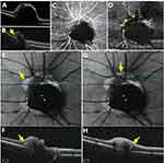
PHOMS appeared ovoid in shape on B-scan (cross-sectional) OCT image and appeared peripapillary hyperreflective bright areas on en-face Min-IP image corresponding to PHOMS on B-scan OCT image. PHOMS showed that blood flow signal of the deep capillary plexus(DCP) was decreased on OCTA. (Figure 2)
A diagnosis of optic disc melanocytoma with PHOMS was established based on its characteristic location and clinical features, prompting the patient to be advised for regular follow-up.
Discussion
Optic Disc Melanocytoma is an infrequent, benign, unilateral primary tumor of the optic disc characterized by its highly pigmented nature and distinct leafy margins.1 The occurrence of optic disc melanocytoma with PHOMS is exceptionally uncommon.
The SD-OCT characteristics of ODMC have been described as a dome-shaped lesion exhibiting hyper-reflectivity on its anterior surface and dense posterior shadowing, resembling an optical cavity.2 OCTA offers a noninvasive, safe, and efficient technique for assessing various neoplasms, including the growth and vascularity in ODMC. Moreover, OCTA has the potential to enhance the evaluation of vascular abnormalities in tumors while demonstrating that melanin’s impact on OCTA beam penetration is not statistically significant.3 Fundus autofluorescence imaging serves as a non-invasive adjunctive tool for distinguishing optic disc melanocytoma from juxtapapillary choroidal nevus and juxtapapillary uveal melanoma.4
Differential diagnosis of the peripapillary lesion encompassed optic disc melanocytoma with secondary CNV, choroidal melanoma, choroidal nevus, congenital hypertrophy of the retinal pigmented epithelium (CHRPE), and combined hamartoma of retina and retinal pigmented epithelium (RPE). Ultrasound features such as a thickness less than 2 mm and base smaller than 5 mm, high internal reflectivity, and absence of acoustic hollowness (a characteristic feature of melanoma), in conjunction with its juxtapapillary location and the patient’s age, indicated a benign pigmented lesion.5
The widespread adoption of SD-OCT and EDI-OCT (enhanced depth imaging-optical coherence tomography) has resulted in the recognition of PHOMS as a prevalent neuro-ophthalmological finding using OCT technology.6 It was postulated that PHOMS could potentially be attributed to a lateral herniation of distended retinal ganglion cell axons into the peripapillary region, firmly ensconced between the peripapillary nerve fiber layer and Bruch’s membrane, thereby giving rise to a torus or doughnut-shaped structure encircling the disc margin.7 PHOMS refers to a condition characterized by the lateral bulging herniation of distended axons into the peripapillary region above Bruch’s membrane opening. It should be noted that PHOMS is not simply synonymous with buried optic disc drusen (ODD), as previously described. The frequent coexistence of PHOMS with ODD, papilloedema, anterior ischaemic optic neuropathy, tilted optic disc syndrome, inflammatory demyelinating disorders, and other diseases associated with axoplasmic stasis offers valuable insights into its underlying pathophysiology.8
The present study examines the role of en-face Min-IP image in investigating the existing literature on the correlation between PHOMS and common neuro-ophthalmic conditions. Min-IP OCT is an innovative algorithm that identifies and presents the minimum optical intensity observed between the internal limiting membrane and retinal pigment epithelium. Initially designed to generate an en face fundus image, this software highlights fluid as dark regions. In a healthy retina, the outer nuclear layer exhibits the minimum intensity signal.9 In this study, peripapillary hyperreflective bright areas corresponding to PHOMS on B-scan OCT image were observed as evident on en-face Min-IP image.
Furthermore, despite its benign nature, it harbors the potential for malignancy. Therefore, regular annual assessments are imperative to identify any alterations in the texture, dimensions, and morphological characteristics of the ODMC.10 On B-scan ultrasonography, Gologorsky et al utilized high-resolution B-scan imaging to evaluate benign lesions for potential signs of malignant transformation, such as an increase in size, vascularity, and the development of a nodular appearance.2 The multimodal imaging and en face Min-IP image possess significant clinical diagnostic value.
Data Sharing Statement
The original contributions presented in the study are included in the article, further inquiries can be directed to the corresponding author.
Ethics Statement
Written informed consent was obtained from the individual for the publication of any potentially identifiable images or data included in this article.
This study adhered to the tenets of the Declaration of Helsinki. Institutional review board approval and informed consent from patients was obtained.
Disclosure
The author declares that the research was conducted in the absence of any commercial or financial relationships that could be construed as a potential conflict of interest.
References
1. Mughal J, Javed A, Arshad O, Nasir J, Chatni MH, Usman M. Optic disc melanocytoma; a rare entity. J Ayub Med Coll Abbottabad. 2020;32(4):580–582.
2. Gologorsky D, Schefler A, Ehlies FJ, et al. Clinical imaging and high-resolution ultrasonography in melanocytoma management. Clin Ophthalmol. 2010;4:855–859. doi:10.2147/opth.s11891
3. Zhou N, Xu X, Wei W. Optical coherence tomography angiography characteristics of optic disc melanocytoma. BMC Ophthalmol. 2020;20(1):429. doi:10.1186/s12886-020-01676-7
4. Salvanos P, Utheim TP, Moe MC, Eide N, Bragadόttir R. Autofluorescence imaging in the differential diagnosis of optic disc melanocytoma. Acta Ophthalmol. 2015;93(5):476–480. doi:10.1111/aos.12725
5. Pinheiro RL, Proença RB, Fonseca C. An atypical presentation of optic disc melanocytoma: a case report. Korean J Ophthalmol. 2023;37(2):192–194. doi:10.3341/kjo.2022.0153
6. Li B, Li H, Huang Q, Zheng Y. Peripapillary hyper-reflective ovoid mass-like structures (PHOMS): clinical significance, associations, and prognostic implications in ophthalmic conditions. Front Neurol. 2023;14:1190279. doi:10.3389/fneur.2023.1190279
7. Fraser JA, Sibony PA, Petzold A, Thaung C, Hamann S. Peripapillary hyper‐reflective ovoid mass‐like structure (PHOMS): an optical coherence tomography marker of Axoplasmic stasis in the optic nerve head. J Neuroophthalmol. 2021;41(4):431–441. doi:10.1097/WNO.0000000000001203
8. Heath Jeffery RC, Chen FK. Peripapillary hyperreflective ovoid mass‐like structures: multimodal imaging—A review. Clin Exp Ophthalmol. 2023;51(1):67–80. doi:10.1111/ceo.14182
9. Benjamin P, Nicholson MD, Divya Nigam BA, et al. Effect of ranibizumab on high-speed indocyanine green angiography and minimum intensity projection optical coherence tomography findings in neovascular age-related macular degeneration. Retina. 2015;35(1):58–68. doi:10.1097/IAE.0000000000000260
10. Badawi A, Al-Ghadeer H. Bilateral congenital ptosis associated with optic disc melanocytoma. Middle East Afr J Ophthalmol. 2021;28(1):60–62. doi:10.4103/meajo.MEAJO_543_20
 © 2024 The Author(s). This work is published and licensed by Dove Medical Press Limited. The full terms of this license are available at https://www.dovepress.com/terms.php and incorporate the Creative Commons Attribution - Non Commercial (unported, v3.0) License.
By accessing the work you hereby accept the Terms. Non-commercial uses of the work are permitted without any further permission from Dove Medical Press Limited, provided the work is properly attributed. For permission for commercial use of this work, please see paragraphs 4.2 and 5 of our Terms.


